
The vocal cords, also known as vocal folds, are folds of tissue in the throat that are key in creating sounds through vocalization. The size of vocal cords affects the pitch of voice. Open when breathing and vibrating for speech or singing, the folds are controlled via the vagus nerve.
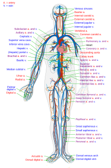
The blood vessels are the part of the circulatory system, and microcirculation, that transports blood throughout the human body. These vessels are designed to transport nutrients and oxygen to the tissues of the body. They also take waste and carbon dioxide and carry them away from the tissues and back to the heart. Blood vessels are needed to sustain life as all of the body’s tissues rely on their functionality.There are three major types of blood vessels: the arteries, which carry the blood away from the heart; the capillaries, which enable the actual exchange of water and chemicals between the blood and the tissues; and the veins, which carry blood from the capillaries back toward the heart. The word vascular, meaning relating to the blood vessels, is derived from the Latin vas, meaning vessel. Some structures -- such as cartilage, the epithelium, and the lens and cornea of the eye -- do not contain blood vessels and are labeled avascular.

A mucous membrane or mucosa is a membrane that lines various cavities in the body and covers the surface of internal organs. It consists of one or more layers of epithelial cells overlying a layer of loose connective tissue. It is mostly of endodermal origin and is continuous with the skin at various body openings such as the eyes, ears, inside the nose, inside the mouth, lip, vagina, the urethral opening and the anus. Some mucous membranes secrete mucus, a thick protective fluid. The function of the membrane is to stop pathogens and dirt from entering the body and to prevent bodily tissues from becoming dehydrated.
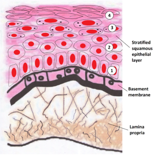
The lamina propria is a thin layer of connective tissue that forms part of the moist linings known as mucous membranes or mucosa, which line various tubes in the body, such as the respiratory tract, the gastrointestinal tract, and the urogenital tract.

A renal corpuscle is the blood-filtering component of the nephron of the kidney. It consists of a glomerulus - a tuft of capillaries composed of endothelial cells, and a glomerular capsule known as Bowman's capsule.

The nuclear lamina is a dense fibrillar network inside the nucleus of most cells. It is composed of intermediate filaments and membrane associated proteins. Besides providing mechanical support, the nuclear lamina regulates important cellular events such as DNA replication and cell division. Additionally, it participates in chromatin organization and it anchors the nuclear pore complexes embedded in the nuclear envelope.

Loose connective tissue is a category of connective tissue which includes areolar tissue, reticular tissue, and adipose tissue. Loose connective tissue is the most common type of connective tissue in vertebrates. It holds organs in place and attaches epithelial tissue to other underlying tissues. For example, it forms telae, such as the tela submucosa and tela subserosa, which connect mucous and serous membranes to the muscular layer. It also surrounds the blood vessels and nerves. Cells called fibroblasts are widely dispersed in this tissue; they are irregular branching cells that secrete strong fibrous proteins and proteoglycans as an extracellular matrix. The cells of this type of tissue are generally separated by quite some distance by a gelatinous substance primarily made up of collagenous and elastic fibers.

The basement membrane is a thin, fibrous, extracellular matrix of tissue that separates the lining of an internal or external body surface from underlying connective tissue in metazoans. This surface may be epithelium, mesothelium and endothelium
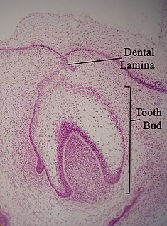
The dental lamina is a band of epithelial tissue seen in histologic sections of a developing tooth. The dental lamina is first evidence of tooth development and begins at the sixth week in utero or three weeks after the rupture of the buccopharyngeal membrane. It is formed when cells of the oral ectoderm proliferate faster than cells of other areas. Best described as an in-growth of oral ectoderm, the dental lamina is frequently distinguished from the vestibular lamina, which develops concurrently. This dividing tissue is surrounded by and, some would argue, stimulated by ectomesenchymal growth. When it is present, the dental lamina connects the developing tooth bud to the epithelium of the oral cavity. Eventually, the dental lamina disintegrates into small clusters of epithelium and is resorbed. In situations when the clusters are not resorbed, eruption cysts are formed over the developing tooth and delay its eruption into the oral cavity. This invagination of ectodermal tissues is the progenitor to the later ameloblasts and enamel while the ectomesenchyme is responsible for the dental papilla and later odontoblasts.
The oral mucosa is the mucous membrane lining the inside of the mouth and consists of stratified squamous epithelium termed oral epithelium and an underlying connective tissue termed lamina propria. The oral cavity has sometimes been described as a mirror that reflects the health of the individual. Changes indicative of disease are seen as alterations in the oral mucosa lining the mouth, which can reveal systemic conditions, such as diabetes or vitamin deficiency, or the local effects of chronic tobacco or alcohol use. The oral mucosa tends to heal faster and with less scar formation compared to the skin. The underlying mechanism remains unknown but research suggest that extracellular vesicles might be involved.
The lateral periodontal cyst is a non-inflammatory developmental cyst that arises from the epithelial post-functional dental lamina, which is a remnant from odontogenesis. It is more common in middle-aged males. Usually asymptomatic, it presents as a regular well-corticated radiolucency on the side of a mandibular canine or premolar root. Histologically, the cyst appears similar to the gingival cyst of the adult, having a non-keratinized squamous epithelial lining. The involved tooth is usually vital and has no indication for root canal treatment unless the signs of non-vital or necrotic pulpal tissue were confirmed. The cysts arise from epithelial rest cells in the periodontal ligament, although it is unknown whether from the cell rests of Malassez, reduced enamel epithelium or dental lamina remnants, and are generally treated by surgical enucleation.

The glomerular basement membrane (GBM) of the kidney is the basal lamina layer of the glomerulus. The glomerular endothelial cells, the GBM and the filtration slits between the podocytes perform the filtration function of the glomerulus, separating the blood in the capillaries from the filtrate that forms in Bowman's capsule. The GBM is a fusion of the endothelial cell and podocyte basal laminas.
The junctional epithelium (JE) is that epithelium which lies at, and in health also defines, the base of the gingival sulcus. The probing depth of the gingival sulcus is measured by a calibrated periodontal probe. In a healthy-case scenario, the probe is gently inserted, slides by the sulcular epithelium (SE), and is stopped by the epithelial attachment (EA). However, the probing depth of the gingival sulcus may be considerably different from the true histological gingival sulcus depth.
The lamina lucida is a component of the basement membrane which is found between the epithelium and underlying connective tissue. It is a roughly 40 nanometre wide electron-lucent zone between the plasma membrane of the basal cells and the (electron-dense) lamina densa of the basement membrane.
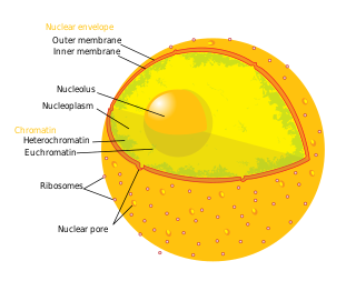
The nuclear envelope, also known as the nuclear membrane, is made up of two lipid bilayer membranes which in eukaryotic cells surrounds the nucleus, which encases the genetic material.

Medullary laminae of thalamus are layers of myelinated fibres that appear on cross sections of the thalamus. They also are commonly referred to as laminae medullares thalami or medullary layers of thalamus. The specific layers are:

The internal elastic lamina or internal elastic lamella is a layer of elastic tissue that forms the outermost part of the tunica intima of blood vessels. It separates tunica intima from tunica media.

The reticular membrane is a thin, stiff lamina that extends from the outer hair cells to the Hensen's cells. The RM is composed of "minute-fiddle-shaped cuticular structures" called the phalangeal extensions of the outer hair cells, interspaced with extensions coming from the outer phalangeal cells. The RM separates endolymph in the cochlear duct from underlying corticolymph and perilymph of the scala tympani.

The gastrointestinal wall surrounding the lumen of the gastrointestinal tract is made up of four layers of specialised tissue – from the lumen outwards:

