The fixation reflex is that concerned with attracting the eye on a peripheral object. For example, when a light shines in the periphery, the eyes shift gaze on it. It is controlled by the occipital lobe of the cerebral cortex, corroborated by three main tests:
Removal of cortex causes shutdown of this reflex
Drawing a figure on the cortex surface will cause eye movements in the direction traveled
Detecting an image by recording the actual signals from the eyes
Older research declares that a motor pathway from the occipital cortex to the brainstem motor neurons was via the superior colliculi. This is the case in lower animals, but in humans, the theory that eye-muscle nuclei aside from the superior colliculi of the midbrain is now generally held.
When an object is focused directly at an object but the eyes drift off their target, the fixation reflex keeps the eyes focused on the original object, albeit moving itself.
A saccade is a quick, simultaneous movement of both eyes between two or more phases of fixation in the same direction. In contrast, in smooth-pursuit movements, the eyes move smoothly instead of in jumps. The phenomenon can be associated with a shift in frequency of an emitted signal or a movement of a body part or device. Controlled cortically by the frontal eye fields (FEF), or subcortically by the superior colliculus, saccades serve as a mechanism for fixation, rapid eye movement, and the fast phase of optokinetic nystagmus. The word appears to have been coined in the 1880s by French ophthalmologist Émile Javal, who used a mirror on one side of a page to observe eye movement in silent reading, and found that it involves a succession of discontinuous individual movements.
The visual system is the physiological basis of visual perception. The system detects, transduces and interprets information concerning light within the visible range to construct an image and build a mental model of the surrounding environment. The visual system is associated with the eye and functionally divided into the optical system and the neural system.
The parietal lobe is one of the four major lobes of the cerebral cortex in the brain of mammals. The parietal lobe is positioned above the temporal lobe and behind the frontal lobe and central sulcus.
The occipital lobe is one of the four major lobes of the cerebral cortex in the brain of mammals. The name derives from its position at the back of the head, from the Latin ob, 'behind', and caput, 'head'.
The midbrain or mesencephalon is the rostral-most portion of the brainstem connecting the diencephalon and cerebrum with the pons. It consists of the cerebral peduncles, tegmentum, and tectum.
The vestibulo-ocular reflex (VOR) is a reflex acting to stabilize gaze during head movement, with eye movement due to activation of the vestibular system. The reflex acts to stabilize images on the retinas of the eye during head movement. Gaze is held steadily on a location by producing eye movements in the direction opposite that of head movement. For example, when the head moves to the right, the eyes move to the left, meaning the image a person sees stays the same even though the head has turned. Since slight head movement is present all the time, VOR is necessary for stabilizing vision: people with an impaired reflex find it difficult to read using print, because the eyes do not stabilise during small head tremors, and also because damage to reflex can cause nystagmus.
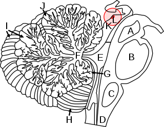
In neuroanatomy, the superior colliculus is a structure lying on the roof of the mammalian midbrain. In non-mammalian vertebrates, the homologous structure is known as the optic tectum or optic lobe. The adjective form tectal is commonly used for both structures.
The accommodation reflex is a reflex action of the eye, in response to focusing on a near object, then looking at a distant object, comprising coordinated changes in vergence, lens shape (accommodation) and pupil size. It is dependent on cranial nerve II, superior centers (interneuron) and cranial nerve III. The change in the shape of the lens is controlled by ciliary muscles inside the eye. Changes in contraction of the ciliary muscles alter the focal distance of the eye, causing nearer or farther images to come into focus on the retina; this process is known as accommodation. The reflex, controlled by the parasympathetic nervous system, involves three responses: pupil constriction, lens accommodation, and convergence.
In neuroanatomy, the pretectal area, or pretectum, is a midbrain structure composed of seven nuclei and comprises part of the subcortical visual system. Through reciprocal bilateral projections from the retina, it is involved primarily in mediating behavioral responses to acute changes in ambient light such as the pupillary light reflex, the optokinetic reflex, and temporary changes to the circadian rhythm. In addition to the pretectum's role in the visual system, the anterior pretectal nucleus has been found to mediate somatosensory and nociceptive information.
Eye movement includes the voluntary or involuntary movement of the eyes. Eye movements are used by a number of organisms to fixate, inspect and track visual objects of interests. A special type of eye movement, rapid eye movement, occurs during REM sleep.
The extraocular muscles, or extrinsic ocular muscles, are the seven extrinsic muscles of the human eye. Six of the extraocular muscles, the four recti muscles, and the superior and inferior oblique muscles, control movement of the eye and the other muscle, the levator palpebrae superioris, controls eyelid elevation. The actions of the six muscles responsible for eye movement depend on the position of the eye at the time of muscle contraction.
Oscillopsia is a visual disturbance in which objects in the visual field appear to oscillate. The severity of the effect may range from a mild blurring to rapid and periodic jumping. Oscillopsia is an incapacitating condition experienced by many patients with neurological disorders. It may be the result of ocular instability occurring after the oculomotor system is affected, no longer holding images steady on the retina. A change in the magnitude of the vestibulo-ocular reflex due to vestibular disease can also lead to oscillopsia during rapid head movements. Oscillopsia may also be caused by involuntary eye movements such as nystagmus, or impaired coordination in the visual cortex and is one of the symptoms of superior canal dehiscence syndrome. Those affected may experience dizziness and nausea. Oscillopsia can also be used as a quantitative test to document aminoglycoside toxicity. Permanent oscillopsia can arise from an impairment of the ocular system that serves to maintain ocular stability. Paroxysmal oscillopsia can be due to an abnormal hyperactivity in the peripheral ocular or vestibular system.
Cortical blindness is the total or partial loss of vision in a normal-appearing eye caused by damage to the brain's occipital cortex. Cortical blindness can be acquired or congenital, and may also be transient in certain instances. Acquired cortical blindness is most often caused by loss of blood flow to the occipital cortex from either unilateral or bilateral posterior cerebral artery blockage and by cardiac surgery. In most cases, the complete loss of vision is not permanent and the patient may recover some of their vision. Congenital cortical blindness is most often caused by perinatal ischemic stroke, encephalitis, and meningitis. Rarely, a patient with acquired cortical blindness may have little or no insight that they have lost vision, a phenomenon known as Anton–Babinski syndrome.
The corneal reflex, also known as the blink reflex or eyelid reflex, is an involuntary blinking of the eyelids elicited by stimulation of the cornea, though it could result from any peripheral stimulus. Stimulation should elicit both a direct and consensual response. The reflex occurs at a rapid rate of 0.1 seconds. The purpose of this reflex is to protect the eyes from foreign bodies and bright lights. The blink reflex also occurs when sounds greater than 40–60 dB are made.
In the scientific study of vision, smooth pursuit describes a type of eye movement in which the eyes remain fixated on a moving object. It is one of two ways that visual animals can voluntarily shift gaze, the other being saccadic eye movements. Pursuit differs from the vestibulo-ocular reflex, which only occurs during movements of the head and serves to stabilize gaze on a stationary object. Most people are unable to initiate pursuit without a moving visual signal. The pursuit of targets moving with velocities of greater than 30°/s tends to require catch-up saccades. Smooth pursuit is asymmetric: most humans and primates tend to be better at horizontal than vertical smooth pursuit, as defined by their ability to pursue smoothly without making catch-up saccades. Most humans are also better at downward than upward pursuit. Pursuit is modified by ongoing visual feedback.
Attentional shift occurs when directing attention to a point increases the efficiency of processing of that point and includes inhibition to decrease attentional resources to unwanted or irrelevant inputs. Shifting of attention is needed to allocate attentional resources to more efficiently process information from a stimulus. Research has shown that when an object or area is attended, processing operates more efficiently. Task switching costs occur when performance on a task suffers due to the increased effort added in shifting attention. There are competing theories that attempt to explain why and how attention is shifted as well as how attention is moved through space.
Eye–hand coordination is the coordinated motor control of eye movement with hand movement and the processing of visual input to guide reaching and grasping along with the use of proprioception of the hands to guide the eyes, a modality of multisensory integration. Eye–hand coordination has been studied in activities as diverse as the movement of solid objects such as wooden blocks, archery, sporting performance, music reading, computer gaming, copy-typing, and even tea-making. It is part of the mechanisms of performing everyday tasks; in its absence, most people would not be able to carry out even the simplest of actions such as picking up a book from a table.
The menace response is one of three forms of blink reflex. It includes the reflexive blinking that occurs specifically in response to the rapid approach of an object. The menace response comprises blinking of the eyelids, in order to protect the eyes from potential damage, but may also include turning of the head, neck, or even the trunk away from the optical stimulus that triggers the reflex.
Transsaccadic memory is the neural process that allows humans to perceive their surroundings as a seamless, unified image despite rapid changes in fixation points. Transsaccadic memory is a relatively new topic of interest in the field of psychology. Conflicting views and theories have spurred several types of experiments intended to explain transsaccadic memory and the neural mechanisms involved.
Cerebral diplopia or polyopia describes seeing two or more images arranged in ordered rows, columns, or diagonals after fixation on a stimulus. The polyopic images occur monocular bilaterally and binocularly, differentiating it from ocular diplopia or polyopia. The number of duplicated images can range from one to hundreds. Some patients report difficulty in distinguishing the replicated images from the real images, while others report that the false images differ in size, intensity, or color. Cerebral polyopia is sometimes confused with palinopsia, in which multiple images appear while watching an object. However, in cerebral polyopia, the duplicated images are of a stationary object which are perceived even after the object is removed from the visual field. Movement of the original object causes all of the duplicated images to move, or the polyopic images disappear during motion. In palinoptic polyopia, movement causes each polyopic image to leave an image in its wake, creating hundreds of persistent images (entomopia).
This page is based on this Wikipedia article Text is available under the CC BY-SA 4.0 license; additional terms may apply. Images, videos and audio are available under their respective licenses.