
Förster resonance energy transfer (FRET), fluorescence resonance energy transfer, resonance energy transfer (RET) or electronic energy transfer (EET) is a mechanism describing energy transfer between two light-sensitive molecules (chromophores). A donor chromophore, initially in its electronic excited state, may transfer energy to an acceptor chromophore through nonradiative dipole–dipole coupling. The efficiency of this energy transfer is inversely proportional to the sixth power of the distance between donor and acceptor, making FRET extremely sensitive to small changes in distance.
Fluorescence-lifetime imaging microscopy or FLIM is an imaging technique based on the differences in the exponential decay rate of the photon emission of a fluorophore from a sample. It can be used as an imaging technique in confocal microscopy, two-photon excitation microscopy, and multiphoton tomography.
In particle physics, the quantum yield of a radiation-induced process is the number of times a specific event occurs per photon absorbed by the system.

Allophycocyanin is a protein from the light-harvesting phycobiliprotein family, along with phycocyanin, phycoerythrin and phycoerythrocyanin. It is an accessory pigment to chlorophyll. All phycobiliproteins are water-soluble and therefore cannot exist within the membrane like carotenoids, but aggregate, forming clusters that adhere to the membrane called phycobilisomes. Allophycocyanin absorbs and emits red light, and is readily found in Cyanobacteria, and red algae. Phycobilin pigments have fluorescent properties that are used in immunoassay kits. In flow cytometry, it is often abbreviated APC. To be effectively used in applications such as FACS, High-Throughput Screening (HTS) and microscopy, APC needs to be chemically cross-linked.

In chemistry, quenching refers to any process which decreases the fluorescent intensity of a given substance. A variety of processes can result in quenching, such as excited state reactions, energy transfer, complex-formation and collisions. As a consequence, quenching is often heavily dependent on pressure and temperature. Molecular oxygen, iodine ions and acrylamide are common chemical quenchers. The chloride ion is a well known quencher for quinine fluorescence. Quenching poses a problem for non-instant spectroscopic methods, such as laser-induced fluorescence.
Fluorescence anisotropy or fluorescence polarization is the phenomenon where the light emitted by a fluorophore has unequal intensities along different axes of polarization. Early pioneers in the field include Aleksander Jablonski, Gregorio Weber, and Andreas Albrecht. The principles of fluorescence polarization and some applications of the method are presented in Lakowicz's book.
Förster coupling is the resonant energy transfer between excitons within adjacent QD's. The first studies of Forster were performed in the context of the sensitized luminescence of solids. Here, an excited sensitizer atom can transfer its excitation to a neighbouring acceptor atom, via an intermediate virtual photon. This same mechanism has also been shown to be responsible for exciton transfer between QD's and within molecular systems and biosystems, all of which may be treated in a similar formulation.

Fluorescence is used in the life sciences generally as a non-destructive way of tracking or analysing biological molecules. Some proteins or small molecules in cells are naturally fluorescent, which is called intrinsic fluorescence or autofluorescence. Alternatively, specific or general proteins, nucleic acids, lipids or small molecules can be "labelled" with an extrinsic fluorophore, a fluorescent dye which can be a small molecule, protein or quantum dot. Several techniques exist to exploit additional properties of fluorophores, such as fluorescence resonance energy transfer, where the energy is passed non-radiatively to a particular neighbouring dye, allowing proximity or protein activation to be detected; another is the change in properties, such as intensity, of certain dyes depending on their environment allowing their use in structural studies.
Single-molecule fluorescence resonance energy transfer is a biophysical technique used to measure distances at the 1-10 nanometer scale in single molecules, typically biomolecules. It is an application of FRET wherein a pair of donor and acceptor fluorophores are excited and detected at a single molecule level. In contrast to "ensemble FRET" which provides the FRET signal of a high number of molecules, single-molecule FRET is able to resolve the FRET signal of each individual molecule. The variation of the smFRET signal is useful to reveal kinetic information that an ensemble measurement cannot provide, especially when the system is under equilibrium with no ensemble/bulk signal change. Heterogeneity among different molecules can also be observed. This method has been applied in many measurements of intramolecular dynamics such as DNA/RNA/protein folding/unfolding and other conformational changes, and intermolecular dynamics such as reaction, binding, adsorption, and desorption that are particularly useful in chemical sensing, bioassays, and biosensing.
Douglas Charles Youvan is an American scientist.
Richard Paul Haugland was an American scientist noted for his work in researching and commercializing fluorescent dyes. He completed his PhD at Stanford in 1970 under Lubert Stryer, showing in a now widely cited and classic paper that Förster resonance energy transfer (FRET) can be used as a "spectroscopic ruler" to measure distances in macromolecules. Haugland founded Molecular Probes in 1975 and continued as its president after the corporation was bought by Invitrogen in 2003. He is the original author of the authoritative volume on molecular probes, The Molecular Probes Handbook, now in its 11th edition.
Fluorescent glucose biosensors are devices that measure the concentration of glucose in diabetic patients by means of sensitive protein that relays the concentration by means of fluorescence, an alternative to amperometric sension of glucose. Due to the prevalence of diabetes, it is the prime drive in the construction of fluorescent biosensors. A recent development has been approved by the FDA allowing a new continuous glucose monitoring system called EverSense, which is a 90-day glucose monitor using fluorescent biosensors.
There are many methods to investigate protein–protein interactions which are the physical contacts of high specificity established between two or more protein molecules involving electrostatic forces and hydrophobic effects. Each of the approaches has its own strengths and weaknesses, especially with regard to the sensitivity and specificity of the method. A high sensitivity means that many of the interactions that occur are detected by the screen. A high specificity indicates that most of the interactions detected by the screen are occurring in reality.
Time-resolved fluorescence energy transfer (TR-FRET) is the practical combination of time-resolved fluorometry (TRF) with Förster resonance energy transfer (FRET) that offers a powerful tool for drug discovery researchers. TR-FRET combines the low background aspect of TRF with the homogeneous assay format of FRET. The resulting assay provides an increase in flexibility, reliability and sensitivity in addition to higher throughput and fewer false positive/false negative results. FRET involves two fluorophores, a donor and an acceptor. Excitation of the donor by an energy source produces an energy transfer to the acceptor if the two are within a given proximity to each other. The acceptor in turn emits light at its characteristic wavelength.
A ligand binding assay (LBA) is an assay, or an analytic procedure, which relies on the binding of ligand molecules to receptors, antibodies or other macromolecules. A detection method is used to determine the presence and extent of the ligand-receptor complexes formed, and this is usually determined electrochemically or through a fluorescence detection method. This type of analytic test can be used to test for the presence of target molecules in a sample that are known to bind to the receptor.
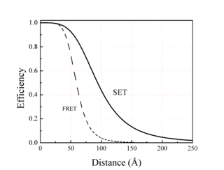
Surface energy transfer (SET) is a dipole-surface energy transfer process involving a metallic surface and a molecular dipole.
Lanthanide probes are a non-invasive analytical tool commonly used for biological and chemical applications. Lanthanides are metal ions which have their 4f energy level filled and generally refer to elements cerium to lutetium in the periodic table. The fluorescence of lanthanide salts is weak because the energy absorption of the metallic ion is low; hence chelated complexes of lanthanides are most commonly used. The term chelate derives from the Greek word for “claw,” and is applied to name ligands, which attach to a metal ion with two or more donor atoms through dative bonds. The fluorescence is most intense when the metal ion has the oxidation state of 3+. Not all lanthanide metals can be used and the most common are: Sm(III), Eu(III), Tb(III), and Dy(III).

Small molecule sensors are an effective way to detect the presence of metal ions in solution. Although many types exist, most small molecule sensors comprise a subunit that selectively binds to a metal that in turn induces a change in a fluorescent subunit. This change can be observed in the small molecule sensor's spectrum, which can be monitored using a detection system such as a microscope or a photodiode. Different probes exist for a variety of applications, each with different dissociation constants with respect to a particular metal, different fluorescent properties, and sensitivities. They show great promise as a way to probe biological processes by monitoring metal ions at low concentrations in biological systems. Since they are by definition small and often capable of entering biological systems, they are conducive to many applications for which other more traditional bio-sensing are less effective or not suitable.
Ammasi Periasamy is an Indian American biophysicist who is a professor at the University of Virginia. He works on light microscopy, including the molecular imaging of living cells. He has developed a range of imaging systems, including confocal, multi-photon and fluorescence-lifetime imaging microscopy base devices.