Related Research Articles

The human teeth function to mechanically break down items of food by cutting and crushing them in preparation for swallowing and digesting. Humans have four types of teeth: incisors, canines, premolars, and molars, which each have a specific function. The incisors cut the food, the canines tear the food and the molars and premolars crush the food. The roots of teeth are embedded in the maxilla or the mandible and are covered by gums. Teeth are made of multiple tissues of varying density and hardness.
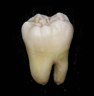
The molars or molar teeth are large, flat teeth at the back of the mouth. They are more developed in mammals. They are used primarily to grind food during chewing. The name molar derives from Latin, molaris dens, meaning "millstone tooth", from mola, millstone and dens, tooth. Molars show a great deal of diversity in size and shape across mammal groups. The third molar of humans is a vestigial organ, as it has lost its original function.

Tooth enamel is one of the four major tissues that make up the tooth in humans and many other animals, including some species of fish. It makes up the normally visible part of the tooth, covering the crown. The other major tissues are dentin, cementum, and dental pulp. It is a very hard, white to off-white, highly mineralised substance that acts as a barrier to protect the tooth but can become susceptible to degradation, especially by acids from food and drink. Calcium hardens the tooth enamel. In rare circumstances enamel fails to form, leaving the underlying dentin exposed on the surface.

Dentin or dentine is a calcified tissue of the body and, along with enamel, cementum, and pulp, is one of the four major components of teeth. It is usually covered by enamel on the crown and cementum on the root and surrounds the entire pulp. By volume, 45% of dentin consists of the mineral hydroxyapatite, 33% is organic material, and 22% is water. Yellow in appearance, it greatly affects the color of a tooth due to the translucency of enamel. Dentin, which is less mineralized and less brittle than enamel, is necessary for the support of enamel. Dentin rates approximately 3 on the Mohs scale of mineral hardness. There are two main characteristics which distinguish dentin from enamel: firstly, dentin forms throughout life; secondly, dentin is sensitive and can become hypersensitive to changes in temperature due to the sensory function of odontoblasts, especially when enamel recedes and dentin channels become exposed.
Cosmetic dentistry is generally used to refer to any dental work that improves the appearance of teeth, gums and/or bite. It primarily focuses on improvement in dental aesthetics in color, position, shape, size, alignment and overall smile appearance. Many dentists refer to themselves as "cosmetic dentists" regardless of their specific education, specialty, training, and experience in this field. This has been considered unethical with a predominant objective of marketing to patients. The American Dental Association does not recognize cosmetic dentistry as a formal specialty area of dentistry. However, there are still dentists that promote themselves as cosmetic dentists.
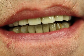
A crown, or dental cap, is a type of dental restoration which completely caps or encircles a tooth or dental implant. A crown may be needed when a large cavity threatens the health of a tooth. They are typically bonded to the tooth by dental cement. Crowns can be made from many materials, which are usually fabricated using indirect methods. Crowns are used to improve the strength or appearance of teeth and to halt deterioration. While beneficial to dental health, the procedure and materials can be costly.
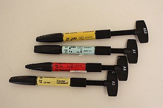
Dental composite resins are dental cements made of synthetic resins. Synthetic resins evolved as restorative materials since they were insoluble, of good tooth-like appearance, insensitive to dehydration, easy to manipulate and reasonably inexpensive. Composite resins are most commonly composed of Bis-GMA and other dimethacrylate monomers, a filler material such as silica and in most current applications, a photoinitiator. Dimethylglyoxime is also commonly added to achieve certain physical properties such as flow-ability. Further tailoring of physical properties is achieved by formulating unique concentrations of each constituent.
An enamel rod is the basic unit of tooth enamel. Measuring 4 μm wide to 8 μm high, an enamel rod is a tightly packed, highly organized mass of hydroxyapatite crystals, which are hexagonal in shape and provide rigidity to the rods and strengthen the enamel. In cross section, it is best compared to a keyhole with the top, or head, oriented toward the crown of the tooth and the bottom, or tail, oriented toward the root of the tooth.
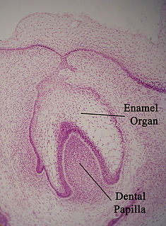
The enamel organ, also known as the dental organ, is a cellular aggregation seen in a developing tooth and it lies above the dental papilla. The enamel organ is responsible for the formation of enamel, initiation of dentine formation, establishment of the shape of a tooth's crown, and establishment of the dentoenamel junction.

Tooth development or odontogenesis is the complex process by which teeth form from embryonic cells, grow, and erupt into the mouth. For human teeth to have a healthy oral environment, all parts of the tooth must develop during appropriate stages of fetal development. Primary (baby) teeth start to form between the sixth and eighth week of prenatal development, and permanent teeth begin to form in the twentieth week. If teeth do not start to develop at or near these times, they will not develop at all, resulting in hypodontia or anodontia.
Rod sheath is an area identified in histologic sections of a tooth. It is found where enamel rods, the functional unit of enamel, meet interrod enamel. The crystals of both types of enamel meet at sharp angles and form the appearance of a space called the rod sheath. As a result of this space, the rod sheath consists of more protein than other areas of enamel. For this reason, the rod sheath is characterized as being hypomineralized in comparison to the rest of the highly mineralized enamel.

In dentistry, a veneer is a layer of material placed over a tooth. Veneers can improve the aesthetics of a smile and protect the tooth's surface from damage.

Dentinogenesis imperfecta (DI) is a genetic disorder of tooth development. This condition is a type of dentin dysplasia that causes teeth to be discolored and translucent giving teeth an opalescent sheen. Although genetic factors are the main contributor for the disease, any environmental or systemic upset that impedes calcification or metabolisation of calcium can also result in anomalous dentine.
Dens invaginatus (DI), also known as tooth within a tooth, is a rare dental malformation found in teeth where there is an infolding of enamel into dentine. The prevalence of condition is 0.3 - 10%, affecting more males than females. The condition is presented in two forms, coronal and radicular, with the coronal form being more common.
Dens evaginatus is a rare odontogenic developmental anomaly that is found in teeth where the outer surface appears to form an extra bump or cusp.

Talon cusp is a rare dental anomaly resulting in an extra cusp or cusp-like projection on an anterior tooth, located on the inside surface of the affected tooth.

Dental attrition is a type of tooth wear caused by tooth-to-tooth contact, resulting in loss of tooth tissue, usually starting at the incisal or occlusal surfaces. Tooth wear is a physiological process and is commonly seen as a normal part of aging. Advanced and excessive wear and tooth surface loss can be defined as pathological in nature, requiring intervention by a dental practitioner. The pathological wear of the tooth surface can be caused by bruxism, which is clenching and grinding of the teeth. If the attrition is severe, the enamel can be completely worn away leaving underlying dentin exposed, resulting in an increased risk of dental caries and dentin hypersensitivity. It is best to identify pathological attrition at an early stage to prevent unnecessary loss of tooth structure as enamel does not regenerate.
Dental anatomy is a field of anatomy dedicated to the study of human tooth structures. The development, appearance, and classification of teeth fall within its purview. Tooth formation begins before birth, and the teeth's eventual morphology is dictated during this time. Dental anatomy is also a taxonomical science: it is concerned with the naming of teeth and the structures of which they are made, this information serving a practical purpose in dental treatment.

Enamel tufts are hypomineralized ribbon-like structures that run longitudinally to the tooth axis and extend from the dentinoenamel junction (DEJ) one fifth to a third into the enamel. They are called ‘‘tufts’’ due to their wavy look within the enamel microstructure.
Hard tissue is tissue which is mineralized and has a firm intercellular matrix. The hard tissues of humans are bone, tooth enamel, dentin, and cementum. The term is in contrast to soft tissue.
References
- Cate, A.R. Ten. Oral Histology: development, structure, and function. 5th ed. 1998. ISBN 0-8151-2952-1.
| | This dentistry article is a stub. You can help Wikipedia by expanding it. |