In neurophysiology, long-term depression (LTD) is an activity-dependent reduction in the efficacy of neuronal synapses lasting hours or longer following a long patterned stimulus. LTD occurs in many areas of the CNS with varying mechanisms depending upon brain region and developmental progress.
Eyeblink conditioning (EBC) is a form of classical conditioning that has been used extensively to study neural structures and mechanisms that underlie learning and memory. The procedure is relatively simple and usually consists of pairing an auditory or visual stimulus with an eyeblink-eliciting unconditioned stimulus (US). Naïve organisms initially produce a reflexive, unconditioned response (UR) that follows US onset. After many CS-US pairings, an association is formed such that a learned blink, or conditioned response (CR), occurs and precedes US onset. The magnitude of learning is generally gauged by the percentage of all paired CS-US trials that result in a CR. Under optimal conditions, well-trained animals produce a high percentage of CRs. The conditions necessary for, and the physiological mechanisms that govern, eyeblink CR learning have been studied across many mammalian species, including mice, rats, guinea pigs, rabbits, ferrets, cats, and humans. Historically, rabbits have been the most popular research subjects.

The inferior olivary nucleus (ION), is a structure found in the medulla oblongata underneath the superior olivary nucleus. In vertebrates, the ION is known to coordinate signals from the spinal cord to the cerebellum to regulate motor coordination and learning. These connections have been shown to be tightly associated, as degeneration of either the cerebellum or the ION results in degeneration of the other.

Basket cells are inhibitory GABAergic interneurons of the brain, found throughout different regions of the cortex and cerebellum.
Climbing fibers are the name given to a series of neuronal projections from the inferior olivary nucleus located in the medulla oblongata.

Purkinje cells, or Purkinje neurons, are a class of GABAergic neurons located in the cerebellum. They are named after their discoverer, Czech anatomist Jan Evangelista Purkyně, who characterized the cells in 1839.

The cerebellum has four deep cerebellar nuclei embedded in the white matter in its center.
In neuroscience, Golgi cells are inhibitory interneurons found within the granular layer of the cerebellum. They were first identified as inhibitory by Eccles et al. in 1964. It was also the first example of an inhibitory feed back network, where the inhibitory interneuron was identified anatomically. These cells synapse onto the dendrite of granule cells and unipolar brush cells. They receive excitatory input from mossy fibres, also synapsing on granule cells, and parallel fibers, which are long granule cell axons. Thereby this circuitry allows for feed-forward and feed-back inhibition of granule cells.

The fastigial nucleus is located in the cerebellum. It is one of the four deep cerebellar nuclei, and is grey matter embedded in the white matter of the cerebellum.
Mossy fibers are one of the major inputs to cerebellum. There are many sources of this pathway, the largest of which is the cerebral cortex, which sends input to the cerebellum via the pontocerebellar pathway. Other contributors include the vestibular nerve and nuclei, the spinal cord, the reticular formation, and feedback from deep cerebellar nuclei. Axons enter the cerebellum via the middle and inferior cerebellar peduncles, where some branch to make contact with deep cerebellar nuclei. They ascend into the white matter of the cerebellum, where each axon branches to innervate granule cells in several cerebellar folia.

The anatomy of the cerebellum can be viewed at three levels. At the level of gross anatomy, the cerebellum consists of a tightly folded and crumpled layer of cortex, with white matter underneath, several deep nuclei embedded in the white matter, and a fluid-filled ventricle in the middle. At the intermediate level, the cerebellum and its auxiliary structures can be broken down into several hundred or thousand independently functioning modules or "microzones". At the microscopic level, each module consists of the same small set of neuronal elements, laid out with a highly stereotyped geometry.
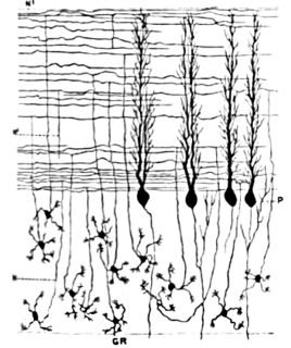
Cerebellar granule cells form the thick granular layer of the cerebellar cortex and are among the smallest neurons in the brain. Cerebellar granule cells are also the most numerous neurons in the brain: in humans, estimates of their total number average around 50 billion, which means that they constitute about 3/4 of the brain's neurons.
The name granule cell has been used for a number of different types of neuron whose only common feature is that they all have very small cell bodies. Granule cells are found within the granular layer of the cerebellum, the dentate gyrus of the hippocampus, the superficial layer of the dorsal cochlear nucleus, the olfactory bulb, and the cerebral cortex.

The rhombic lip is a posterior section of the developing metencephalon which can be recognized transiently within the vertebrate embryo. It extends posteriorly from the roof of the fourth ventricle to dorsal neuroepithelial cells. The rhombic lip can be divided into eight structural units based on rhombomeres 1-8 (r1-r8), which can be recognized at early stages of hindbrain development. Producing granule cells and five brainstem nuclei, the rhombic lip plays an important role in developing a complex cerebellar neural system.
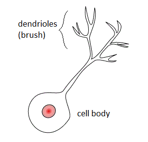
Unipolar brush cells (UBCs) are a class of excitatory glutamatergic interneuron found in the granular layer of the cerebellar cortex and also in the granule cell domain of the cochlear nucleus.
Cartwheel cells are neurons of the dorsal cochlear nucleus (DCN) where they greatly outnumber the other inhibitory interneurons of the DCN. Their somas lie on the superficial side of the pyramidal layer of the DCN, and their dendrites receive input from the parallel fibres of the granule cell layer. Their axons do not extend beyond the dorsal cochlear nucleus but synapse with other cartwheel cells and pyramidal cells within the DCN releasing GABA and glycine onto their targets.
The granular layer is the innermost layer of the cerebellar cortex. The cerebellar cortex is composed of three layers: molecular layer, purkinje cells and granular layer, respectively from outermost to innermost. The cerebellum is mainly responsible for managing the coordination of skeletal muscles and body movements.

The cerebellar glomerulus is a small, intertwined mass of nerve fiber terminals in the granular layer of the cerebellar cortex. It consists of post-synaptic granule cell dendrites and pre-synaptic Golgi cell axon terminals surrounding the pre-synaptic terminals of mossy fibers.


