Hallmarks of Cancer (Activating Invasion and Metastasis)

Last updated

Invasion and metastasis are fundamental hallmarks of cancer, representing the ability of the cancer cells to spread from their site of origin to distant tissues and organs. [1] These processes are central to cancer's lethality, accounting for the majority of cancer - related deaths, and marking an important barrier to effective treatment.

Contents

These processes enable cancers to establish secondary tumors, contributing to the majority of cancer-related deaths. Understanding and targeting the mechanisms of invasion and metastasis remain pivotal in cancer therapy and research.

Mechanisms of Invasion

Epithelial-Mesenchymal Transition (EMT)

Epithelial-mesenchymal transition (EMT) is a process where epithelial cells lose their adhesive properties and gain a more mobile, invasive phenotype. [2] This change is regulated by signaling pathways like TGF-B, Wnt, and Notch as well as transcription factors such TWIST, SNAIL, AND ZEB. [3] EMT allows cancer cells to invade surrounding tissues and spread more effectively.

Degradation of the Extracellular Matrix (ECM)

To invade nearby tissues, cancer cells cells secrete enzymes such as matrix matalloproteinases (MMPs) that degrade the ECM and basement membrane. [4] This breakdown creates pathways for cancel cells to migrate and infiltrate new areas.

Tumor Microenvironment

The tumor microenvironment, composed of stromal cells, immune cells and singaling molecules, supports invasion by creating good and favorable conditions for tumor cell migration. [5] For example, cancer-associated fibroblasts (CAFs) produce substances that remodel the ECM and promote cancer progression [5]

Local Tissue Invasion

Epithelial-to-mesenchymal transition (EMT) is a biological process in which epithelial cells lose their polarity and cell-cell adhesion properties and acquire mesenchymal traits, such as enhanced motility and invasiveness. This transformation plays a critical for various physiological processes, such as embryonic development, wound healing, and tissue regeneration. However, in cancer, EMT is often hijacked to promote tumor progression and metastasis. During EMT, epithelial markers like E-cadherin are downregulated, while mesenchymal markers such as N-cadherin and vimentin are upregulated. This change enables cancer cells to detach from the primary tumor, invade surrounding tissues, and ultimately spread to distant locations in the body by entering the bloodstream or lymphatic pathways. [6]

Cancer cells undergo several changes that enable them to invade surrounding tissues. A primary mechanism is the downregulation of E-cadherin which is an epithelial adhesion molecule that helps maintain cell-cell adhesion. Loss of E-cadherin reduces cellular cohesion, allowing cancer cells to detach from the primary tumor. This is a hallmark of EMT, during which cancer cells acquire mesenchymal traits, such as increased motility and invasiveness. [7] [8]

Mechanisms of Metastasis

Intravastation

Intravasation is the process where tumor cells enter blood or lymphatic blood vessels, allowing them to travel to distant parts of the body. This step is important in the metastatic journey as it enables tumor cells to leave their original site and circulate through the body. pro-angiogenic factors like VEGF, [9] along with interactions between cancer calls and the vessel walls, make it easier for tumor cells to penetrate into the bloodstream or lymphatic system. By gaining access to these transport networks, cancer cells increase their ability to spread and form new tumors in distant tissues.

Circulating Tumor cells (CTCs)

When cancer cells enter the bloodstream, they are known as circulating tumor cells (CTCs). To protect themselves from being detected and destroyed by the immune system, these cells often group together in clusters or cover themselves with platelets. This protective strategy increases their chances of survival and makes it easier for them to spread to other parts of the body. [10]

Extravasation and Colonization

Extravasation occurs when circulating tumor cells leave the bloodstream and invade new tissues, guided by molecules like integrins. [11] Integrins help the cells attach and move into their new environment. Once settled, cancer cells form a metastatic niche that helps them grow and establish a new tumor in a new location.

Roles of Biomarkers In Tumor Metastasis

E-cadherin

E-cadherin is an epithelial adhesion protein that plays an essential role in maintaining tissue structure by facilitating cell-cell adhesion. Its downregulation is a major feature of EMT transition which is a process that is important for cancer metastasis. The loss of E-cadherin disrupts cellular adhesion, allowing tumor cells to detach from the primary site and invade surrounding tissues. This type of suppression is often mediated by EMT transcription factors such as ZEB1, Snail, and Twist, is then repress E-cadherin gene expression. Furthermore, when E-cadherin is reduced, it facilitates interactions with the extracellular matrix (ECM) which in then enhances the ability of cancer cells to migrate and invade surrounding tissues. By promoting these interactions, E-cadherin is able to support cellular motility and aid tumor cells navigate the tissue structures which drives metastasis. [12] [13] Research emphasizes E-cadherin as a major biomarker in metastatic cancers such as breast and colorectal cancers. Low levels of E-cadherin are often linked to poor clinical outcomes, therapy resistance and aggressive tumor phenotypes. [14]

Related Research Articles

Morphogenesis is the biological process that causes a cell, tissue or organism to develop its shape. It is one of three fundamental aspects of developmental biology along with the control of tissue growth and patterning of cellular differentiation.

<span class="mw-page-title-main">Metastasis</span> Spreading of a disease inside a body

Metastasis is a pathogenic agent's spreading from an initial or primary site to a different or secondary site within the host's body; the term is typically used when referring to metastasis by a cancerous tumor. The newly pathological sites, then, are metastases (mets). It is generally distinguished from cancer invasion, which is the direct extension and penetration by cancer cells into neighboring tissues.

<span class="mw-page-title-main">Catenin</span> Type of protein

Catenins are a family of proteins found in complexes with cadherin cell adhesion molecules of animal cells. The first two catenins that were identified became known as α-catenin and β-catenin. α-Catenin can bind to β-catenin and can also bind filamentous actin (F-actin). β-Catenin binds directly to the cytoplasmic tail of classical cadherins. Additional catenins such as γ-catenin and δ-catenin have been identified. The name "catenin" was originally selected because it was suspected that catenins might link cadherins to the cytoskeleton.

The epithelial–mesenchymal transition (EMT) is a process by which epithelial cells lose their cell polarity and cell–cell adhesion, and gain migratory and invasive properties to become mesenchymal stem cells; these are multipotent stromal cells that can differentiate into a variety of cell types. EMT is essential for numerous developmental processes including mesoderm formation and neural tube formation. EMT has also been shown to occur in wound healing, in organ fibrosis and in the initiation of metastasis in cancer progression.

Intravasation is the invasion of cancer cells through the basement membrane into a blood or lymphatic vessel. Intravasation is one of several carcinogenic events that initiate the escape of cancerous cells from their primary sites. Other mechanisms include invasion through basement membranes, extravasation, and colonization of distant metastatic sites. Cancer cell chemotaxis also relies on this migratory behavior to arrive at a secondary destination designated for cancer cell colonization.

<span class="mw-page-title-main">Mesenchyme</span> Type of animal embryonic connective tissue

Mesenchyme is a type of loosely organized animal embryonic connective tissue of undifferentiated cells that give rise to most tissues, such as skin, blood or bone. The interactions between mesenchyme and epithelium help to form nearly every organ in the developing embryo.

<span class="mw-page-title-main">MMP2</span> Protein-coding gene in humans

72 kDa type IV collagenase also known as matrix metalloproteinase-2 (MMP-2) and gelatinase A is an enzyme that in humans is encoded by the MMP2 gene. The MMP2 gene is located on chromosome 16 at position 12.2.

<span class="mw-page-title-main">Basal-like carcinoma</span> Breast cancer subtype

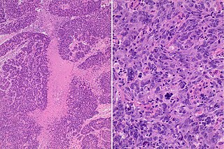
The basal-like carcinoma is a recently proposed subtype of breast cancer defined by its gene expression and protein expression profile.

<span class="mw-page-title-main">Epithelial cell adhesion molecule</span> Transmembrane glycoprotein

Epithelial cell adhesion molecule (EpCAM), also known as CD326 among other names, is a transmembrane glycoprotein mediating Ca2+-independent homotypic cell–cell adhesion in epithelia. EpCAM is also involved in cell signaling, migration, proliferation, and differentiation. Additionally, EpCAM has oncogenic potential via its capacity to upregulate c-myc, e-fabp, and cyclins A & E. Since EpCAM is expressed exclusively in epithelia and epithelial-derived neoplasms, EpCAM can be used as diagnostic marker for various cancers. It appears to play a role in tumorigenesis and metastasis of carcinomas, so it can also act as a potential prognostic marker and as a potential target for immunotherapeutic strategies.

<span class="mw-page-title-main">SNAI1</span> Protein

Zinc finger protein SNAI1 is a protein that in humans is encoded by the SNAI1 gene. Snail is a family of transcription factors that promote the repression of the adhesion molecule E-cadherin to regulate epithelial to mesenchymal transition (EMT) during embryonic development.

<span class="mw-page-title-main">SCRIB</span> Protein-coding gene in the species Homo sapiens

SCRIB, also known as Scribble, SCRIBL, or Scribbled homolog (Drosophila), is a scaffold protein which in humans is encoded by the SCRIB gene. It was originally isolated in Drosophila melanogaster in a pathway (also known as the Scribble complex) with DLGAP5 (Discs large) and LLGL1 (Lethal giant larvae) as a tumor suppressor. In humans, SCRIB is found as a membrane protein and is involved in cell migration, cell polarity, and cell proliferation in epithelial cells. There is also strong evidence that SCRIB may play a role in cancer progression because of its strong homology to the Drosophila protein.

<span class="mw-page-title-main">Cadherin-1</span> Human protein-coding gene

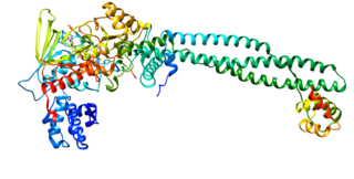
Cadherin-1 or Epithelial cadherin(E-cadherin), is a protein that in humans is encoded by the CDH1 gene. Mutations are correlated with gastric, breast, colorectal, thyroid, and ovarian cancers. CDH1 has also been designated as CD324. It is a tumor suppressor gene.

<span class="mw-page-title-main">Metastatic breast cancer</span> Type of cancer

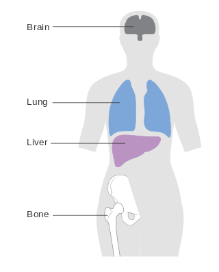
Metastatic breast cancer, also referred to as metastases, advanced breast cancer, secondary tumors, secondaries or stage IV breast cancer, is a stage of breast cancer where the breast cancer cells have spread to distant sites beyond the axillary lymph nodes. There is no cure for metastatic breast cancer; there is no stage after IV.

A mesenchymal–epithelial transition (MET) is a reversible biological process that involves the transition from motile, multipolar or spindle-shaped mesenchymal cells to planar arrays of polarized cells called epithelia. MET is the reverse process of epithelial–mesenchymal transition (EMT) and it has been shown to occur in normal development, induced pluripotent stem cell reprogramming, cancer metastasis and wound healing.

mir-200 RNA family

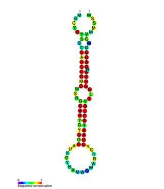
In molecular biology, the miR-200 microRNA is a short RNA molecule. MicroRNAs function to regulate the expression levels of other genes by binding and cleaving mRNAs or inhibiting translation. The miR-200 family contains miR-200a, miR-200b, miR-200c, miR-141, and miR-429. There is growing evidence to suggest that miR-200 microRNAs are involved in cancer metastasis.

<span class="mw-page-title-main">The Hallmarks of Cancer</span> 2000 paper by Hanahan and Weinberg

The hallmarks of cancer were originally six biological capabilities acquired during the multistep development of human tumors and have since been increased to eight capabilities and two enabling capabilities. The idea was coined by Douglas Hanahan and Robert Weinberg in their paper "The Hallmarks of Cancer" published January 2000 in Cell.

<span class="mw-page-title-main">Vasculogenic mimicry</span>

Vasculogenic mimicry (VM) is a strategy used by tumors to ensure sufficient blood supply is brought to its cells through establishing new tumor vascularization. This process is similar to tumor angiogenesis; on the other hand vascular mimicry is unique in that this process occurs independent of endothelial cells. Vasculature is instead developed de novo by cancer cells, which under stress conditions such as hypoxia, express similar properties to stem cells, capable of differentiating to mimic the function of endothelial cells and form vasculature-like structures. The ability of tumors to develop and harness nearby vasculature is considered one of the hallmarks of cancer disease development and is thought to be closely linked to tumor invasion and metastasis. Vascular mimicry has been observed predominantly in aggressive and metastatic cancers and has been associated with negative tumor characteristics such as increased metastasis, increased tissue invasion, and overall poor outcomes for patient survival. Vascular mimicry poses a serious problem for current therapeutic strategies due to its ability to function in the presence of Anti-angiogenic therapeutic agents. In fact, such therapeutics have been found to actually drive VM formation in tumors, causing more aggressive and difficult to treat tumors to develop.

Breast cancer is the most prevalent type of cancer among women globally, with 685,000 deaths recorded worldwide in 2020. The most commonly used treatment methods for breast cancer include surgery, radiotherapy and chemotherapy. Some of these treated patients experience disease relapse and metastasis. The aggressive progression and recurrence of this disease has been attributed the presence of a subset of tumor cells known as breast cancer stem cells (BCSCs). These cells possess the abilities of self-renewal and tumor initiation, allowing them to be drivers of metastases and tumor growth. The microenvironment in which these cells reside is filled with residential inflammatory cells that provide the needed signaling cues for BCSC-mediated self-renewal and survival. The production of cytokines allows these cells to escape from the primary tumor and travel through the circulation to distant organs, commencing the process of metastasis. Due to their significant role in driving disease progression, BCSCs represent a new target by which to treat the tumor at the source of metastasis.

<span class="mw-page-title-main">Invasion (cancer)</span> Direct extension and penetration by cancer cells into neighboring tissues

Invasion is the process by which cancer cells directly extend and penetrate into neighboring tissues in cancer. It is generally distinguished from metastasis, which is the spread of cancer cells through the circulatory system or the lymphatic system to more distant locations. Yet, lymphovascular invasion is generally the first step of metastasis.

<span class="mw-page-title-main">Collective–amoeboid transition</span>

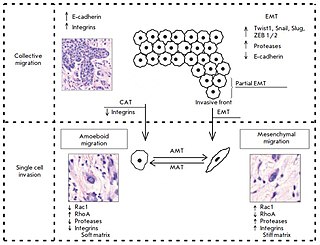
The collective–amoeboid transition (CMT) is a process by which collective multicellular groups dissociate into amoeboid single cells following the down-regulation of integrins. CMTs contrast with epithelial–mesenchymal transitions (EMT) which occur following a loss of E-cadherin. Like EMTs, CATs are involved in the invasion of tumor cells into surrounding tissues, with amoeboid movement more likely to occur in soft extracellular matrix (ECM) and mesenchymal movement in stiff ECM. Although once differentiated, cells typically do not change their migration mode, EMTs and CMTs are highly plastic with cells capable of interconverting between them depending on intracelluar regulatory signals and the surrounding ECM.

References

  1. Hanahan, Douglas; Weinberg, Robert A (2011-03-04). "Hallmarks of Cancer: The Next Generation". Cell. 144 (5): 646–674. doi:10.1016/j.cell.2011.02.013. PMID   21376230.
  2. Thiery, Jean Paul; Acloque, Hervé; Huang, Ruby Y.J.; Nieto, M. Angela (November 2009). "Epithelial-Mesenchymal Transitions in Development and Disease". Cell. 139 (5): 871–890. doi:10.1016/j.cell.2009.11.007. PMID   19945376.
  3. Nieto, M. Angela; Huang, Ruby Yun Ju; Jackson, Rebecca A; Thiery, Jean Paul (June 2016). "EMT: 2016". Cell. 166 (1): 21–45. doi:10.1016/j.cell.2016.06.028. PMID   27368099.
  4. Egeblad, Mikala; Nakasone, Elizabeth S.; Werb, Zena (June 2010). "Tumors as Organs: Complex Tissues that Interface with the Entire Organism". Developmental Cell. 18 (6): 884–901. doi:10.1016/j.devcel.2010.05.012. PMC   2905377 . PMID   20627072.
  5. 1 2 Kalluri, Raghu (September 2016). "The biology and function of fibroblasts in cancer". Nature Reviews Cancer. 16 (9): 582–598. doi:10.1038/nrc.2016.73. ISSN   1474-175X. PMID   27550820.
  6. Celià-Terrassa, Toni; Kang, Yibin (2024-02-07). "How important is EMT for cancer metastasis?". PLOS Biology. 22 (2): e3002487. doi: 10.1371/journal.pbio.3002487 . ISSN   1545-7885. PMC   10849258 . PMID   38324529.
  7. Liu, Qiu-Luo; Luo, Maochao; Huang, Canhua; Chen, Hai-Ning; Zhou, Zong-Guang (2021-04-29). "Epigenetic Regulation of Epithelial to Mesenchymal Transition in the Cancer Metastatic Cascade: Implications for Cancer Therapy". Frontiers in Oncology. 11. doi: 10.3389/fonc.2021.657546 . ISSN   2234-943X. PMC   8117142 . PMID   33996581.
  8. Sznurkowska, Magdalena K.; Aceto, Nicola (August 2022). "The gate to metastasis: key players in cancer cell intravasation". The FEBS Journal. 289 (15): 4336–4354. doi:10.1111/febs.16046. ISSN   1742-464X. PMC   9546053 . PMID   34077633.
  9. Valastyan, Scott; Weinberg, Robert A. (October 2010). "Tumor Metastasis: Molecular Insights and Evolving Paradigms". Cell. 147 (2): 275–292. doi:10.1016/j.cell.2011.09.024. PMC   3261217 . PMID   22000009.
  10. Aceto, Nicola; Bardia, Aditya; Miyamoto, David T.; Donaldson, Maria C.; Wittner, Ben S.; Spencer, Joel A.; Yu, Min; Pely, Adam; Engstrom, Amanda; Zhu, Huili; Brannigan, Brian W.; Kapur, Ravi; Stott, Shannon L.; Shioda, Toshi; Ramaswamy, Sridhar (August 2014). "Circulating Tumor Cell Clusters Are Oligoclonal Precursors of Breast Cancer Metastasis". Cell. 158 (5): 1110–1122. doi:10.1016/j.cell.2014.07.013. PMC   4149753 . PMID   25171411.
  11. Massagué, Joan; Obenauf, Anna C. (January 2016). "Metastatic colonization by circulating tumour cells". Nature. 529 (7586): 298–306. Bibcode:2016Natur.529..298M. doi:10.1038/nature17038. ISSN   0028-0836. PMC   5029466 . PMID   26791720.
  12. Cell 100:59
  13. Evan, GI; Vousden, KH (17 May 2001). "Proliferation, cell cycle and apoptosis in cancer". Nature. 411 (6835): 342–8. Bibcode:2001Natur.411..342E. doi:10.1038/35077213. PMID   11357141. S2CID   4414024.
  14. Kunst, Claudia; Haderer, Marika; Heckel, Sebastian; Schlosser, Sophie; Müller, Martina (December 2016). "The p53 family in hepatocellular carcinoma". Translational Cancer Research. 5 (6): 632–638. doi: 10.21037/tcr.2016.11.79 . ISSN   2219-6803.