Related Research Articles

A finger is a prominent digit on the forelimbs of most tetrapod vertebrate animals, especially those with prehensile extremities such as humans and other primates. Most tetrapods have five digits (pentadactyly), and short digits are typically referred to as toes, while those that are notably elongated are called fingers. In humans, the fingers are flexibly articulated and opposable, serving as an important organ of tactile sensation and fine movements, which are crucial to the dexterity of the hands and the ability to grasp and manipulate objects.
The flexor digitorum profundus is a muscle in the forearm of humans that flexes the fingers. It is considered an extrinsic hand muscle because it acts on the hand while its muscle belly is located in the forearm.
Flexor digitorum superficialis is an extrinsic flexor muscle of the fingers at the proximal interphalangeal joints.

Tenosynovitis is the inflammation of the fluid-filled sheath that surrounds a tendon, typically leading to joint pain, swelling, and stiffness. Tenosynovitis can be either infectious or noninfectious. Common clinical manifestations of noninfectious tenosynovitis include de Quervain tendinopathy and stenosing tenosynovitis

Trigger finger, also known as stenosing tenosynovitis, is a disorder characterized by catching or locking of the involved finger in full or near full flexion, typically with force. There may be tenderness in the palm of the hand near the last skin crease. The name "trigger finger" may refer to the motion of "catching" like a trigger on a gun. The ring finger and thumb are most commonly affected.
In anatomy, a vinculum is a band of connective tissue, similar to a ligament, that connects a flexor tendon to a phalanx bone. They contain tiny vessels which supply blood to the tendon. In vertebrate anatomy, they are referred to as mesotendons.

Psoriatic arthritis (PsA) is a long-term inflammatory arthritis that occurs in people affected by the autoimmune disease psoriasis. The classic feature of psoriatic arthritis is swelling of entire fingers and toes with a sausage-like appearance. This often happens in association with changes to the nails such as small depressions in the nail (pitting), thickening of the nails, and detachment of the nail from the nailbed. Skin changes consistent with psoriasis frequently occur before the onset of psoriatic arthritis but psoriatic arthritis can precede the rash in 15% of affected individuals. It is classified as a type of seronegative spondyloarthropathy.

Achilles tendon rupture is when the Achilles tendon, at the back of the ankle, breaks. Symptoms include the sudden onset of sharp pain in the heel. A snapping sound may be heard as the tendon breaks and walking becomes difficult.

The extensor digitorum muscle is a muscle of the posterior forearm present in humans and other animals. It extends the medial four digits of the hand. Extensor digitorum is innervated by the posterior interosseous nerve, which is a branch of the radial nerve.
In human anatomy, the extensor pollicis longus muscle (EPL) is a skeletal muscle located dorsally on the forearm. It is much larger than the extensor pollicis brevis, the origin of which it partly covers and acts to stretch the thumb together with this muscle.
The flexor pollicis longus is a muscle in the forearm and hand that flexes the thumb. It lies in the same plane as the flexor digitorum profundus. This muscle is unique to humans, being either rudimentary or absent in other primates. A meta-analysis indicated accessory flexor pollicis longus is present in around 48% of the population.

In human anatomy, the adductor pollicis muscle is a muscle in the hand that functions to adduct the thumb. It has two heads: transverse and oblique.

The palmar aponeurosis invests the muscles of the palm, and consists of central, lateral, and medial portions.

The interphalangeal joints of the hand are the hinge joints between the phalanges of the fingers that provide flexion towards the palm of the hand.
The knee examination, in medicine and physiotherapy, is performed as part of a physical examination, or when a patient presents with knee pain or a history that suggests a pathology of the knee joint.
The posterior compartment of the forearm contains twelve muscles which primarily extend the wrist and digits. It is separated from the anterior compartment by the interosseous membrane between the radius and ulna.

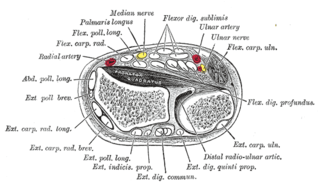
In the human body, the carpal tunnel or carpal canal is a flattened body cavity on the flexor (palmar/volar) side of the wrist, bounded by the carpal bones and flexor retinaculum. It forms the passageway that transmits the median nerve and the tendons of the extrinsic flexor muscles of the hand from the forearm to the hand. The median artery is an anatomical variant. When present it lies between the radial artery, and the ulnar artery and runs with the median nerve supplying the same structures innervated.

The common flexor sheath of hand or the ulnar bursa is a synovial sheath in the carpal tunnel of the human hand.
Jersey finger, also known as rugby finger, is a finger-related tendon injury that is common in sport and can result in permanent loss of flexion of the end of the finger if not surgically repaired. The injury is common when one player grabs another's jersey with the tips of one or more fingers while that player is pulling or running away. It is the most common closed flexor tendon injury and occurs in the ring finger in 75% of cases.

Extensor tendon compartments of the wrist are anatomical tunnels on the back of the wrist that contain tendons of muscles that extend the wrist and the digits.
References
- ↑ "Medscape" . Retrieved 8 November 2014.
- ↑ "Wheeless Online" . Retrieved 8 November 2014.
- ↑ Infection, An Issue of Orthopedic Clinics. Elsevier Health Sciences. 2017. ISBN 9780323524186.
| | This medical sign article is a stub. You can help Wikipedia by expanding it. |