Related Research Articles
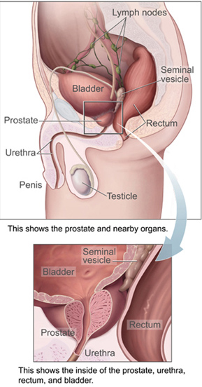
The prostate is both an accessory gland of the male reproductive system and a muscle-driven mechanical switch between urination and ejaculation. It is found in all male mammals. It differs between species anatomically, chemically, and physiologically. Anatomically, the prostate is found below the bladder, with the urethra passing through it. It is described in gross anatomy as consisting of lobes and in microanatomy by zone. It is surrounded by an elastic, fibromuscular capsule and contains glandular tissue, as well as connective tissue.

A lymph node, or lymph gland, is a kidney-shaped organ of the lymphatic system and the adaptive immune system. A large number of lymph nodes are linked throughout the body by the lymphatic vessels. They are major sites of lymphocytes that include B and T cells. Lymph nodes are important for the proper functioning of the immune system, acting as filters for foreign particles including cancer cells, but have no detoxification function.

The ureters are tubes made of smooth muscle that propel urine from the kidneys to the urinary bladder. In a human adult, the ureters are usually 20–30 cm (8–12 in) long and around 3–4 mm (0.12–0.16 in) in diameter. The ureter is lined by urothelial cells, a type of transitional epithelium, and has an additional smooth muscle layer that assists with peristalsis in its lowest third.

Bladder cancer is any of several types of cancer arising from the tissues of the urinary bladder. Symptoms include blood in the urine, pain with urination, and low back pain. It is caused when epithelial cells that line the bladder become malignant.

In vertebrates, the gallbladder, also known as the cholecyst, is a small hollow organ where bile is stored and concentrated before it is released into the small intestine. In humans, the pear-shaped gallbladder lies beneath the liver, although the structure and position of the gallbladder can vary significantly among animal species. It receives and stores bile, produced by the liver, via the common hepatic duct, and releases it via the common bile duct into the duodenum, where the bile helps in the digestion of fats.

The lamina propria is a thin layer of connective tissue that forms part of the moist linings known as mucous membranes or mucosae, which line various tubes in the body, such as the respiratory tract, the gastrointestinal tract, and the urogenital tract.
Courvoisier's principle states that a painless palpably enlarged gallbladder accompanied with mild jaundice is unlikely to be caused by gallstones. Usually, the term is used to describe the physical examination finding of the right-upper quadrant of the abdomen. This sign implicates possible malignancy of the gallbladder or pancreas and the swelling is unlikely due to gallstones.
Cancer staging is the process of determining the extent to which a cancer has developed by growing and spreading. Contemporary practice is to assign a number from I to IV to a cancer, with I being an isolated cancer and IV being a cancer that has spread to the limit of what the assessment measures. The stage generally takes into account the size of a tumor, whether it has invaded adjacent organs, how many regional (nearby) lymph nodes it has spread to, and whether it has appeared in more distant locations (metastasized).
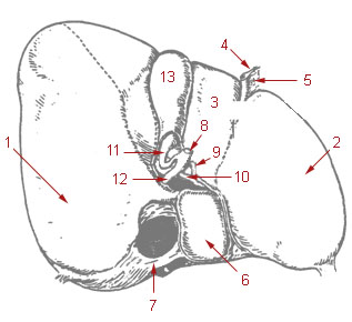
The common hepatic duct is the first part of the biliary tract. It joins the cystic duct coming from the gallbladder to form the common bile duct.

Cystectomy is a medical term for surgical removal of all or part of the urinary bladder. It may also be rarely used to refer to the removal of a cyst. The most common condition warranting removal of the urinary bladder is bladder cancer.

Prostatectomy is the surgical removal of all or part of the prostate gland. This operation is done for benign conditions that cause urinary retention, as well as for prostate cancer and for other cancers of the pelvis.

The sentinel lymph node is the hypothetical first lymph node or group of nodes draining a cancer. In case of established cancerous dissemination it is postulated that the sentinel lymph nodes are the target organs primarily reached by metastasizing cancer cells from the tumor.

Pelvic exenteration is a radical surgical treatment that removes all organs from a person's pelvic cavity. It is used to treat certain advanced or recurrent cancers. The urinary bladder, urethra, rectum, and anus are removed. In women, the vagina, cervix, uterus, Fallopian tubes, ovaries and, in some cases, the vulva are removed. In men, the prostate is removed. The procedure leaves the person with a permanent colostomy and urinary diversion.

Radical retropubic prostatectomy is a surgical procedure in which the prostate gland is removed through an incision in the abdomen. It is most often used to treat individuals who have early prostate cancer. Radical retropubic prostatectomy can be performed under general, spinal, or epidural anesthesia and requires blood transfusion less than one-fifth of the time. Radical retropubic prostatectomy is associated with complications such as urinary incontinence and impotence, but these outcomes are related to a combination of individual patient anatomy, surgical technique, and the experience and skill of the surgeon.

The cystohepatic triangle is an anatomic space bordered by the cystic duct inferiorly, the common hepatic duct medially, and the inferior surface of the liver superiorly. The cystic artery lies within the hepatobiliary triangle, which is used to locate it during a laparoscopic cholecystectomy.

The inferior mesenteric lymph nodes consist of:

Supraclavicular lymph nodes are lymph nodes found above the clavicle, that can be felt in the supraclavicular fossa. The supraclavicular lymph nodes on the left side are called Virchow's nodes. It leads to an appreciable mass that can be recognized clinically, called Troisier sign.
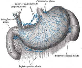
The celiac lymph nodes are associated with the branches of the celiac artery. Other lymph nodes in the abdomen are associated with the superior and inferior mesenteric arteries. The celiac lymph nodes are grouped into three sets: the gastric, hepatic and splenic lymph nodes. They receive lymph from the stomach, duodenum, pancreas, spleen, liver, and gall bladder.
The hepatic lymph nodes consist of the following groups:

Jean-François Calot was a French surgeon best known for describing treatment of curvature of the spine in Pott's disease. He also described a method of treating tuberculous abscesses and defined Calot's triangle.
References
- ↑ Williams, Austin D.; Gefen, Jonathan; Mann, Barry D. (2019-07-03). Surgery Morning Report: Beyond the Pearls E-Book. Elsevier Health Sciences. p. 62. ISBN 978-0-323-59760-9.
- ↑ CLAVIEN, PIERRE-ALAIN; Sarr, Michael G.; Fong, Yuman; Miyazaki, Masaru (2015-09-16). Atlas of Upper Gastrointestinal and Hepato-Pancreato-Biliary Surgery. Springer. p. 724. ISBN 978-3-662-46546-2.
| | This article related to the lymphatic system is a stub. You can help Wikipedia by expanding it. |