Luteinizing hormone/choriogonadotropin receptor

Last updated
LHCGR
Available structures
PDB Ortholog search: PDBe RCSB
Identifiers
Aliases LHCGR , HHG, LCGR, LGR2, LH/CG-R, LH/CGR, LHR, LHRHR, LSH-R, ULG5, Luteinizing hormone/choriogonadotropin receptor
External IDs OMIM: 152790 MGI: 96783 HomoloGene: 37276 GeneCards: LHCGR
Orthologs
SpeciesHumanMouse
Entrez
Ensembl
UniProt
RefSeq (mRNA)

NM_000233

NM_013582
NM_001364898

RefSeq (protein)

NP_000224

NP_038610
NP_001351827

Location (UCSC) Chr 2: 48.69 – 48.76 Mb Chr 17: 89.02 – 89.1 Mb
PubMed search [3] [4]
Wikidata
View/Edit Human View/Edit Mouse

The luteinizing hormone/choriogonadotropin receptor (LHCGR), also lutropin/choriogonadotropin receptor (LCGR) or luteinizing hormone receptor (LHR), is a transmembrane receptor found predominantly in the ovary and testis, but also many extragonadal organs such as the uterus and breasts. The receptor interacts with both luteinizing hormone (LH) and chorionic gonadotropins (such as hCG in humans) and represents a G protein-coupled receptor (GPCR). Its activation is necessary for the hormonal functioning during reproduction.

Contents

LHCGR gene

The gene for the LHCGR is found on chromosome 2 p21 in humans, close to the FSH receptor gene. It consists of 70 kbp (versus 54 kpb for the FSHR). [5] The gene is similar to the gene for the FSH receptor and the TSH receptor.

Receptor structure

The LHCGR consists of 674 amino acids and has a molecular mass of about 85–95 kDA based on the extent of glycosylation. [6]

Like other GPCRs, the LHCG receptor possess seven membrane-spanning domains or transmembrane helices. [7] The extracellular domain of the receptor is heavily glycosylated. These transmembrane domains contain two highly conserved cysteine residues, which build disulfide bonds to stabilize the receptor structure. The transmembrane part is highly homologous with other members of the rhodopsin family of GPCRs. [8] The C-terminal domain is intracellular and brief, rich in serine and threonine residues for possible phosphorylation.

Ligand binding and signal transduction

Upon binding of LH to the external part of the membrane spanning receptor, a transduction of the signal takes place. This process results in the activation of a heterotrimeric G protein. Binding of LH to the receptor shifts its conformation. The activated receptor promotes the binding of GTP to the G protein and its subsequent activation. After binding GTP, the G protein heterotrimer detaches from the receptor and disassembles. The alpha-subunit Gs binds adenylate cyclase and activates the cAMP system. [9]

It is believed that a receptor molecule exists in a conformational equilibrium between active and inactive states. The binding of LH (or CG) to the receptor shifts the equilibrium towards the active form of the receptor. For a cell to respond to LH only a small percentage (≈1%) of receptor sites need to be activated.

Phosphorylation by cAMP-dependent protein kinases

Cyclic AMP-dependent protein kinases (protein kinase A) are activated by the signal cascade originated by the activation of the G protein Gs by the LHCG-receptor. Activated Gs binds the enzyme adenylate cyclase and this leads to the production of cyclic AMP (cAMP). Cyclin AMP-dependent protein kinases are present as tetramers with two regulatory subunits and two catalytic subunits. Upon binding of cAMP to the regulatory subunits, the catalytic units are released and initiate the phosphorylation of proteins leading to the physiologic action. Cyclic AMP is degraded by phosphodiesterase and release 5’AMP. One of the targets of protein kinase A is the Cyclic AMP Response Element Binding Protein, CREB, which binds DNA in the cell nucleus via direct interactions with specific DNA sequences called cyclic AMP response elements (CRE); this process results in the activation or inactivation of gene transcription. [5]

The signal is amplified by the involvement of cAMP and the resulting phosphorylation. The process is modified by prostaglandins. Other cellular regulators that participate are the intracellular calcium concentration regulated by phospholipase C activation, nitric oxide, and other growth factors.

Other pathways of signaling exist for the LHCGR. [6]

Action

Luteinizing hormone up-regulates cholesterol side chain cleaving enzyme in sensitive tissues, the first step of all human steroidogenesis. Steroidogenesis.svg
Luteinizing hormone up-regulates cholesterol side chain cleaving enzyme in sensitive tissues, the first step of all human steroidogenesis.

The LHCG receptor's main function is the regulation of steroidogenesis. This is accomplished by increasing the intracellular levels of the enzyme cholesterol side chain cleaving enzyme, a member of the cytochrome P450 family. This leads to increased conversion of cholesterol into androgen precursors required to make many steroid hormones, including testosterone and estrogens. [10]

Ovary

In the ovary, the LHCG receptor is necessary for follicular maturation and ovulation, as well as luteal function. Its expression requires appropriate hormonal stimulation by FSH and estradiol. The LHCGR is present on granulosa cells, theca cells, luteal cells, and interstitial cells [6] The LCGR is restimulated by increasing levels of chorionic gonadotropins in case a pregnancy is developing. In turn, luteal function is prolonged and the endocrine milieu is supportive of the nascent pregnancy.

Testis

In the male the LHCGR has been identified on the Leydig cells that are critical for testosterone production, and support spermatogenesis.

Normal LHCGR functioning is critical for male fetal development, as the fetal Leydig cells produce androstenedione which is converted to testosterone in fetal Sertoli cells to induce masculinization.

Extragonadal

LHCGR have been found in many types of extragonadal tissues, and the physiologic role of some has remained largely unexplored. Thus receptors have been found in the uterus, sperm, seminal vesicles, prostate, skin, breast, adrenals, thyroid, neural retina, neuroendocrine cells, and (rat) brain. [6]

Receptor regulation

The seven transmembrane a-helix structure of a G protein-coupled receptor such as LHCGR 7TM receptor.png
The seven transmembrane α-helix structure of a G protein-coupled receptor such as LHCGR

Upregulation

Upregulation refers to the increase in the number of receptor sites on the membrane. Estrogen and FSH upregulate LHCGR sites in preparation for ovulation. After ovulation, the luteinized ovary maintains LHCGR s that allow activation in case there is an implantation. Upregulation in males requires gene transcription to synthesize LH receptors within the cell cytoplasm. Some reasons as to why downregulated LH receptors are not upregulated are: lack of gene transcription, lack of RNA to protein conversion and lack of cell membrane targeted shipments from Golgi.

Desensitization

The LHCGRs become desensitized when exposed to LH for some time. A key reaction of this downregulation is the phosphorylation of the intracellular (or cytoplasmic) receptor domain by protein kinases. This process uncouples Gs protein from the LHCGR.

Downregulation

Downregulation refers to the decrease in the number of receptor molecules. This is usually the result of receptor endocytosis. In this process, the bound LCGR-hormone complex binds arrestin and concentrates in clathrin coated pits. Clathrin coated pits recruit dynamin and pinch off from the cell surface, becoming clathrin-coated vesicles. Clathrin-coated vesicles are processed into endosomes, some of which are recycled to the cell surface while others are targeted to lysosomes. Receptors targeted to lysosomes are degraded. Use of long-acting agonists will downregulate the receptor population by promoting their endocytosis.

Modulators

Antibodies to LHCGR can interfere with LHCGR activity.

LHCGR antagonists and agonists

In 2019, the discovery of potent, and selective antagonists of the Luteinizing Hormone Receptor (BAY-298 and BAY-899) were reported which were able to reduce sex hormone levels in vivo. [11] The latter fulfils the quality criteria for a 'Donated Chemical Probe' as defined by the Structural Genomics Consortium. [12]

A series of thienopyr(im)idine-based compounds [13] leading to optimized Org 43553 were described as the first Luteinizing Hormone Receptor agonists. [14] [15]

LHCGR abnormalities

Loss-of-function mutations in females can lead to infertility. In 46, XY individuals severe inactivation can cause male pseudohermaphroditism, as fetal Leydig cells during may not respond and thus interfere with masculinization. [16] Less severe inactivation can result in hypospadias or a micropenis. [6]

History

Alfred G. Gilman and Martin Rodbell received the 1994 Nobel Prize in Medicine and Physiology for the discovery of the G Protein System.

Interactions

Luteinizing hormone/choriogonadotropin receptor has been shown to interact with GIPC1. [17]

Related Research Articles

<span class="mw-page-title-main">Luteinizing hormone</span> Gonadotropin secreted by the adenohypophysis

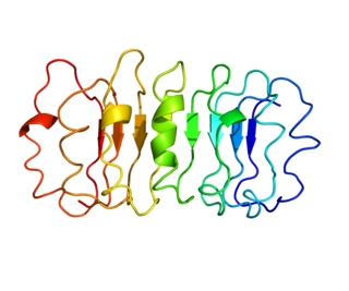
Luteinizing hormone is a hormone produced by gonadotropic cells in the anterior pituitary gland. The production of LH is regulated by gonadotropin-releasing hormone (GnRH) from the hypothalamus. In females, an acute rise of LH known as an LH surge, triggers ovulation and development of the corpus luteum. In males, where LH had also been called interstitial cell–stimulating hormone (ICSH), it stimulates Leydig cell production of testosterone. It acts synergistically with follicle-stimulating hormone (FSH).

<span class="mw-page-title-main">Follicle-stimulating hormone</span> Gonadotropin that regulates the development of reproductive processes

Follicle-stimulating hormone (FSH) is a gonadotropin, a glycoprotein polypeptide hormone. FSH is synthesized and secreted by the gonadotropic cells of the anterior pituitary gland and regulates the development, growth, pubertal maturation, and reproductive processes of the body. FSH and luteinizing hormone (LH) work together in the reproductive system.

<span class="mw-page-title-main">Gonadotropin-releasing hormone</span> Mammalian protein found in Homo sapiens

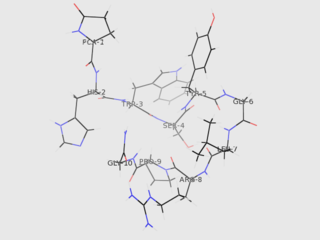
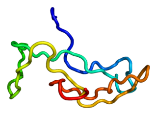
Gonadotropin-releasing hormone (GnRH) is a releasing hormone responsible for the release of follicle-stimulating hormone (FSH) and luteinizing hormone (LH) from the anterior pituitary. GnRH is a tropic peptide hormone synthesized and released from GnRH neurons within the hypothalamus. The peptide belongs to gonadotropin-releasing hormone family. It constitutes the initial step in the hypothalamic–pituitary–gonadal axis.

Gonadotropins are glycoprotein hormones secreted by gonadotropic cells of the anterior pituitary of vertebrates. This family includes the mammalian hormones follicle-stimulating hormone (FSH) and luteinizing hormone (LH), the placental/chorionic gonadotropins, human chorionic gonadotropin (hCG) and equine chorionic gonadotropin (eCG), as well as at least two forms of fish gonadotropins. These hormones are central to the complex endocrine system that regulates normal growth, sexual development, and reproductive function. LH and FSH are secreted by the anterior pituitary gland, while hCG and eCG are secreted by the placenta in pregnant humans and mares, respectively. The gonadotropins act on the gonads, controlling gamete and sex hormone production.

<span class="mw-page-title-main">Estrogen receptor</span> Proteins activated by the hormone estrogen

Estrogen receptors (ERs) are a group of proteins found inside cells. They are receptors that are activated by the hormone estrogen (17β-estradiol). Two classes of ER exist: nuclear estrogen receptors, which are members of the nuclear receptor family of intracellular receptors, and membrane estrogen receptors (mERs), which are mostly G protein-coupled receptors. This article refers to the former (ER).

The gonadotropin-releasing hormone receptor (GnRHR), also known as the luteinizing hormone releasing hormone receptor (LHRHR), is a member of the seven-transmembrane, G-protein coupled receptor (GPCR) family. It is the receptor of gonadotropin-releasing hormone (GnRH). The GnRHR is expressed on the surface of pituitary gonadotrope cells as well as lymphocytes, breast, ovary, and prostate.

<span class="mw-page-title-main">Follicle-stimulating hormone receptor</span> Protein-coding gene in the species Homo sapiens

The follicle-stimulating hormone receptor or FSH receptor (FSHR) is a transmembrane receptor that interacts with the follicle-stimulating hormone (FSH) and represents a G protein-coupled receptor (GPCR). Its activation is necessary for the hormonal functioning of FSH. FSHRs are found in the ovary, testis, and uterus.

<span class="mw-page-title-main">Thyrotropin receptor</span> Mammalian protein found in Homo sapiens

The thyrotropin receptor is a receptor that responds to thyroid-stimulating hormone and stimulates the production of thyroxine (T4) and triiodothyronine (T3). The TSH receptor is a member of the G protein-coupled receptor superfamily of integral membrane proteins and is coupled to the Gs protein.

<span class="mw-page-title-main">Kisspeptin</span> Mammalian protein

Kisspeptins are proteins encoded by the KISS1 gene in humans. Kisspeptins are ligands of the G-protein coupled receptor, GPR54. Kiss1 was originally identified as a human metastasis suppressor gene that has the ability to suppress melanoma and breast cancer metastasis. Kisspeptin-GPR54 signaling has an important role in initiating secretion of gonadotropin-releasing hormone (GnRH) at puberty, the extent of which is an area of ongoing research. Gonadotropin-releasing hormone is released from the hypothalamus to act on the anterior pituitary triggering the release of luteinizing hormone (LH), and follicle stimulating hormone (FSH). These gonadotropic hormones lead to sexual maturation and gametogenesis. Disrupting GPR54 signaling can cause hypogonadotrophic hypogonadism in rodents and humans. The Kiss1 gene is located on chromosome 1. It is transcribed in the brain, adrenal gland, and pancreas.

<span class="mw-page-title-main">ACTH receptor</span> Mammalian protein found in Homo sapiens

The adrenocorticotropic hormone receptor or ACTH receptor also known as the melanocortin receptor 2 or MC2 receptor is a type of melanocortin receptor (type 2) which is specific for ACTH. A G protein–coupled receptor located on the external cell plasma membrane, it is coupled to Gαs and upregulates levels of cAMP by activating adenylyl cyclase. The ACTH receptor plays a role in immune function and glucose metabolism.

<span class="mw-page-title-main">GNRHR</span> Protein-coding gene in the species Homo sapiens

Gonadotropin-releasing hormone receptor is a protein that in humans is encoded by the GNRHR gene.

<span class="mw-page-title-main">KiSS1-derived peptide receptor</span> Mammalian protein found in Homo sapiens

The KiSS1-derived peptide receptor is a G protein-coupled receptor which binds the peptide hormone kisspeptin (metastin). Kisspeptin is encoded by the metastasis suppressor gene KISS1, which is expressed in a variety of endocrine and gonadal tissues. Activation of the kisspeptin receptor is linked to the phospholipase C and inositol trisphosphate second messenger cascades inside the cell.

<span class="mw-page-title-main">Parathyroid hormone 1 receptor</span> Protein-coding gene in the species Homo sapiens

Parathyroid hormone/parathyroid hormone-related peptide receptor, also known as parathyroid hormone 1 receptor (PTH1R), is a protein that in humans is encoded by the PTH1R gene. PTH1R functions as a receptor for parathyroid hormone (PTH) and for parathyroid hormone-related protein (PTHrP), also called parathyroid hormone-like hormone (PTHLH).

Progonadoliberin-2 is a protein that in humans is encoded by the GNRH2 gene.

<span class="mw-page-title-main">CGB1</span> Protein-coding gene in the species Homo sapiens

Choriogonadotropin subunit beta variant 1 is a protein that in humans is encoded by the CGB1 gene.

<span class="mw-page-title-main">CGB2 (gene)</span> Protein-coding gene in the species Homo sapiens

Choriogonadotropin subunit beta variant 2 is a protein that in humans is encoded by the CGB2 gene.

<span class="mw-page-title-main">Luteinizing hormone beta polypeptide</span> Protein-coding gene in the species Homo sapiens

Luteinizing hormone subunit beta also known as lutropin subunit beta or LHβ is a polypeptide that in association with an alpha subunit common to all gonadotropin hormones forms the reproductive signaling molecule luteinizing hormone. In humans it is encoded by the LHB gene.

<span class="mw-page-title-main">Chorionic gonadotropin beta</span> Protein-coding gene in the species Homo sapiens

Choriogonadotropin subunit beta (CG-beta) also known as chorionic gonadotrophin chain beta is a protein that in humans is encoded by the CGB gene.

<span class="mw-page-title-main">Leydig cell hypoplasia</span> Medical condition

Leydig cell hypoplasia (LCH), also known as Leydig cell agenesis, is a rare autosomal recessive genetic and endocrine syndrome affecting an estimated 1 in 1,000,000 genetic males. It is characterized by an inability of the body to respond to luteinizing hormone (LH), a gonadotropin which is normally responsible for signaling Leydig cells of the testicles to produce testosterone and other androgen sex hormones. The condition manifests itself as pseudohermaphroditism, hypergonadotropic hypogonadism, reduced or absent puberty, and infertility.

Hypogonadotropic hypogonadism (HH), is due to problems with either the hypothalamus or pituitary gland affecting the hypothalamic-pituitary-gonadal axis. Hypothalamic disorders result from a deficiency in the release of gonadotropic releasing hormone (GnRH), while pituitary gland disorders are due to a deficiency in the release of gonadotropins from the anterior pituitary. GnRH is the central regulator in reproductive function and sexual development via the HPG axis. GnRH is released by GnRH neurons, which are hypothalamic neuroendocrine cells, into the hypophyseal portal system acting on gonadotrophs in the anterior pituitary. The release of gonadotropins, LH and FSH, act on the gonads for the development and maintenance of proper adult reproductive physiology. LH acts on Leydig cells in the male testes and theca cells in the female. FSH acts on Sertoli cells in the male and follicular cells in the female. Combined this causes the secretion of gonadal sex steroids and the initiation of folliculogenesis and spermatogenesis. The production of sex steroids forms a negative feedback loop acting on both the anterior pituitary and hypothalamus causing a pulsatile secretion of GnRH. GnRH neurons lack sex steroid receptors and mediators such as kisspeptin stimulate GnRH neurons for pulsatile secretion of GnRH.

References

  1. 1 2 3 GRCh38: Ensembl release 89: ENSG00000138039 - Ensembl, May 2017
  2. 1 2 3 GRCm38: Ensembl release 89: ENSMUSG00000024107 - Ensembl, May 2017
  3. "Human PubMed Reference:". National Center for Biotechnology Information, U.S. National Library of Medicine.
  4. "Mouse PubMed Reference:". National Center for Biotechnology Information, U.S. National Library of Medicine.
  5. 1 2 Simoni M, Gromoll J, Nieschlag E (Dec 1997). "The follicle-stimulating hormone receptor: biochemistry, molecular biology, physiology, and pathophysiology". Endocrine Reviews. 18 (6): 739–73. doi: 10.1210/edrv.18.6.0320 . PMID   9408742.
  6. 1 2 3 4 5 Ascoli M, Fanelli F, Segaloff DL (Apr 2002). "The lutropin/choriogonadotropin receptor, a 2002 perspective". Endocrine Reviews. 23 (2): 141–74. doi: 10.1210/edrv.23.2.0462 . PMID   11943741.
  7. Dufau ML (1998). "The luteinizing hormone receptor". Annual Review of Physiology. 60: 461–96. doi:10.1146/annurev.physiol.60.1.461. PMID   9558473.
  8. Jiang X, Dias JA, He X (Jan 2014). "Structural biology of glycoprotein hormones and their receptors: insights to signaling". Molecular and Cellular Endocrinology. 382 (1): 424–51. doi: 10.1016/j.mce.2013.08.021 . PMID   24001578.
  9. Ryu KS, Gilchrist RL, Koo YB, Ji I, Ji TH (Apr 1998). "Gene, interaction, signal generation, signal divergence and signal transduction of the LH/CG receptor". International Journal of Gynaecology and Obstetrics. 60 (Suppl 1): S9-20. doi:10.1016/S0020-7292(98)80001-5. PMID   9833610. S2CID   4798893.
  10. Dufau ML, Cigorraga S, Baukal AJ, Sorrell S, Bator JM, Neubauer JF, Catt KJ (Dec 1979). "Androgen biosynthesis in Leydig cells after testicular desensitization by luteinizing hormone-releasing hormone and human chorionic gonadotropin". Endocrinology. 105 (6): 1314–21. doi:10.1210/endo-105-6-1314. PMID   227658.
  11. Wortmann L, Lindenthal B, Muhn P, et al. (2019). "Discovery of BAY-298 and BAY-899: Tetrahydro-1,6-naphthyridine-Based, Potent, and Selective Antagonists of the Luteinizing Hormone Receptor Which Reduce Sex Hormone Levels in Vivo". Journal of Medicinal Chemistry. 62 (22): 10321–10341. doi: 10.1021/acs.jmedchem.9b01382 . PMID   31670515. S2CID   204967109.
  12. "Donated Chemical Probes". thesgc.org. Retrieved July 31, 2023.
  13. van Straten NC, Schoonus-Gerritsma GG, van Someren RG, Draaijer J, Adang AE, Timmers CM, Hanssen RG, van Boeckel CA (2002-10-04). "The First Orally Active Low Molecular Weight Agonists for the LH Receptor: Thienopyr(im)idines with Therapeutic Potential for Ovulation Induction". ChemBioChem. 3 (10): 1023–1026. doi:10.1002/1439-7633(20021004)3:10<1023::AID-CBIC1023>3.0.CO;2-9. PMID   12362369. S2CID   8732411.
  14. Heitman LH, Oosterom J, Bonger KM, Timmers CM, Wiegerinck PH, IJzerman AP (2008-02-01). "[3H]Org 43553, the First Low-Molecular-Weight Agonistic and Allosteric Radioligand for the Human Luteinizing Hormone Receptor". Molecular Pharmacology. 73 (2): 518–524. doi:10.1124/mol.107.039875. hdl: 1887/3209412 . ISSN   0026-895X. PMID   17989351. S2CID   34584880.
  15. van de Lagemaat R, Timmers CM, Kelder J, van Koppen C, Mosselman S, Hanssen RG (March 2009). "Induction of ovulation by a potent, orally active, low molecular weight agonist (Org 43553) of the luteinizing hormone receptor". Human Reproduction (Oxford, England). 24 (3): 640–648. doi: 10.1093/humrep/den412 . ISSN   1460-2350. PMID   19088107.
  16. Wu SM, Chan WY (1999). "Male pseudohermaphroditism due to inactivating luteinizing hormone receptor mutations". Archives of Medical Research. 30 (6): 495–500. doi: 10.1016/S0188-4409(99)00074-0 . PMID   10714363.
  17. Hirakawa T, Galet C, Kishi M, Ascoli M (Dec 2003). "GIPC binds to the human lutropin receptor (hLHR) through an unusual PDZ domain binding motif, and it regulates the sorting of the internalized human choriogonadotropin and the density of cell surface hLHR". The Journal of Biological Chemistry. 278 (49): 49348–57. doi: 10.1074/jbc.M306557200 . PMID   14507927.

Further reading