Magnex history
Since 1982 Magnex has grown to become the World Leader in NMR Magnet Technology. Below is a brief description of the last 25 successful years
1982 Magnex Formed
1983 First Whole Body 0.5 tesla MRI Magnet System produced for Elscint Ltd.
1984 First Whole Body 2.0 tesla MRI/MRS Magnet System produced for Elscint Ltd.
1985 Magnex moves to Abingdon Business Park
Magnex develops World's first 4.7 tesla 400 mm Horizontal Bore Magnet System for Bruker GmbH
1986 Magnex develops the first Super Wide-Bore 400 MHz High Resolution Magnet System (400 MHz 104 mm)
1987 Full range of Horizontal Bore Spectroscopy Magnet Systems developed (2.0-7.0 tesla, 20–40 cm bore)
1988 Magnex develops the World's first compact 3.0 tesla 800 mm Shielded MRS Magnet System for Henry Ford Hospital Detroit
1989 Magnex Inc established
Magnex develops 500 MHz High Resolution Magnet Systems
1990 Magnex develops 600 MHz High Resolution Magnet Systems
1991 Delivery and installation of Magnex's first 600 MHz 54 mm and 500 MHz 89 mm High Resolution Magnet Systems
1992 Delivery and installation of the World's first 550 MHz Wide-Bore(89mm) High Resolution Magnet System
Delivery of Magnex's first compact 1.5 tesla MRI Magnet System
1993 Magnex develops and install the World's first 9.4 tesla 310 mm Horizontal bore Spectroscopy Magnet System
Delivery and installation of the World's first 7.0 tesla 400 mm Horizontal Bore Spectroscopy Magnet System
Delivery of Magnex's first Compact Active Screen 0.5 tesla MRI Magnet System
1994 Delivery and installation of the first Magnex/JMT 750 MHz 54 mm Magnet System at an Academic Institute (in conjunction with Bruker)
Delivery and installation by Magnex of the World's first Super-Wide Bore 500 MHz/125mm High Resolution Magnet System
Installation of the World's first dedicated fMRI Magnet System (3.0 teslas) at Pittsburgh with GE Medical Systems
1995 Completion of the World's first Actively Shielded High Resolution Magnet Systems
Delivery of the World's first 750 MHz 62 mm Magnet System
Delivery of the World's first compact Shielded FTMS Magnet System
1996 Delivery of the World's first Actively Shielded 600 MHz 52 mm Magnet System
1997 Delivery of the World's first 800 MHz 52 mm Magnet System operating at 4.2 K
1998 Delivery of the World's first ultra high field (8 T) clinical research magnet.
1999 Delivery of the World's second ultra high field (7 T) clinical research magnet.
Delivery of Europe's highest field (4.7 T) clinical research magnet.
2000 Delivery and installation of the world's first actively shielded 500 MHz 125 mm high resolution magnet system.
Delivery of the World's first 500 MHz 40 cm Horizontal bore Magnet System.
Sale of clinical business to GE Medical Systems.
2001 Relocated to The Magnet Technology Centre, Yarnton UK
Demonstration of the World's first 7.0 tesla 600 mm Vertical Bore fMRI Magnet System
2002 Installation of the company's 500th NMR/MRI Magnet System.
Completion of the World's first 12.0 tesla actively shielded FTMS Magnet System.
2003 Installation of World's first actively shielded 600 MHz 89 mm bore magnet system.
Delivery of the world first 9.4 T 65 cm research magnet.
2004 Delivery and installation of the world's first 800 MHz 89 mm Magnet System.
Varian, Inc. acquisition of Magnex Scientific.
2005 Delivery of Premium Shielded 500 MHz and 600 MHz magnets
2006 Introduction of 9.4/820 and 7T/680 actively shielded clinical MRI systems. European MR Application’s lab opened in B4
2007 Introduction of 16.4T/260 and 14.1/260 in-vivo MRS systems
2008 Introduction of PremiumCOMPACT 800MHz NMR magnet. Shipment and commissioning of World’s first 9.4 Tesla/900mm MRI magnet

Magnetic resonance imaging (MRI) is a medical imaging technique used in radiology to form pictures of the anatomy and the physiological processes of the body. MRI scanners use strong magnetic fields, magnetic field gradients, and radio waves to generate images of the organs in the body. MRI does not involve X-rays or the use of ionizing radiation, which distinguishes it from CT and PET scans. MRI is a medical application of nuclear magnetic resonance (NMR) which can also be used for imaging in other NMR applications, such as NMR spectroscopy.
Some of the technological applications of superconductivity include:

A superconducting magnet is an electromagnet made from coils of superconducting wire. They must be cooled to cryogenic temperatures during operation. In its superconducting state the wire has no electrical resistance and therefore can conduct much larger electric currents than ordinary wire, creating intense magnetic fields. Superconducting magnets can produce greater magnetic fields than all but the strongest non-superconducting electromagnets and can be cheaper to operate because no energy is dissipated as heat in the windings. They are used in MRI machines in hospitals, and in scientific equipment such as NMR spectrometers, mass spectrometers, fusion reactors and particle accelerators. They are also used for levitation, guidance and propulsion in a magnetic levitation (maglev) railway system being constructed in Japan.

Oxford Instruments plc is a United Kingdom manufacturing and research company that designs and manufactures tools and systems for industry and research. The company is headquartered in Abingdon, Oxfordshire, England, with sites in the United Kingdom, United States, Europe, and Asia. It is listed on the London Stock Exchange and is a constituent of the FTSE 250 Index.
The tesla is a derived unit of the magnetic B-field strength in the International System of Units.
In nuclear magnetic resonance (NMR) spectroscopy, the chemical shift is the resonant frequency of an atomic nucleus relative to a standard in a magnetic field. Often the position and number of chemical shifts are diagnostic of the structure of a molecule. Chemical shifts are also used to describe signals in other forms of spectroscopy such as photoemission spectroscopy.

Nuclear magnetic resonance spectroscopy, most commonly known as NMR spectroscopy or magnetic resonance spectroscopy (MRS), is a spectroscopic technique to observe local magnetic fields around atomic nuclei. The sample is placed in a magnetic field and the NMR signal is produced by excitation of the nuclei sample with radio waves into nuclear magnetic resonance, which is detected with sensitive radio receivers. The intramolecular magnetic field around an atom in a molecule changes the resonance frequency, thus giving access to details of the electronic structure of a molecule and its individual functional groups. As the fields are unique or highly characteristic to individual compounds, in modern organic chemistry practice, NMR spectroscopy is the definitive method to identify monomolecular organic compounds.

The National High Magnetic Field Laboratory (MagLab) is a facility at Florida State University, the University of Florida, and Los Alamos National Laboratory in New Mexico, that performs magnetic field research in physics, biology, bioengineering, chemistry, geochemistry, biochemistry. It is the only such facility in the US, and is among twelve high magnetic facilities worldwide. The lab is supported by the National Science Foundation and the state of Florida, and works in collaboration with private industry.

Magnetic resonance microscopy is magnetic resonance imaging (MRI) at a microscopic level down to the scale of microns. The first definition of MRM was MRI having voxel resolutions of better than 100 μm.

Interventional magnetic resonance imaging, also Interventional MRI or IMRI, is the use of magnetic resonance imaging (MRI) to do interventional radiology procedures.

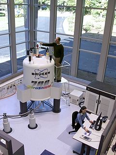
Bruker Corporation is an American manufacturer of scientific instruments for molecular and materials research, as well as for industrial and applied analysis. It is headquartered in Billerica, Massachusetts, and is the publicly traded parent company of Bruker Scientific Instruments and Bruker Energy & Supercon Technologies (BEST) divisions.
Nuclear magnetic resonance (NMR) in the geomagnetic field is conventionally referred to as Earth's field NMR (EFNMR). EFNMR is a special case of low field NMR.
Low field NMR spans a range of different nuclear magnetic resonance (NMR) modalities, going from NMR conducted in permanent magnets, supporting magnetic fields of a few tesla (T), all the way down to zero field NMR, where the Earth's field is carefully shielded such that magnetic fields of nanotesla (nT) are achieved where nuclear spin precession is close to zero. In a broad sense, Low-field NMR is the branch of NMR that is not conducted in superconducting high-field magnets. Low field NMR also includes Earth's field NMR where simply the Earth's magnetic field is exploited to cause nuclear spin-precession which is detected. With magnetic fields on the order of μT and below magnetometers such as SQUIDs or atomic magnetometers are used as detectors. "Normal" high field NMR relies on the detection of spin-precession with inductive detection with a simple coil. However, this detection modality becomes less sensitive as the magnetic field and the associated frequencies decrease. Hence the push toward alternative detection methods at very low fields.
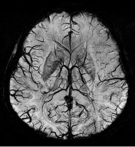
Susceptibility weighted imaging (SWI), originally called BOLD venographic imaging, is an MRI sequence that is exquisitely sensitive to venous blood, hemorrhage and iron storage. SWI uses a fully flow compensated, long echo, gradient recalled echo (GRE) pulse sequence to acquire images. This method exploits the susceptibility differences between tissues and uses the phase image to detect these differences. The magnitude and phase data are combined to produce an enhanced contrast magnitude image. The imaging of venous blood with SWI is a blood-oxygen-level dependent (BOLD) technique which is why it was referred to as BOLD venography. Due to its sensitivity to venous blood SWI is commonly used in traumatic brain injuries (TBI) and for high resolution brain venographies but has many other clinical applications. SWI is offered as a clinical package by Philips and Siemens but can be run on any manufacturer’s machine at field strengths of 1.0 T, 1.5 T, 3.0 T and higher.

Phosphorus-31 NMR spectroscopy is an analytical chemistry technique that uses nuclear magnetic resonance (NMR) to study chemical compounds that contain phosphorus. Phosphorus is commonly found in organic compounds and coordination complexes, making it useful to measure 31P NMR spectra routinely. Solution 31P-NMR is one of the more routine NMR techniques because 31P has an isotopic abundance of 100% and a relatively high gyromagnetic ratio. The 31P nucleus also has a spin of 1⁄2, making spectra relatively easy to interpret. The only other highly sensitive NMR-active nuclei spin 1⁄2 that are monoisotopic are 1H and 19F.
Nuclear magnetic resonance (NMR) is a physical phenomenon in which nuclei in a strong constant magnetic field are perturbed by a weak oscillating magnetic field and respond by producing an electromagnetic signal with a frequency characteristic of the magnetic field at the nucleus. This process occurs near resonance, when the oscillation frequency matches the intrinsic frequency of the nuclei, which depends on the strength of the static magnetic field, the chemical environment, and the magnetic properties of the isotope involved; in practical applications with static magnetic fields up to ca. 20 tesla, the frequency is similar to VHF and UHF television broadcasts (60–1000 MHz). NMR results from specific magnetic properties of certain atomic nuclei. Nuclear magnetic resonance spectroscopy is widely used to determine the structure of organic molecules in solution and study molecular physics and crystals as well as non-crystalline materials. NMR is also routinely used in advanced medical imaging techniques, such as in magnetic resonance imaging (MRI).
Preclinical imaging is the visualization of living animals for research purposes, such as drug development. Imaging modalities have long been crucial to the researcher in observing changes, either at the organ, tissue, cell, or molecular level, in animals responding to physiological or environmental changes. Imaging modalities that are non-invasive and in vivo have become especially important to study animal models longitudinally. Broadly speaking, these imaging systems can be categorized into primarily morphological/anatomical and primarily molecular imaging techniques. Techniques such as high-frequency micro-ultrasound, magnetic resonance imaging (MRI) and computed tomography (CT) are usually used for anatomical imaging, while optical imaging, positron emission tomography (PET), and single photon emission computed tomography (SPECT) are usually used for molecular visualizations.

The physics of magnetic resonance imaging (MRI) concerns fundamental physical considerations of MRI techniques and technological aspects of MRI devices. MRI is a medical imaging technique mostly used in radiology and nuclear medicine in order to investigate the anatomy and physiology of the body, and to detect pathologies including tumors, inflammation, neurological conditions such as stroke, disorders of muscles and joints, and abnormalities in the heart and blood vessels among others. Contrast agents may be injected intravenously or into a joint to enhance the image and facilitate diagnosis. Unlike CT and X-ray, MRI uses no ionizing radiation and is, therefore, a safe procedure suitable for diagnosis in children and repeated runs. Patients with specific non-ferromagnetic metal implants, cochlear implants, and cardiac pacemakers nowadays may also have an MRI in spite of effects of the strong magnetic fields. This does not apply on older devices, details for medical professionals are provided by the device's manufacturer.
A Benchtop nuclear magnetic resonance spectrometer refers to a Fourier transform nuclear magnetic resonance (FT-NMR) spectrometer that is significantly more compact and portable than the conventional equivalents, such that it is portable and can reside on a laboratory benchtop. This convenience comes from using permanent magnets, which have a lower magnetic field and decreased sensitivity compared to the much larger and more expensive cryogen cooled superconducting NMR magnets. Instead of requiring dedicated infrastructure, rooms and extensive installations these benchtop instruments can be placed directly on the bench in a lab and moved as necessary. These spectrometers offer improved workflow, even for novice users, as they are simpler and easy to use. They differ from relaxometers in that they can be used to measure high resolution NMR spectra and are not limited to the determination of relaxation or diffusion parameters.
Magritek is a scientific instrument company based in Wellington, New Zealand, and Aachen, Germany, that was established in 2004 and specialises in compact, portable and benchtop nuclear magnetic resonance (NMR) and magnetic resonance imaging (MRI) products. The technology was originally developed to enable NMR measurements in Antarctica by scientists at Massey and Victoria Universities in New Zealand, including Dr Robin Dykstra. This was combined with compact, handheld NMR magnet technology developed by researchers at RWTH University in Aachen.
