The Netherlands Institute for Innovative Ocular Surgery (NIOS) is an institute providing products for extraocular implants and extraocular cosmetic surgery.
The Netherlands Institute for Innovative Ocular Surgery (NIOS) is an institute providing products for extraocular implants and extraocular cosmetic surgery.
| | This Netherlands-related article is a stub. You can help Wikipedia by expanding it. |

Ophthalmology is a surgical subspecialty within medicine that deals with the diagnosis and treatment of eye disorders.
An evisceration is the removal of the eye's contents, leaving the scleral shell and extraocular muscles intact. The procedure is usually performed to reduce pain or improve cosmesis in a blind eye, as in cases of endophthalmitis unresponsive to antibiotics. An ocular prosthetic can be fitted over the eviscerated eye in order to improve cosmesis.

Strabismus is a vision disorder in which the eyes do not properly align with each other when looking at an object. The eye that is focused on an object can alternate. The condition may be present occasionally or constantly. If present during a large part of childhood, it may result in amblyopia, or lazy eyes, and loss of depth perception. If onset is during adulthood, it is more likely to result in double vision.

Eye surgery, also known as ophthalmic or ocular surgery, is surgery performed on the eye or its adnexa, by an ophthalmologist. Eye surgery is part of ophthalmology. The eye is a fragile organ, and requires due care before, during, and after a surgical procedure to minimize or prevent further damage. An eye surgeon is responsible for selecting the appropriate surgical procedure for the patient, and for taking the necessary safety precautions. Mentions of eye surgery can be found in several ancient texts dating back as early as 1800 BC, with cataract treatment starting in the fifth century BC. It continues to be a widely practiced class of surgery, with various techniques having been developed for treating eye problems.

An eye examination is a series of tests performed to assess vision and ability to focus on and discern objects. It also includes other tests and examinations pertaining to the eyes. Eye examinations are primarily performed by an optometrist, ophthalmologist, or an orthoptist. Health care professionals often recommend that all people should have periodic and thorough eye examinations as part of routine primary care, especially since many eye diseases are asymptomatic.

The medial rectus muscle is a muscle in the orbit near the eye. It is one of the extraocular muscles. It originates from the common tendinous ring, and inserts into the anteromedial surface of the eye. It is supplied by the inferior division of the oculomotor nerve (III). It rotates the eye medially (adduction).
The inferior oblique muscle or obliquus oculi inferior is a thin, narrow muscle placed near the anterior margin of the floor of the orbit. The inferior oblique is one of the extraocular muscles, and is attached to the maxillary bone (origin) and the posterior, inferior, lateral surface of the eye (insertion). The inferior oblique is innervated by the inferior branch of the oculomotor nerve.

The extraocular muscles, or extrinsic ocular muscles, are the seven extrinsic muscles of the human eye. Six of the extraocular muscles, the four recti muscles, and the superior and inferior oblique muscles, control movement of the eye and the other muscle, the levator palpebrae superioris, controls eyelid elevation. The actions of the six muscles responsible for eye movement depend on the position of the eye at the time of muscle contraction.

Enucleation is the removal of the eye that leaves the eye muscles and remaining orbital contents intact. This type of ocular surgery is indicated for a number of ocular tumors, in eyes that have sustained severe trauma, and in eyes that are otherwise blind and painful.
Ocular myasthenia gravis (MG) is a disease of the neuromuscular junction resulting in hallmark variability in muscle weakness and fatigability. MG is an autoimmune disease where anomalous antibodies are produced against the naturally occurring acetylcholine receptors in voluntary muscles. MG may be limited to the muscles of the eye, leading to abrupt onset of weakness/fatigability of the eyelids or eye movement. MG may also involve other muscle groups.
An extraocular implant is a cosmetic implant involving a tiny piece of decorative jewelry which is implanted within the superficial, interpalpebral conjunctiva or sclera of the human eye.

Hypertropia is a condition of misalignment of the eyes (strabismus), whereby the visual axis of one eye is higher than the fellow fixating eye. Hypotropia is the similar condition, focus being on the eye with the visual axis lower than the fellow fixating eye. Dissociated vertical deviation is a special type of hypertropia leading to slow upward drift of one or rarely both eyes, usually when the patient is inattentive.
A retrobulbar block is a regional anesthetic nerve block in the retrobulbar space, the area located behind the globe of the eye. Injection of local anesthetic into this space constitutes the retrobulbar block. This injection provides akinesia of the extraocular muscles by blocking cranial nerves II, III, and VI, thereby preventing movement of the globe. Cranial nerve IV lies outside the muscle cone, and therefore is not affected by the local anesthesia. As a result, intorsion of the eye is still possible. It also provides sensory anesthesia of the conjunctiva, cornea and uvea by blocking the ciliary nerves. This block is most commonly employed for cataract surgery, but also provides anesthesia for other intraocular surgeries.

Strabismus surgery is surgery on the extraocular muscles to correct strabismus, the misalignment of the eyes. Strabismus surgery is a one-day procedure that is usually performed under general anesthesia most commonly by either a neuro- or pediatric ophthalmologist. The patient spends only a few hours in the hospital with minimal preoperative preparation. After surgery, the patient should expect soreness and redness but is generally free to return home.
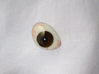
An ocular prosthesis, artificial eye or glass eye is a type of craniofacial prosthesis that replaces an absent natural eye following an enucleation, evisceration, or orbital exenteration. The prosthesis fits over an orbital implant and under the eyelids. Though often referred to as a glass eye, the ocular prosthesis roughly takes the shape of a convex shell and is made of medical grade plastic acrylic. A few ocular prostheses today are made of cryolite glass. A variant of the ocular prosthesis is a very thin hard shell known as a scleral shell which can be worn over a damaged or eviscerated eye. Makers of ocular prosthetics are known as ocularists. An ocular prosthesis does not provide vision; this would be a visual prosthesis. Someone with an ocular prosthesis is altogether blind on the affected side and has monocular vision.
Burt Kushner is an American pediatric ophthalmologist specializing in the diagnosis and treatment of strabismus. Kushner's contributions include demonstration of improved visual fields of patients following strabismus surgery, elucidation of torsional contribution to patients with diplopia, corticosteroid treatment of periocular capillary hemangioma, and novel hypotheses on the mechanism of "overacting" extraocular muscles.
Chronic progressive external ophthalmoplegia (CPEO) is a type of eye disorder characterized by slowly progressive inability to move the eyes and eyebrows. It is often the only feature of mitochondrial disease, in which case the term CPEO may be given as the diagnosis. In other people suffering from mitochondrial disease, CPEO occurs as part of a syndrome involving more than one part of the body, such as Kearns–Sayre syndrome. Occasionally CPEO may be caused by conditions other than mitochondrial diseases.
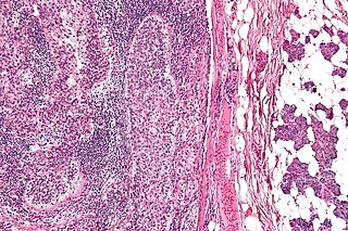
Sebaceous carcinoma, also known as sebaceous gland carcinoma (SGc), sebaceous cell carcinoma, and meibomian gland carcinoma is an uncommon malignant cutaneous tumor. Most are typically about 1.4 cm at presentation. SGc originates from sebaceous glands in the skin and, therefore, may originate anywhere in the body where these glands are found. SGc can be divided into 2 types: periocular and extraocular. The periocular region is rich in sebaceous glands making it a common site of origin. The cause of these lesions in the vast majority of cases is unknown. Occasional cases may be associated with Muir-Torre syndrome. SGc accounts for approximately 0.7% of all skin cancers, and the incidence of SGc is highest in Caucasian, Asian, and Indian populations. Due to the rarity of this tumor and variability in clinical and histological presentation, SGc is often misdiagnosed as an inflammatory condition or a more common neoplasm. SGc is commonly treated with wide local excision or Mohs micrographic surgery, and the relative survival rates at 5 and 10 years are 92.72 and 86.98%, respectively.
Ocular surgery may be performed under topical, local or general anesthesia. Local anaesthesia is more preferred because it is economical, easy to perform and the risk involved is less. Local anaesthesia has a rapid onset of action and provides a dilated pupil with low intraocular pressure.
Botulinum toxin therapy of strabismus is a medical technique used sometimes in the management of strabismus, in which botulinum toxin is injected into selected extraocular muscles in order to reduce the misalignment of the eyes. The injection of the toxin to treat strabismus, reported upon in 1981, is considered to be the first ever use of botulinum toxin for therapeutic purposes. Today, the injection of botulinum toxin into the muscles that surround the eyes is one of the available options in the management of strabismus. Other options for strabismus management are vision therapy and occlusion therapy, corrective glasses and prism glasses, and strabismus surgery.