Related Research Articles

Mastectomy is the medical term for the surgical removal of one or both breasts, partially or completely. A mastectomy is usually carried out to treat breast cancer. In some cases, people believed to be at high risk of breast cancer have the operation as a preventive measure. Alternatively, some people can choose to have a wide local excision, also known as a lumpectomy, an operation in which a small volume of breast tissue containing the tumor and a surrounding margin of healthy tissue is removed to conserve the breast.

The spleen is an organ found in virtually all vertebrates. Similar in structure to a large lymph node, it acts primarily as a blood filter. The word spleen comes from Ancient Greek σπλήν (splḗn).

Chest pain is pain or discomfort in the chest, typically the front of the chest. It may be described as sharp, dull, pressure, heaviness or squeezing. Associated symptoms may include pain in the shoulder, arm, upper abdomen or jaw or nausea, sweating or shortness of breath. It can be divided into heart-related and non-heart-related pain. Pain due to insufficient blood flow to the heart is also called angina pectoris. Those with diabetes or the elderly may have less clear symptoms.

Portal hypertension is hypertension in the hepatic portal system – made up of the portal vein and its branches, that drain from most of the intestine to the liver. Portal hypertension is defined as a hepatic venous pressure gradient. Cirrhosis is the most common cause of portal hypertension; other, less frequent causes are therefore grouped as non-cirrhotic portal hypertension. When it becomes severe enough to cause symptoms or complications, treatment may be given to decrease portal hypertension itself or to manage its complications.

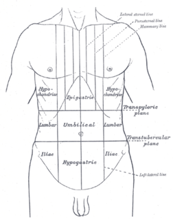
Splenomegaly is an enlargement of the spleen. The spleen usually lies in the left upper quadrant (LUQ) of the human abdomen. Splenomegaly is one of the four cardinal signs of hypersplenism which include: some reduction in number of circulating blood cells affecting granulocytes, erythrocytes or platelets in any combination; a compensatory proliferative response in the bone marrow; and the potential for correction of these abnormalities by splenectomy. Splenomegaly is usually associated with increased workload, which suggests that it is a response to hyperfunction. It is therefore not surprising that splenomegaly is associated with any disease process that involves abnormal red blood cells being destroyed in the spleen. Other common causes include congestion due to portal hypertension and infiltration by leukemias and lymphomas. Thus, the finding of an enlarged spleen, along with caput medusae, is an important sign of portal hypertension.
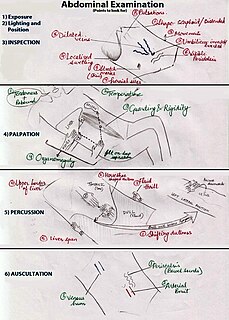
An abdominal examination is a portion of the physical examination which a physician or nurse uses to clinically observe the abdomen of a patient for signs of disease. The physical examination typically occurs after a thorough medical history is taken, that is, after the physician asks the patient the course of their symptoms. The abdominal examination is conventionally split into four different stages: first, inspection of the patient and the visible characteristics of their abdomen. Auscultation (listening) of the abdomen with a stethoscope. Palpation of the patient's abdomen. Finally, percussion (tapping) of the patient's abdomen and abdominal organs. Depending on the need to test for specific diseases such as ascites, special tests may be performed as a part of the physical examination. An abdominal examination may be performed because the physician suspects a disease of the organs inside the abdominal cavity, or simply as a part of a complete physical examination for other conditions. In a complete physical examination, the abdominal exam classically follows the respiratory examination and cardiovascular examination.
The abdominojugular test, also known as abdominojugular reflux (AJR), is a physical examination test useful in diagnosing right ventricle dysfunction, particularly right ventricular failure.

Costovertebral angle (CVA) tenderness is pain that results from touching the region inside of the costovertebral angle. The CVA is formed by the 12th rib and the spine. Assessing for CVA tenderness is part of the abdominal exam, and CVA tenderness indicates kidney pathology.
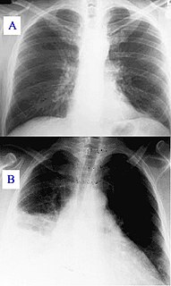
A pulmonary consolidation is a region of normally compressible lung tissue that has filled with liquid instead of air. The condition is marked by induration of a normally aerated lung. It is considered a radiologic sign. Consolidation occurs through accumulation of inflammatory cellular exudate in the alveoli and adjoining ducts. The liquid can be pulmonary edema, inflammatory exudate, pus, inhaled water, or blood. Consolidation must be present to diagnose pneumonia: the signs of lobar pneumonia are characteristic and clinically referred to as consolidation.
Traube's (semilunar) space is an anatomic space of some clinical importance. It is a crescent-shaped space, encompassed by the lower edge of the left lung, the anterior border of the spleen, the left costal margin and the inferior margin of the left lobe of the liver. Thus, its surface markings are respectively the left sixth rib superiorly, the left mid axillary line laterally, and the left costal margin inferiorly.

Castell's sign is a medical sign assessed to evaluate splenomegaly and typically part of an abdominal examination. It is an alternative physical examination maneuver to percussion over Traube's space.
Mohs surgery, developed in 1938 by a general surgeon, Frederic E. Mohs, is microscopically controlled surgery used to treat common types of skin cancer. During the surgery, after each removal of tissue and while the patient waits, the tissue is examined for cancer cells. That examination dictates the decision for additional tissue removal. Mohs surgery is the gold standard method for obtaining complete margin control during removal of a skin cancer using frozen section histology. CCPDMA or Mohs surgery allows for the removal of a skin cancer with very narrow surgical margin and a high cure rate.
Felty's syndrome, also called Felty syndrome, (FS) is rare autoimmune disease characterized by the triad of rheumatoid arthritis, enlargement of the spleen and too few neutrophils in the blood. The condition is more common in those aged 50–70 years, specifically more prevalent in females than males, and more so in Caucasians than those of African descent. It is a deforming disease that causes many complications for the individual.

Abdominal ultrasonography is a form of medical ultrasonography to visualise abdominal anatomical structures. It uses transmission and reflection of ultrasound waves to visualise internal organs through the abdominal wall. For this reason, the procedure is also called a transabdominal ultrasound, in contrast to endoscopic ultrasound, the latter combining ultrasound with endoscopy through visualize internal structures from within hollow organs.
Diaphragmatic excursion is the movement of the thoracic diaphragm during breathing.

Hemopericardium refers to blood in the pericardial sac of the heart. It is clinically similar to a pericardial effusion, and, depending on the volume and rapidity with which it develops, may cause cardiac tamponade.

Cirrhosis, also known as liver cirrhosis or hepatic cirrhosis, is a condition in which the liver does not function properly due to long-term damage. This damage is characterized by the replacement of normal liver tissue by scar tissue. Typically, the disease develops slowly over months or years. Early on, there are often no symptoms. As the disease worsens, a person may become tired, weak, itchy, have swelling in the lower legs, develop yellow skin, bruise easily, have fluid buildup in the abdomen, or develop spider-like blood vessels on the skin. The fluid build-up in the abdomen may become spontaneously infected. Other serious complications include hepatic encephalopathy, bleeding from dilated veins in the esophagus or dilated stomach veins, and liver cancer. Hepatic encephalopathy results in confusion and may lead to unconsciousness.
Unnecessary health care is health care provided with a higher volume or cost than is appropriate. In the United States, where health care costs are the highest as a percentage of GDP, overuse was the predominant factor in its expense, accounting for about a third of its health care spending in 2012.

Eliglustat is a treatment for Gaucher's disease discovered at the University of Michigan and developed by Genzyme Corp that was approved by the FDA August 2014. Commonly used as the tartrate salt, the compound is believed to work by inhibition of glucosylceramide synthase. According to an article in Journal of the American Medical Association the oral substrate reduction therapy resulted in "significant improvements in spleen volume, hemoglobin level, liver volume, and platelet count" in untreated adults with Gaucher disease Type 1.
Spleen pain is a pain felt from the left upper quadrant of the abdomen or epigastrium where the human spleen is located or neighboring.
References
- ↑ Grover SA; Barkun AN; Sackett DL (1993-11-10). "Does this patient have splenomegaly?". JAMA. 270 (18): 2218–2221. doi:10.1001/jama.1993.03510180088040. ISSN 0098-7484.
| This medical sign article is a stub. You can help Wikipedia by expanding it. |