Orbital fissure may refer to:
| This disambiguation page lists articles associated with the title Orbital fissure. If an internal link led you here, you may wish to change the link to point directly to the intended article. |
Orbital fissure may refer to:
| This disambiguation page lists articles associated with the title Orbital fissure. If an internal link led you here, you may wish to change the link to point directly to the intended article. |

In the human skull, the zygomatic bone is a paired irregular bone which articulates with the maxilla, the temporal bone, the sphenoid bone and the frontal bone. It is situated at the upper and lateral part of the face and forms the prominence of the cheek, part of the lateral wall and floor of the orbit, and parts of the temporal fossa and the infratemporal fossa. It presents a malar and a temporal surface; four processes, and four borders.
In anatomy, a fissure is a groove, natural division, deep furrow, elongated cleft, or tear in various parts of the body also generally called a sulcus, or in the brain a sulcus.

In anatomy, the orbit is the cavity or socket of the skull in which the eye and its appendages are situated. Anatomical term created by Gerard of Cremona. "Orbit" can refer to the bony socket, or it can also be used to imply the contents. In the adult human, the volume of the orbit is 30 millilitres, of which the eye occupies 6.5 ml. The orbital contents comprise the eye, the orbital and retrobulbar fascia, extraocular muscles, cranial nerves II, III, IV, V, and VI, blood vessels, fat, the lacrimal gland with its sac and nasolacrimal duct, the eyelids, medial and lateral palpebral ligaments, check ligaments, the suspensory ligament, septum, ciliary ganglion and short ciliary nerves.
The inferior rectus muscle is a muscle in the orbit.

The medial rectus muscle is a muscle in the orbit.

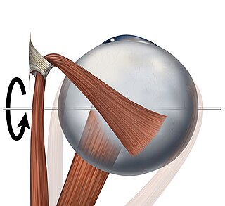
The superior orbital fissure is a foramen in the skull, although strictly it is more of a cleft, lying between the lesser and greater wings of the sphenoid bone.

The maxillary nerve (CN V2) is one of the three branches or divisions of the trigeminal nerve, the fifth (V) cranial nerve. It comprises the principal functions of sensation from the maxillary, nasal cavity, sinuses, the palate and subsequently that of the mid-face, and is intermediate, both in position and size, between the ophthalmic nerve and the mandibular nerve.

The ophthalmic nerve is the first branch of the trigeminal nerve. The ophthalmic nerve is a sensory nerve mostly carrying general somatic afferent fibers that transmit sensory information to the CNS from structures of the eyeball, the skin of the upper face and anterior scalp, the lining of the upper part of the nasal cavity and air cells, and the meninges of the anterior cranial fossa. Some of ophthalmic nerve branches also convey parasympathetic fibers.

In human anatomy, the pterygopalatine fossa is a fossa in the skull. A human skull contains two pterygopalatine fossae—one on the left side, and another on the right side. Each fossa is a cone-shaped paired depression deep to the infratemporal fossa and posterior to the maxilla on each side of the skull, located between the pterygoid process and the maxillary tuberosity close to the apex of the orbit. It is the indented area medial to the pterygomaxillary fissure leading into the sphenopalatine foramen. It communicates with the nasal and oral cavities, infratemporal fossa, orbit, pharynx, and middle cranial fossa through eight foramina.

The greater wing of the sphenoid bone, or alisphenoid, is a bony process of the sphenoid bone; there is one on each side, extending from the side of the body of the sphenoid and curving upward, laterally, and backward.
The lesser wings of the sphenoid or orbito-sphenoids are two thin triangular plates, which arise from the upper and anterior parts of the body, and, projecting lateralward, end in sharp points [Fig. 1].

The zygomatic nerve is not to be confused with the zygomatic branches of the facial nerve.

The lateral wall and the floor of the orbit are separated posteriorly by the inferior orbital fissure which transmits the zygomatic branch of the maxillary nerve and the ascending branches from the pterygopalatine ganglion. The infraorbital vessels are found in the inferior orbital fissure, and travel down the infraorbital groove into the infraorbital canal and exit through the infraorbital foramen. Inferior division of ophthalmic vein passes through the inferior orbital fissure.

The inferior ophthalmic vein begins in a venous network at the forepart of the floor and medial wall of the orbit; it receives some vorticose veins and other veins from the inferior rectus muscle, inferior oblique muscle, lacrimal sac and eyelids, runs backward in the lower part of the orbit lying above the inferior rectus and divides into two branches.
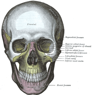
In human anatomy, the infraorbital foramen is an opening in the maxillary bone of the skull located below the infraorbital margin of the orbit. It transmits the infraorbital artery and vein, and the infraorbital nerve, a branch of the maxillary nerve. It is typically 6.10 to 10.9 mm from the infraorbital margin.

The pterygomaxillary fissure is a fissure of the human skull. It is vertical, and descends at right angles from the medial end of the inferior orbital fissure. It is a triangular interval, formed by the divergence of the maxilla from the pterygoid process of the sphenoid.
The infratemporal fossa is an irregularly shaped cavity, situated below and medial to the zygomatic arch. It is not fully enclosed by bone in all directions, and it contains superficial muscles that are visible during dissection after removing skin and fascia: namely, the lower part of the temporalis muscle, the lateral pterygoid, and the medial pterygoid.

The orbital process of the palatine bone is placed on a higher level than the sphenoidal, and is directed upward and lateralward from the front of the vertical part, to which it is connected by a constricted neck. It presents five surfaces, which enclose an air cell. Of these surfaces, three are articular and two non-articular.

Not to be confused with the inferior orbital fissure, which is just lateral to the infraorbital groove.

Tolosa–Hunt syndrome (THS) is a rare disorder characterized by severe and unilateral headaches with orbital pain, along with weakness and paralysis (ophthalmoplegia) of certain eye muscles.