Look up ostium in Wiktionary, the free dictionary.
An ostium (pl.: ostia) in anatomy is a small opening or orifice.
Contents
Ostia , not as a plural, is also the name of a number of places.
Ostium or ostia may refer to:
An ostium (pl.: ostia) in anatomy is a small opening or orifice.
Ostia , not as a plural, is also the name of a number of places.
Ostium or ostia may refer to:
OS, O.S., Os, O's, or os may refer to:

Paranasal sinuses are a group of four paired air-filled spaces that surround the nasal cavity. The maxillary sinuses are located under the eyes; the frontal sinuses are above the eyes; the ethmoidal sinuses are between the eyes and the sphenoidal sinuses are behind the eyes. The sinuses are named for the facial bones and sphenoid bone in which they are located. Their role is disputed.
Sinus may refer to:
In anatomy and osteology, a foramen is an opening or enclosed gap within the dense connective tissue of extant and extinct amniote animals, typically to allow passage of nerves, arteries, veins or other soft tissue structures from one body compartment to another.

Atrial septal defect (ASD) is a congenital heart defect in which blood flows between the atria of the heart. Some flow is a normal condition both pre-birth and immediately post-birth via the foramen ovale; however, when this does not naturally close after birth it is referred to as a patent (open) foramen ovale (PFO). It is common in patients with a congenital atrial septal aneurysm (ASA).
Porocytes are tubular cells which make up the pores of a sponge known as ostia.
In the fetal heart, the foramen ovale, also foramen Botalli or the ostium secundum of Born, allows blood to enter the left atrium from the right atrium. It is one of two fetal cardiac shunts, the other being the ductus arteriosus. Another similar adaptation in the fetus is the ductus venosus. In most individuals, the foramen ovale closes at birth. It later forms the fossa ovalis.

The interatrial septum is the wall of tissue that separates the right and left atria of the heart.

The coronary sinus is the largest vein of the heart. It drains over half of the deoxygenated blood from the heart muscle into the right atrium. It begins on the backside of the heart, in between the left atrium, and left ventricle; it begins at the junction of the great cardiac vein, and oblique vein of the left atrium. It receives multiple tributaries. It passes across the backside of the heart along a groove between left atrium and left ventricle, then drains into the right atrium at the orifice of the coronary sinus.

The superior sagittal sinus, within the human head, is an unpaired dural venous sinus lying along the attached margin of the falx cerebri. It allows blood to drain from the lateral aspects of anterior cerebral hemispheres to the confluence of sinuses. Cerebrospinal fluid drains through arachnoid granulations into the superior sagittal sinus and is returned to venous circulation.
In the anatomy of the heart, the valve of the coronary sinus is a valve located at the orifice of the coronary sinus where the coronary sinus drains into the right atrium. It prevents blood from flowing backwards into the coronary sinus during contraction of the heart.

The primitive atrium is a stage in the embryonic development of the human heart. It grows rapidly and partially encircles the bulbus cordis; the groove against which the bulbus cordis lies is the first indication of a division into right and left atria.

During heart development of a human embryo, the single primitive atrium becomes divided into right and left by a septum, the septum primum. The septum primum grows downward into the single atrium.
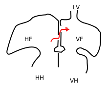
In the developing heart, the atria are initially open to each other, with the opening known as the primary interatrial foramen or ostium primum. The foramen lies beneath the edge of septum primum and the endocardial cushions. It progressively decreases in size as the septum grows downwards, and disappears with the formation of the atrial septum.
The valve of the inferior vena cava is a venous valve that lies at the junction of the inferior vena cava and right atrium.
Coronary artery anomalies are variations of the coronary circulation, affecting <1% of the general population. Symptoms include chest pain, shortness of breath and syncope, although cardiac arrest may be the first clinical presentation. Several varieties are identified, with a different potential to cause sudden cardiac death.
Ostia may refer to:

In anatomy, the term nasal meatus can refer to any of the three meatuses (passages) through the skull's nasal cavity: the superior meatus, middle meatus, and inferior meatus.

Heart development, also known as cardiogenesis, refers to the prenatal development of the heart. This begins with the formation of two endocardial tubes which merge to form the tubular heart, also called the primitive heart tube. The heart is the first functional organ in vertebrate embryos.
The heart is a muscular organ situated in the mediastinum. It consists of four chambers, four valves, two main arteries, and the conduction system. The left and right sides of the heart have different functions: the right side receives de-oxygenated blood through the superior and inferior venae cavae and pumps blood to the lungs through the pulmonary artery, and the left side receives saturated blood from the lungs.