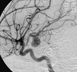
Intracranial aneurysm, also known as brain aneurysm, is a cerebrovascular disorder in which weakness in the wall of a cerebral artery or vein causes a localized dilation or ballooning of the blood vessel.
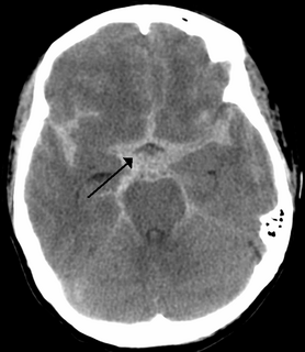
Subarachnoid hemorrhage (SAH) is bleeding into the subarachnoid space—the area between the arachnoid membrane and the pia mater surrounding the brain. Symptoms may include a severe headache of rapid onset, vomiting, decreased level of consciousness, fever, and sometimes seizures. Neck stiffness or neck pain are also relatively common. In about a quarter of people a small bleed with resolving symptoms occurs within a month of a larger bleed.
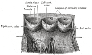
Aneurysm of the aortic sinus, also known as the sinus of Valsalva, is comparatively rare. When present, it is usually in either the right (65–85%) or in the noncoronary (10–30%) sinus, rarely in the left sinus.

A pseudoaneurysm, also known as a false aneurysm, is a collection of blood that forms between the two outer layers of an artery, the tunica media and the tunica adventitia. It is usually caused by a penetrating injury to the vessel, which then bleeds, but forms a space between the above two layers, rather than exiting the vessel. It may be pulsatile and can resemble a true aneurysm. A true aneurysm involves all three layers of the blood vessel. A dissecting aneurysm is when blood from the vessel lumen tracks between the two inner layers, the intima and the tunica media. This can cause blockage of the flow. A perivascular hematoma is a collection of blood that is external to the three vessel layers. Due to being close to the vessel, it can also be pulsatile, and can be mistaken for a pseudoaneurysm or aneurysm.

The abdominal aorta is the largest artery in the abdominal cavity. As part of the aorta, it is a direct continuation of the descending aorta.
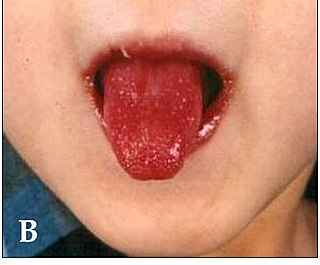
Kawasaki disease, also known as mucocutaneous lymph node syndrome, is a disease in which blood vessels throughout the body become inflamed. The most common symptoms include a fever that lasts for more than five days not affected by usual medications, large lymph nodes in the neck, a rash in the genital area, and red eyes, lips, palms or soles of the feet. Other symptoms include sore throat and diarrhea. Within three weeks of the onset of symptoms, the skin from the hands and feet may peel. Recovery then typically occurs. In some children, coronary artery aneurysms may form in the heart after 1–2 years.

Cerebral angiography is a form of angiography which provides images of blood vessels in and around the brain, thereby allowing detection of abnormalities such as arteriovenous malformations and aneurysms. It was pioneered in 1927 by the Portuguese neurologist Egas Moniz at the University of Lisbon, who also helped develop thorotrast for use in the procedure.

The splenic artery is the blood vessel that supplies oxygenated blood to the spleen. It branches from the celiac artery, and follows a course superior to the pancreas.

An arteriovenous fistula is an abnormal connection or passageway between an artery and a vein. It may be congenital, surgically created for hemodialysis treatments, or acquired due to pathologic process, such as trauma or erosion of an arterial aneurysm.

In human anatomy, the left and right posterior communicating arteries are arteries at the base of the brain that form part of the circle of Willis. Each posterior communicating artery connects the three cerebral arteries of the same side. Anteriorly, it connects to the internal carotid artery (ICA) prior to the terminal bifurcation of the ICA into the anterior cerebral artery and middle cerebral artery. Posteriorly, it communicates with the posterior cerebral artery.
In human anatomy, the anterior communicating artery is a blood vessel of the brain that connects the left and right anterior cerebral arteries.
ICD-10 is an international statistical classification used in health care and related industries.
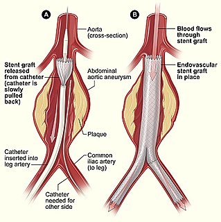
Endovascular aneurysm repair, is a type of endovascular surgery used to treat pathology of the aorta, most commonly an abdominal aortic aneurysm (AAA). When used to treat thoracic aortic disease, the procedure is then specifically termed TEVAR for "thoracic endovascular aortic/aneurysm repair." The procedure involves the placement of an expandable stent graft within the aorta to treat aortic disease without operating directly on the aorta. In 2003, EVAR surpassed open aortic surgery as the most common technique for repair of AAA, and in 2010, EVAR accounted for 78% of all intact AAA repair in the United States.

Coronary artery aneurysm is an abnormal dilatation of part of the coronary artery.

Ventricular aneurysms are one of the many complications that may occur after a heart attack. The word aneurysm refers to a bulge or ‘pocketing’ of the wall or lining of a vessel commonly occurring in the blood vessels at the base of the septum, or within the aorta. In the heart, they usually arise from a patch of weakened tissue in a ventricular wall, which swells into a bubble filled with blood. This, in turn, may block the passageways leading out of the heart, leading to severely constricted blood flow to the body. Ventricular aneurysms can be fatal. They are usually non-rupturing because they are lined by scar tissue.

Retroperitoneal bleeding refers to an accumulation of blood found in the retroperitoneal space.

Endovascular coiling, is an endovascular treatment for intracranial aneurysms and bleeding throughout the body. The procedure reduces blood circulation to the aneurysm through the use of microsurgical detachable platinum wires, with the clinician inserting one or more into the aneurysm until it is determined that blood flow is no longer occurring within the space. It is one of two main treatments for cerebral aneurysms, the other being surgical clipping. Clipping is an alternative to stenting for bleeding.
Open aortic surgery (OAS), also known as open aortic repair (OAR), describes a technique whereby an abdominal or retroperitoneal surgical incision is used to visualize and control the aorta for purposes of treatment. OAS is used to treat aneurysms of the abdominal and thoracic aorta, aortic dissection, acute aortic syndrome, and aortic ruptures. Aortobifemoral bypass is also used to treat atherosclerotic disease of the abdominal aorta below the level of the renal arteries. In 2003, OAS was surpassed by endovascular aneurysm repair (EVAR) as the most common technique for repairing abdominal aortic aneurysms in the United States. In OAS for abdominal aortic aneurysm, the aneurysmal portion of the aorta is replaced with a graft, usually made of dacron or PTFE.

A flow diverter is an endovascular prosthesis used to treat intracranial aneurysms. It is placed in the aneurysm's parent artery, covering the neck, in order to divert blood flow and determine a progressive thrombosis of the sac. Flow diverting stents consist of structural Cobalt-chrome or Nitinol alloy wires and often a set of radiopaque wires woven together in a flexible braid.