Periarchicortex

Last updated

Periarchicortex is one of two subtypes of periallocortex, [1] [2] the other being peripaleocortex. [3] It is formed at borders between archicortex (a subtype of allocortex) and isocortex and shows slow histological transition from the four-layered structure typical for archicortex to the six-layered structure typical for isocortex. [1]

Periallocortex is one of three subtypes of allocortex, the other two subtypes being paleocortex and archicortex. The periallocortex is formed at transition areas where any of the other two subtypes of allocortex borders with the neocortex.

Peripaleocortex is one of two subtypes of periallocortex, the other being periarchicortex. Peripaleocortex is formed at borders between isocortex (neocortex) and paleocortex. It shows slow histological transition from the three-layered structure characteristic of paleocortex to the typical six-layered structure characteristic of isocortex. The main peripaleocortex area is anterior insular cortex.

Archicortex Phylogenetically the oldest part of the cerebral cortex or pallium

In anatomy of animals, the archicortex or archipallium is the phylogenetically oldest region of the brain's pallium or cortex.

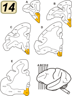
Cortical areas that are generally considered to belong to periarchicortex, include presubiculum, parasubiculum, entorhinal cortex, perirhinal cortex, retrosplenial cortex, periarchicortical part of cingulate cortex, posterior part of subcallosal area, and subgenual area. [1]

In the rodent, the parasubiculum is a retrohippocampal isocortical structure, and a major component of the subicular complex. It receives numerous subcortical and cortical inputs, and sends major projections to the superficial layers of the entorhinal cortex.

Entorhinal cortex Part of the brains cerebral cortex

The entorhinal cortex (EC) is an area of the brain located in the medial temporal lobe and functioning as a hub in a widespread network for memory, navigation and the perception of time. The EC is the main interface between the hippocampus and neocortex. The EC-hippocampus system plays an important role in declarative (autobiographical/episodic/semantic) memories and in particular spatial memories including memory formation, memory consolidation, and memory optimization in sleep. The EC is also responsible for the pre-processing (familiarity) of the input signals in the reflex nictitating membrane response of classical trace conditioning, the association of impulses from the eye and the ear occurs in the entorhinal cortex.

The Perirhinal cortex is a cortical region in the medial temporal lobe that is made up of Brodmann areas 35 and 36. It receives highly processed sensory information from all sensory regions, and is generally accepted to be an important region for memory. It is bordered caudally by postrhinal cortex or parahippocampal cortex and ventrally and medially by entorhinal cortex.

Periarchicortex does not contact immediately at borders with the true isocortex and does not transit directly into it. Instead, another transitional area from the isocortex side, called proisocortex, is formed at such borders. [4] So, at borders between "true" archicortex and true isocortex, there are two transitional areas. One such area, which is anatomically located closer to the archicortex side and histologically more resembling it, is called periarchicortex. [1] Another transitional area, which is anatomically located closer to the true isocortex and histologically more resembling it, is called proisocortex. [4]

Proisocortex or pro-isocortex is one of two subtypes of cortical areas in the areas belonging to the neocortex. The other subtype is termed the true isocortex. Proisocortical areas are transitional areas placed between areas of true isocortex and areas of periallocortex. The histological structure of proisocortex is also transitional between true isocortex and either peripaleocortex or periarchicortex, depending on with which subtype of periallocortex the given proisocortical area borders.

Related Research Articles

Cerebral cortex Part of a mammals brain

The cerebral cortex, also known as the cerebral mantle, is the outer layer of neural tissue of the cerebrum of the brain, in humans and other mammals. It is separated into two cortices, by the longitudinal fissure that divides the cerebrum into the left and right cerebral hemispheres. The two hemispheres are joined beneath the cortex by the corpus callosum. The cerebral cortex is the largest site of neural integration in the central nervous system. It plays a key role in memory, attention, perception, awareness, thought, language, and consciousness.

Brodmann area

A Brodmann area is a region of the cerebral cortex, in the human or other primate brain, defined by its cytoarchitecture, or histological structure and organization of cells.

Neocortex Mammalian structure involved in higher-order brain functions

The neocortex, also called the neopallium and isocortex, is the part of the mammalian brain involved in higher-order brain functions such as sensory perception, cognition, generation of motor commands, spatial reasoning and language.

Brodmann area 14

Brodmann Area 14 is one of Brodmann's subdivisions of the cerebral cortex in the brain. It was defined by Brodmann in the guenon monkey . While Brodmann, writing in 1909, argued that no equivalent structure existed in humans, later work demonstrated that area 14 has a clear homologue in the human ventromedial prefrontal cortex.

The allocortex is one of the two types of cerebral cortex, the other being the neocortex. It is characterized by having just three or four cell layers, in contrast with the six layers of the neocortex, and takes up a much smaller area than the neocortex. There are three subtypes of allocortex: the paleocortex, the archicortex, and the periallocortex – a transitional zone between the neocortex and the allocortex.

Brodmann area 13

Brodmann area 13 is a subdivision of the cerebral cortex as defined on the guenon monkey and on the basis of cytoarchitecture. Brodmann area 13 is found in humans as part of the insula. This structure lies between the lateral and medial layers of the brain. Thus it is sometimes misidentified as not being a Brodmann area.

Brodmann Area 15 is one of Brodmann's subdivisions of the cerebral cortex in the brain.

Cytoarchitecture

Cytoarchitecture, also known as cytoarchitectonics, is the study of the cellular composition of the central nervous system's tissues under the microscope. Cytoarchitectonics is one of the ways to parse the brain, by obtaining sections of the brain using a microtome and staining them with chemical agents which reveal where different neurons are located.

Paleocortex The part of the cerebral cortex or pallium that is phylogenetically younger than archicortex (or archipallium) but older than neocortex (or neopallium)

In anatomy of animals, the paleocortex, or paleopallium is a region within the telencephalon in the brain which is older in an evolutionary sense than the archicortex and the neocortex.

Paralimbic cortex

The paralimbic cortex is an area of three-layered cortex that includes the following regions: the piriform cortex, entorhinal cortex, the parahippocampal cortex on the medial surface of the temporal lobe, and the cingulate cortex just above the corpus callosum.

Percival Sylvester Bailey was an American neuropathologist, neurosurgeon and psychiatrist who was a native of rural southern Illinois.

Pallium (neuroanatomy) layers of nerve cells on the surface of cerebral hemispheres of chordate animals

In neuroanatomy, pallium refers to the layers of grey and white matter that cover the upper surface of the cerebrum in vertebrates. The non-pallial part of the telencephalon builds the subpallium. In basal vertebrates the pallium is a relatively simple three-layered structure, encompassing 3-4 histogenetically distinct domains, plus the olfactory bulb. It used to be thought that pallium equals cortex and subpallium equals telencephalic nuclei, but it has turned out, according to comparative evidence provided by molecular markers, that the pallium develops both cortical structures and pallial nuclei, whereas the subpallium develops striatal, pallidal, diagonal-innominate and preoptic nuclei, plus the corticoid structure of the olfactory tuberculum. In mammals, the cortical part of the pallium registers a definite evolutionary step-up in complexity, forming the cerebral cortex, most of which consists of a progressively expanded six-layered portion isocortex, with simpler three-layered cortical regions allocortex at the margins. The allocortex subdivides into hippocampal allocortex, medially, and olfactory allocortex, laterally.

Line of Gennari

The line of Gennari is a band of myelinated axons that run parallel to the surface of the cerebral cortex on the banks of the calcarine fissure in the occipital lobe. This formation is visible to the naked eye as a white strip running through the cortical grey matter, and is the reason the primate V1 is also referred to as "striate cortex." The line of Gennari is due to dense axonal input from the thalamus to layer IV of visual cortex. The structure is named for its discoverer, Francesco Gennari, who first observed it in 1776 as a medical student at the University of Parma. He described it in a book which he published six years later. Although non-primate species have areas that are designated primary visual cortex, some lack a stria of Gennari.

Agranular insula is a portion of the cerebral cortex defined on the basis of internal structure in the human, the macaque, the rat, and the mouse. Classified as allocortex (periallocortex), it is in primates distinguished from adjacent neocortex (proisocortex) by absence of the external granular layer (II) and of the internal granular layer (IV). It occupies the anterior part of the insula, the posterior portion of the orbital gyri and the medial part of the temporal pole. In rodents it is located on the ventrolateral surface of the cortex rostrally, between the piriform area ventrally and the gustatory area or the visceral area dorsally.

Granular insular cortex refers to a portion of the cerebral cortex defined on the basis of internal structure in the human and macaque, the rat, and the mouse. Classified as neocortex, it is in primates distinguished from adjacent allocortex (periallocortex) by the presence of granular layers – external granular layer (II) and internal granular layer (IV) – and by differentiation of the external pyramidal layer (III) into sublayers. In primates it occupies the posterior part of the insula. In rodents it is located on the lateral surface of the cortex rostrally, dorsal to the gustatory area or, more caudally, dorsal to the agranular insula.

Mesocortex is the transitional areas of the cerebral cortex, formed at borders between true isocortex and true allocortex. Parts of mesocortex that lie closer to the true isocortex and have more resemblance to the isocortex in their cytoarchitectonics and histology, are called proisocortex. Parts of mesocortex that lie closer to the true allocortex and have more resemblance to the allocortex in their cytoarchitectonics and histology, are called periallocortex.

References

  1. 1 2 3 4 "Periarchicortex". BrainInfo. University of Washington. Retrieved 13 October 2017.
  2. "Periallocortex". BrainInfo. University of Washington. Retrieved 13 October 2017.
  3. "Peripaleocortex". BrainInfo. University of Washington. Retrieved 13 October 2017.
  4. 1 2 "Proisocortex". BrainInfo. University of Washington. Retrieved 13 October 2017.