Related Research Articles

Microscopy is the technical field of using microscopes to view objects and areas of objects that cannot be seen with the naked eye. There are three well-known branches of microscopy: optical, electron, and scanning probe microscopy, along with the emerging field of X-ray microscopy.

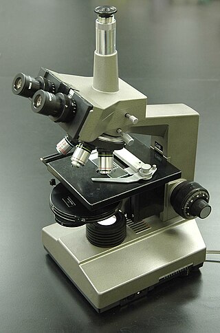
A microscope is a laboratory instrument used to examine objects that are too small to be seen by the naked eye. Microscopy is the science of investigating small objects and structures using a microscope. Microscopic means being invisible to the eye unless aided by a microscope.

Flow cytometry (FC) is a technique used to detect and measure the physical and chemical characteristics of a population of cells or particles.
A total internal reflection fluorescence microscope (TIRFM) is a type of microscope with which a thin region of a specimen, usually less than 200 nanometers can be observed.

A fluorescence microscope is an optical microscope that uses fluorescence instead of, or in addition to, scattering, reflection, and attenuation or absorption, to study the properties of organic or inorganic substances. "Fluorescence microscope" refers to any microscope that uses fluorescence to generate an image, whether it is a simple set up like an epifluorescence microscope or a more complicated design such as a confocal microscope, which uses optical sectioning to get better resolution of the fluorescence image.

Confocal microscopy, most frequently confocal laser scanning microscopy (CLSM) or laser scanning confocal microscopy (LSCM), is an optical imaging technique for increasing optical resolution and contrast of a micrograph by means of using a spatial pinhole to block out-of-focus light in image formation. Capturing multiple two-dimensional images at different depths in a sample enables the reconstruction of three-dimensional structures within an object. This technique is used extensively in the scientific and industrial communities and typical applications are in life sciences, semiconductor inspection and materials science.

Two-photon excitation microscopy is a fluorescence imaging technique that is particularly well-suited to image scattering living tissue of up to about one millimeter in thickness. Unlike traditional fluorescence microscopy, where the excitation wavelength is shorter than the emission wavelength, two-photon excitation requires simultaneous excitation by two photons with longer wavelength than the emitted light. The laser is focused onto a specific location in the tissue and scanned across the sample to sequentially produce the image. Due to the non-linearity of two-photon excitation, mainly fluorophores in the micrometer-sized focus of the laser beam are excited, which results in the spatial resolution of the image. This contrasts with confocal microscopy, where the spatial resolution is produced by the interaction of excitation focus and the confined detection with a pinhole.
High-content screening (HCS), also known as high-content analysis (HCA) or cellomics, is a method that is used in biological research and drug discovery to identify substances such as small molecules, peptides, or RNAi that alter the phenotype of a cell in a desired manner. Hence high content screening is a type of phenotypic screen conducted in cells involving the analysis of whole cells or components of cells with simultaneous readout of several parameters. HCS is related to high-throughput screening (HTS), in which thousands of compounds are tested in parallel for their activity in one or more biological assays, but involves assays of more complex cellular phenotypes as outputs. Phenotypic changes may include increases or decreases in the production of cellular products such as proteins and/or changes in the morphology of the cell. Hence HCA typically involves automated microscopy and image analysis. Unlike high-content analysis, high-content screening implies a level of throughput which is why the term "screening" differentiates HCS from HCA, which may be high in content but low in throughput.

Phase-contrast microscopy (PCM) is an optical microscopy technique that converts phase shifts in light passing through a transparent specimen to brightness changes in the image. Phase shifts themselves are invisible, but become visible when shown as brightness variations.

Stefan Walter Hell is a Romanian-German physicist and one of the directors of the Max Planck Institute for Multidisciplinary Sciences in Göttingen, and of the Max Planck Institute for Medical Research in Heidelberg, both of which are in Germany. He received the Nobel Prize in Chemistry in 2014 "for the development of super-resolved fluorescence microscopy", together with Eric Betzig and William Moerner.

Oxford Instruments Andor Ltd is a global developer and manufacturer of scientific cameras, microscopy systems and spectrographs for academic, government, and industrial applications. Founded in 1989, the company's products play a central role in the advancement of research in the fields of life sciences, physical sciences, and industrial applications. Andor was purchased for £176 million in December 2013 by Oxford Instruments. The company is based in Belfast, Northern Ireland and now employs over 400 staff across the group at its offices in Belfast, Japan, China, Switzerland and the US.
Johan Sebastiaan Ploem is a Dutch microscopist and digital artist. He made significant contribution to the field of fluorescence microscopy, and invented reflection interference contrast microscopy.

Nestor J. Zaluzec is an American scientist and inventor who works at Argonne National Laboratory. He invented and patented the Scanning Confocal Electron Microscope. and the π Steradian Transmission X-ray Detector for Electron-Optical Beam Lines and Microscopes.
Christoph Cremer is a German physicist and emeritus at the Ruprecht-Karls-University Heidelberg, former honorary professor at the University of Mainz and was a former group leader at Institute of Molecular Biology (IMB) at the Johannes Gutenberg University of Mainz, Germany, who has successfully overcome the conventional limit of resolution that applies to light based investigations by a range of different methods. In the meantime, according to his own statement, Christoph Cremer is a member of the Max Planck Institute for Chemistry and the Max Planck Institute for Polymer Research.

The Raman microscope is a laser-based microscopic device used to perform Raman spectroscopy. The term MOLE is used to refer to the Raman-based microprobe. The technique used is named after C. V. Raman, who discovered the scattering properties in liquids.

Microfluorimetry is an adaption of fluorimetry for studying the biochemical and biophysical properties of cells by using microscopy to image cell components tagged with fluorescent molecules. It is a type of microphotometry that gives a quantitative measure of the qualitative nature of fluorescent measurement and therefore, allows for definitive results that would have been previously indiscernible to the naked eye.

Cytometry is the measurement of number and characteristics of cells. Variables that can be measured by cytometric methods include cell size, cell count, cell morphology, cell cycle phase, DNA content, and the existence or absence of specific proteins on the cell surface or in the cytoplasm. Cytometry is used to characterize and count blood cells in common blood tests such as the complete blood count. In a similar fashion, cytometry is also used in cell biology research and in medical diagnostics to characterize cells in a wide range of applications associated with diseases such as cancer and AIDS.

Live-cell imaging is the study of living cells using time-lapse microscopy. It is used by scientists to obtain a better understanding of biological function through the study of cellular dynamics. Live-cell imaging was pioneered in the first decade of the 21st century. One of the first time-lapse microcinematographic films of cells ever made was made by Julius Ries, showing the fertilization and development of the sea urchin egg. Since then, several microscopy methods have been developed to study living cells in greater detail with less effort. A newer type of imaging using quantum dots have been used, as they are shown to be more stable. The development of holotomographic microscopy has disregarded phototoxicity and other staining-derived disadvantages by implementing digital staining based on cells’ refractive index.

Gail McConnell is a Scottish physicist who is Professor of Physics and director of the Centre for Biophotonics at the University of Strathclyde. She is interested in optical microscopy and novel imaging techniques, and leads the Mesolens microscope facility where her research investigates linear and non-linear optics.

J. Paul Robinson is an Australian/American educator, biologist, biomedical engineer, and expert in the applications of flow cytometry. He is a Distinguished Professor of Cytometry in the Purdue University College of Veterinary Medicine, Department of Basic Medical Sciences, a professor of Biomedical Engineering in the Weldon School of Biomedical Engineering, a professor of Computer and Information Management at Purdue University, an adjunct professor of Microbiology & Immunology at West Lafayette Center for Medical Education, Indiana University School of Medicine, and the Director of Purdue University Cytometry Laboratories.
References
- ↑ "Our services".
- ↑ "The Imaging and Cytometry Laboratory".
- ↑ "Council".
- ↑ https://rupress.org/jcb/article/140/1/71/878/Detection-of-Dimers-of-Dimers-of-Human-Leukocyte
- ↑ o'Toole, Peter J.; Morrison, Ian E.G; Cherry, Richard J. (2000). "Investigations of spectrin–lipid interactions using fluoresceinphosphatidylethanolamine as a membrane probe". Biochimica et Biophysica Acta (BBA) - Biomembranes. 1466 (1–2): 39–46. doi: 10.1016/S0005-2736(00)00168-1 . PMID 10825429.
- ↑ https://analyticalscience.wiley.com/content/news-do/peter-o-toole-taking-technology-further
- ↑ "Imaging Scientist - Peter O'Toole".
- ↑ "An interview with Peter O'Toole - part 2". 14 December 2021.
- ↑ Sturmey, R. G.; o'Toole, P. J.; Leese, H. J. (2006). "Fluorescence resonance energy transfer analysis of mitochondrial:lipid association in the porcine oocyte". Reproduction. 132 (6): 829–837. doi:10.1530/REP-06-0073. PMID 17127743.
- ↑ Marrison, Joanne; Räty, Lotta; Marriott, Poppy; O'Toole, Peter (2013). "Ptychography – a label free, high-contrast imaging technique for live cells using quantitative phase information". Scientific Reports. 3: 2369. Bibcode:2013NatSR...3E2369M. doi:10.1038/srep02369. PMC 3734479 . PMID 23917865.
- ↑ Kasprowicz, Richard; Suman, Rakesh; o'Toole, Peter (2017). "Characterising live cell behaviour: Traditional label-free and quantitative phase imaging approaches". The International Journal of Biochemistry & Cell Biology. 84: 89–95. doi:10.1016/j.biocel.2017.01.004. PMID 28111333.
- ↑ Zimmermann, Timo; Marrison, Joanne; Hogg, Karen; o'Toole, Peter (2014). "Clearing up the Signal: Spectral Imaging and Linear Unmixing in Fluorescence Microscopy". Confocal Microscopy. Methods in Molecular Biology. Vol. 1075. pp. 129–148. doi:10.1007/978-1-60761-847-8_5. ISBN 978-1-58829-351-0. PMID 24052349.
- ↑ "Light Microscopy Summer School 2024".
- ↑ "Flow Cytometry Course 2024".
- ↑ https://themicroscopists.bitesizebio.com/
- ↑ "Peter O'Toole becomes new Head of the RMS".
- ↑ "Council".
- ↑ "Under the microscope: An interview with Peter O'Toole". 20 December 2023.