Pterygoid muscle may refer to:
| This disambiguation page lists articles associated with the title Pterygoid muscle. If an internal link led you here, you may wish to change the link to point directly to the intended article. |
Pterygoid muscle may refer to:
| This disambiguation page lists articles associated with the title Pterygoid muscle. If an internal link led you here, you may wish to change the link to point directly to the intended article. |

The mandibular nerve (V3) is the largest of the three divisions of the trigeminal nerve, the fifth cranial nerve (CN V).
There are four classical muscles of mastication. During mastication, three muscles of mastication are responsible for adduction of the jaw, and one helps to abduct it. All four move the jaw laterally. Other muscles, usually associated with the hyoid, such as the mylohyoid muscle, are responsible for opening the jaw in addition to the lateral pterygoid.

In human anatomy, the masseter is one of the muscles of mastication. Found only in mammals, it is particularly powerful in herbivores to facilitate chewing of plant matter. The most obvious muscle of mastication is the masseter muscle, since it is the most superficial and one of the strongest.

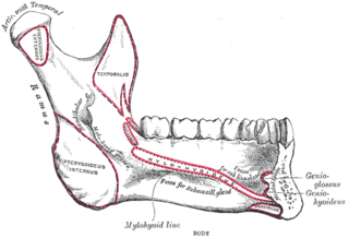
The medial pterygoid, is a thick, quadrilateral muscle of mastication.

The lateral pterygoid or external pterygoid is a muscle of mastication with two heads. It lies superiorly to the medial pterygoid.
Pterygoid, from the Greek for 'winglike', may refer to:

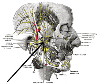
The buccal nerve is a nerve in the face. It is a branch of the mandibular nerve and transmits sensory information from skin over the buccal membrane and from the second and third molar teeth. Not to be confused with the buccal branch of the facial nerve which transmits motor information to the buccinator muscle.
The tensor veli palatini muscle is a broad, thin, ribbon-like muscle in the head that tenses the soft palate.

The pterygoid processes of the sphenoid, one on either side, descend perpendicularly from the regions where the body and the greater wings of the sphenoid bone unite.
The greater wing of the sphenoid bone, or alisphenoid, is a bony process of the sphenoid bone; there is one on each side, extending from the side of the body of the sphenoid and curving upward, laterally, and backward.

The pterygoid plexus is a venous plexus of considerable size, and is situated between the temporalis muscle and lateral pterygoid muscle, and partly between the two pterygoid muscles.

The maxillary artery supplies deep structures of the face. It branches from the external carotid artery just deep to the neck of the mandible.
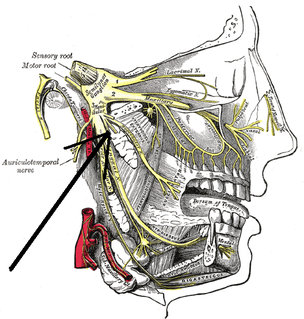
External Pterygoid Nerve : The nerve to the Pterygoideus externus frequently arises in conjunction with the buccinator nerve, but it may be given off separately from the anterior division of the mandibular nerve.

The infratemporal fossa is an irregularly shaped cavity, situated below and medial to the zygomatic arch. It is not fully enclosed by bone in all directions, and it contains superficial muscles that are visible during dissection after removing skin and fascia: namely, the lower part of the temporalis muscle, the lateral pterygoid, and the medial pterygoid.
The medial pterygoid nerve is a branch of the mandibular nerve that innervates the medial pterygoid muscle, tensor veli palatini and tensor tympani.
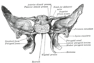
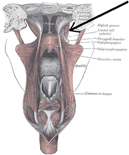
The pterygoid fossa is an anatomical term for the fossa formed by the divergence of the lateral pterygoid plate and the medial pterygoid plate of the sphenoid bone.

Marcus Gunn phenomenon, also known as Marcus Gunn jaw-winking or trigemino-oculomotor synkinesis, is an autosomal dominant condition with incomplete penetrance, in which nursing infants will have rhythmic upward jerking of their upper eyelid. This condition is characterized as a synkinesis: when two or more muscles that are independently innervated have either simultaneous or coordinated movements.

The pterygoid branches of the maxillary artery, irregular in their number and origin, supply the lateral pterygoid muscle and medial pterygoid muscle.
The pterygoid fovea is a concave surface on the uppermost medial side of the ramus of the mandible.
The Infratemporal space is a fascial space of the head and neck. It is a potential space in the side of the head, and is paired on either side. It is located posterior to the maxilla, between the lateral pterygoid plate of the sphenoid bone medially and by the base of skull superiorly. The term is derived from infra- meaning below and temporal which refers to the temporalis muscle.