Lateral pterygoid muscle

Last updated
Lateral pterygoid muscle
Musculuspterygoideuslateralis.png
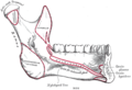
The Pterygoidei; the zygomatic arch and a portion of the ramus of the mandible have been removed (labeled as "pterygoideus externus", visible in pink at center)
Gray311.png
Sagittal section of the temporomandibular joint (labeled as "pterygoideus externus", visible in gray at bottom right)
Details
Origin Superior head: infratemporal surface of sphenoid bone. Inferior head: lateral pterygoid plate
Insertion Superior head: anterior side of the mandibular condyle. Inferior head: pterygoid fovea
Artery pterygoid branches of maxillary artery
Nerve lateral pterygoid nerve from mandibular nerve
Actions depresses and protrudes mandible, side to side movement of mandible
Identifiers
Latin musculus pterygoideus lateralis,
musculus pterygoideus externus
TA98 A04.1.04.006
TA2 2109
FMA 49015
Anatomical terms of muscle

The lateral pterygoid muscle (or external pterygoid muscle) is a muscle of mastication. It has two heads. It lies superior to the medial pterygoid muscle. It is supplied by pterygoid branches of the maxillary artery, and the lateral pterygoid nerve (from the mandibular nerve, CN V3). It depresses and protrudes the mandible. When each muscle works independently, they can move the mandible side to side.

Contents

Structure

The lateral pterygoid muscle has an upper head and a lower head. [1] [2]

It lies superior to the medial pterygoid muscle.

Blood supply

The lateral pterygoid muscle is supplied by pterygoid branches of the maxillary artery.[ citation needed ]

Nerve supply

The lateral pterygoid muscle is supplied by the lateral pterygoid nerve, a branch of the mandibular nerve (CN V3), itself a branch of the trigeminal nerve (CN V).

Function

The primary function of the lateral pterygoid muscle is to pull the head of the condyle out of the mandibular fossa along the articular eminence to protrude the mandible. [2] A concerted effort of the lateral pterygoid muscles helps in lowering the mandible and opening the jaw. Unilateral action of a lateral pterygoid muscle causes contralateral excursion (a form of mastication), usually performed in concert with the medial pterygoids.[ citation needed ] When they work independently, they can move the mandible side to side. [2]

Unlike the other three muscles of mastication, the lateral pterygoid alone can assist in depressing the mandible (opening the jaw). At the beginning of this action it is assisted by the digastric, mylohyoid and geniohyoid muscles.

Clinical significance

The lateral pterygoid muscle may be involved in temporomandibular joint dysfunction. [1] [2]

Additional images

Related Research Articles

<span class="mw-page-title-main">Temporomandibular joint</span> Joints connecting the jawbone to the skull

In anatomy, the temporomandibular joints (TMJ) are the two joints connecting the jawbone to the skull. It is a bilateral synovial articulation between the temporal bone of the skull above and the mandible below; it is from these bones that its name is derived. This joint is unique in that it is a bilateral joint that functions as one unit. Since the TMJ is connected to the mandible, the right and left joints must function together and therefore are not independent of each other.

<span class="mw-page-title-main">Mandibular nerve</span> Branch of the trigeminal nerve responsible for the lower face and jaw

In neuroanatomy, the mandibular nerve (V3) is the largest of the three divisions of the trigeminal nerve, the fifth cranial nerve (CN V). Unlike the other divisions of the trigeminal nerve (ophthalmic nerve, maxillary nerve) which contain only afferent fibers, the mandibular nerve contains both afferent and efferent fibers. These nerve fibers innervate structures of the lower jaw and face, such as the tongue, lower lip, and chin. The mandibular nerve also innervates the muscles of mastication.

<span class="mw-page-title-main">Muscles of mastication</span> Muscles that aid chewing

The four classical muscles of mastication elevate the mandible and move it forward/backward and laterally, facilitating biting and chewing. Other muscles are responsible for opening the jaw, namely the geniohyoid, mylohyoid, and digastric muscles.

<span class="mw-page-title-main">Middle meningeal artery</span> Artery supplying the meninges

The middle meningeal artery is typically the third branch of the first portion of the maxillary artery. After branching off the maxillary artery in the infratemporal fossa, it runs through the foramen spinosum to supply the dura mater and the calvaria. The middle meningeal artery is the largest of the three (paired) arteries that supply the meninges, the others being the anterior meningeal artery and the posterior meningeal artery.

<span class="mw-page-title-main">Masseter muscle</span> One of the masticatory muscles in mammals

In anatomy, the masseter is one of the muscles of mastication. Found only in mammals, it is particularly powerful in herbivores to facilitate chewing of plant matter. The most obvious muscle of mastication is the masseter muscle, since it is the most superficial and one of the strongest.

<span class="mw-page-title-main">Auriculotemporal nerve</span> Branch of the mandibular nerve

The auriculotemporal nerve is a sensory branch of the mandibular nerve (CN V3) that runs with the superficial temporal artery and vein, and provides sensory innervation to parts of the external ear, scalp, and temporomandibular joint. The nerve also conveys post-ganglionic parasympathetic fibres from the otic ganglion to the parotid gland.

<span class="mw-page-title-main">Medial pterygoid muscle</span> Muscle involved in chewing

The medial pterygoid muscle is a thick, quadrilateral muscle of the face. It is supplied by the mandibular branch of the trigeminal nerve (V). It is important in mastication (chewing).

<span class="mw-page-title-main">Pterygopalatine fossa</span> Fossa in the skull

In human anatomy, the pterygopalatine fossa is a fossa in the skull. A human skull contains two pterygopalatine fossae—one on the left side, and another on the right side. Each fossa is a cone-shaped paired depression deep to the infratemporal fossa and posterior to the maxilla on each side of the skull, located between the pterygoid process and the maxillary tuberosity close to the apex of the orbit. It is the indented area medial to the pterygomaxillary fissure leading into the sphenopalatine foramen. It communicates with the nasal and oral cavities, infratemporal fossa, orbit, pharynx, and middle cranial fossa through eight foramina.

<span class="mw-page-title-main">Sphenomandibular ligament</span>

The sphenomandibular ligament is one of the three ligaments of the temporomandibular joint. It is situated medially to - and generally separate from - the articular capsule of the joint. Superiorly, it is attached to the spine of the sphenoid bone; inferiorly, it is attached to the lingula of mandible. The SML acts to limit inferior-ward movement of the mandible.

<span class="mw-page-title-main">Pterygoid processes of the sphenoid</span> Bone plates projecting from the sphenoid bone of the skull

The pterygoid processes of the sphenoid, one on either side, descend perpendicularly from the regions where the body and the greater wings of the sphenoid bone unite.

<span class="mw-page-title-main">Greater wing of sphenoid bone</span> Large part of the skull front behind the eye socket

The greater wing of the sphenoid bone, or alisphenoid, is a bony process of the sphenoid bone, positioned in the skull behind each eye. There is one on each side, extending from the side of the body of the sphenoid and curving upward, laterally, and backward.

<span class="mw-page-title-main">Inferior alveolar artery</span>

The inferior alveolar artery is an artery of the head. It is a branch of the maxillary artery. It descends through the infratemporal fossa as part of a neurovascular bundle with the inferior alveolar nerve and vein to the mandibular foramen where it enters and passes anteriorly inside the mandible, suplying the body of mandible and the dental pulp of the lower molar and premolar teeth. Its terminal incisor branch supplies the rest of the lower teeth. Its mental branch exits the mandibula anteriorly through the mental foramen to supply adjacent lip and skin.

<span class="mw-page-title-main">Maxillary artery</span> Artery supplying face structures in humans

The maxillary artery supplies deep structures of the face. It branches from the external carotid artery just deep to the neck of the mandible.

<span class="mw-page-title-main">Squamous part of temporal bone</span> Front and upper part of the sides of the skull base

The squamous part of temporal bone, or temporal squama, forms the front and upper part of the temporal bone, and is scale-like, thin, and translucent.

<span class="mw-page-title-main">Infratemporal fossa</span> Cavity that is part of the skull

The infratemporal fossa is an irregularly shaped cavity that is a part of the skull. It is situated below and medial to the zygomatic arch. It is not fully enclosed by bone in all directions. It contains superficial muscles, including the lower part of the temporalis muscle, the lateral pterygoid muscle, and the medial pterygoid muscle. It also contains important blood vessels such as the middle meningeal artery, the pterygoid plexus, and the retromandibular vein, and nerves such as the mandibular nerve (CN V3) and its branches.

<span class="mw-page-title-main">Masseteric nerve</span> Nerve of the face

The masseteric nerve is a nerve of the face. It is a branch of the mandibular nerve (CN V3). It passes through the mandibular notch to reach masseter muscle. It provides motor innervation the masseter muscle, and sensory innervation to the temporomandibular joint.

<span class="mw-page-title-main">Mandible</span> Lower jaw bone

In jawed vertebrates, the mandible, lower jaw, or jawbone is a bone that makes up the lower – and typically more mobile – component of the mouth.

Fascial spaces are potential spaces that exist between the fasciae and underlying organs and other tissues. In health, these spaces do not exist; they are only created by pathology, e.g. the spread of pus or cellulitis in an infection. The fascial spaces can also be opened during the dissection of a cadaver. The fascial spaces are different from the fasciae themselves, which are bands of connective tissue that surround structures, e.g. muscles. The opening of fascial spaces may be facilitated by pathogenic bacterial release of enzymes which cause tissue lysis. The spaces filled with loose areolar connective tissue may also be termed clefts. Other contents such as salivary glands, blood vessels, nerves and lymph nodes are dependent upon the location of the space. Those containing neurovascular tissue may also be termed compartments.

<span class="mw-page-title-main">Pterygomandibular space</span>

The pterygomandibular space is a fascial space of the head and neck. It is a potential space in the head and is paired on each side. It is located between the lateral pterygoid muscle and the medial surface of the ramus of the mandible. The pterygomandibular space is one of the four compartments of the masticator space.

<span class="mw-page-title-main">Infratemporal space</span>

The Infratemporal space is a fascial space of the head and neck. It is a potential space in the side of the head, and is paired on either side. It is located posterior to the maxilla, between the lateral pterygoid plate of the sphenoid bone medially and by the base of skull superiorly. The term is derived from infra- meaning below and temporal which refers to the temporalis muscle.

References

  1. 1 2 Murray, Greg M.; Bhutada, Manish; Peck, Christopher C.; Phanachet, Intira; Sae-Lee, Daraporn; Whittle, Terry (1 April 2007). "The human lateral pterygoid muscle". Archives of Oral Biology . Special Issue: International Mastication Symposium, Brisbane, June 2006. 52 (4): 377–380. doi:10.1016/j.archoralbio.2006.10.002. ISSN   0003-9969. PMID   17141177.
  2. 1 2 3 4 Murray, G. M.; Phanachet, I.; Uchida, S.; Whittle, T. (2004). "The human lateral pterygoid muscle: A review of some experimental aspects and possible clinical relevance". Australian Dental Journal. 49 (1): 2–8. doi: 10.1111/j.1834-7819.2004.tb00042.x . ISSN   1834-7819. PMID   15104127.