Superior oblique muscle

Last updated
Superior oblique
Eyemuscles.png
Recti muscles:
2 = superior, 3 = inferior, 4 = medial, 5 = lateral
Oblique muscles: 6 = superior, 8 = inferior
Other muscle: 9 = levator palpebrae superioris
Other structures: 1 = Annulus of Zinn, 7 = Trochlea, 10 = Superior tarsus, 11 = Sclera, 12 = Optic nerve
Gray888.png
Details
Origin Sphenoid bone at the orbital apex, medial to optic canal
Insertion Outer posterior quadrant of the eyeball
Artery Lateral muscular branch of the ophthalmic artery
Nerve Trochlear nerve
Actions Primary action is intorsion (medial rotation); secondary actions are to abduct and depress the eyeball (i.e. it makes the eye move outward and downward)
Identifiers
Latin musculus obliquus superior bulbi
TA98 A15.2.07.016
TA2 2048
FMA 49039
Anatomical terms of muscle

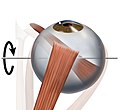
The superior oblique muscle or obliquus oculi superior is a fusiform muscle originating in the upper, medial side of the orbit (i.e. from beside the nose) which abducts, depresses and internally rotates the eye. It is the only extraocular muscle innervated by the trochlear nerve (the fourth cranial nerve).

Contents

Structure

The superior oblique muscle loops through a pulley-like structure (the trochlea of superior oblique) and inserts into the sclera on the posterotemporal surface of the eyeball. It is the pulley system that gives superior oblique its actions, causing depression of the eyeball despite being inserted on the superior surface.

Superior oblique nerve Trochlear and frontal nerves.jpg
Superior oblique nerve

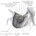
The superior oblique arises immediately above the margin of the optic foramen, superior and medial to the origin of the superior rectus, and, passing forward, ends in a rounded tendon, which plays in a fibrocartilaginous ring or pulley attached to the trochlear fossa of the frontal bone.

The contiguous surfaces of the tendon and ring are lined by a delicate mucous sheath, and enclosed in a thin fibrous investment.

The tendon is reflected caudally, laterally, and inferiorly beneath the superior rectus to the lateral part of the bulb of the eye, and is inserted onto the scleral surface, behind the equator of the eyeball, the insertion of the muscle lying between the superior rectus and lateral rectus.

Function

The primary (main) action of the superior oblique muscle is intorsion (internal rotation), [1] the secondary action is depression (primarily in the adducted position) and the tertiary action is abduction (lateral rotation).

The extraocular muscles rotate the eyeball around vertical, horizontal and antero-posterior axes. Extraocular muscles other than the medial rectus and lateral rectus have more than one action due to the angle they make with the optical axis of the eye while inserting into the eyeball. The superior and inferior oblique muscles make an angle of 51 degrees with the optical axis.[ citation needed ]

The depressing action of superior oblique (making the eye look down towards the mouth) is most effective when the eye is in an adducted position. This is because as the eye is abducted (looks laterally), the contribution made by superior oblique to depression of the eye decreases, as the inferior rectus muscle causes this movement more directly and powerfully. The main muscle for abduction is the lateral rectus, so although superior oblique contributes to a downwards and lateral eye movement, testing this motion would not be specific enough as inferior and lateral recti muscles would also be tested. Therefore, during neurological examinations, the superior oblique is tested by having the patient look inwards and downwards, testing only the depressing action of the muscle. This is a source of confusion on the subject as although clinical testing asks the patient to adduct and depress the eye, anatomically the muscle depresses and abducts it.

The great importance of intorsion and extorsion produced by the two oblique muscles can only be understood when it is considered with regards to the other muscle actions present. The two obliques prevent the eye from rotating about its long axis (retina to pupil) when the superior and inferior rectus muscles contract. This is because the orbit does not face directly forwards- the centre-line of the orbit is a little over 20 degrees out from the mid-line. But because the eyes do face forwards, when acting alone, as well as making the eye look up, superior rectus causes it to rotate slightly about the long axis, so the top of the eye moves medially (intorsion). Similarly, in addition to making the eye look down, inferior rectus would cause the eye to rotate about the long axis so the top of the eye moves slightly laterally (extorsion), if acting alone. Clearly this is undesirable as our vision would rotate when we looked up and down. For this reason, these two rectus muscles work in conjunction with the two obliques. When acting alone, superior oblique causes intorsion, inferior oblique, extorsion. Hence, when inferior rectus contracts so we look down, superior oblique also contracts to prevent extorsion of the eye, and when superior rectus contracts so we look up, inferior oblique contracts to prevent intorsion, thus the undesired rotatory actions of the inferior and superior recti about the long axis of the eye are cancelled out. This keeps our vision horizontally level, irrespective of eye position in the orbit. [2]

A montage of five pictures of the right eye of a male subject with partial heterochromia, demonstrating torsional eye movement Torsional eye movement with partial heterochromia.gif
A montage of five pictures of the right eye of a male subject with partial heterochromia, demonstrating torsional eye movement

Clinical significance

Superior oblique palsy is a common complication of closed head trauma. Restriction of superior oblique movement due to an inelastic tendon is found in Brown syndrome, leading to difficulty elevating the eye in the adducted position.

Superior oblique myokymia is an uncommon neurological condition caused by vascular compression of the trochlear nerve resulting in repeated, brief, involuntary episodes of movement of the eye.

Surgical operations of the superior oblique include tenotomy, recession, silicone expander lengthening, split tendon lengthening, tucking, and the Harada-Ito procedure.

Additional images

Related Research Articles

<span class="mw-page-title-main">Abducens nerve</span> Cranial nerve VI, for eye movements

The abducens nerve or abducent nerve, also known as the sixth cranial nerve, cranial nerve VI, or simply CN VI, is a cranial nerve in humans and various other animals that controls the movement of the lateral rectus muscle, one of the extraocular muscles responsible for outward gaze. It is a somatic efferent nerve.

<span class="mw-page-title-main">Oculomotor nerve</span> Cranial nerve III, for eye movements

The oculomotor nerve, also known as the third cranial nerve, cranial nerve III, or simply CN III, is a cranial nerve that enters the orbit through the superior orbital fissure and innervates extraocular muscles that enable most movements of the eye and that raise the eyelid. The nerve also contains fibers that innervate the intrinsic eye muscles that enable pupillary constriction and accommodation. The oculomotor nerve is derived from the basal plate of the embryonic midbrain. Cranial nerves IV and VI also participate in control of eye movement.

<span class="mw-page-title-main">Trochlear nerve</span> Cranial nerve IV, for eye movements

The trochlear nerve, also known as the fourth cranial nerve, cranial nerve IV, or CN IV, is a cranial nerve that innervates a single muscle - the superior oblique muscle of the eye. Unlike most other cranial nerves, the trochlear nerve is exclusively a motor nerve.

<span class="mw-page-title-main">Orbit (anatomy)</span> Cavity or socket of the skull in which the eye and its appendages are situated

In anatomy, the orbit is the cavity or socket/hole of the skull in which the eye and its appendages are situated. "Orbit" can refer to the bony socket, or it can also be used to imply the contents. In the adult human, the volume of the orbit is 30 millilitres, of which the eye occupies 6.5 ml. The orbital contents comprise the eye, the orbital and retrobulbar fascia, extraocular muscles, cranial nerves II, III, IV, V, and VI, blood vessels, fat, the lacrimal gland with its sac and duct, the eyelids, medial and lateral palpebral ligaments, cheek ligaments, the suspensory ligament, septum, ciliary ganglion and short ciliary nerves.

<span class="mw-page-title-main">Eye movement</span> Movement of the eyes

Eye movement includes the voluntary or involuntary movement of the eyes. Eye movements are used by a number of organisms to fixate, inspect and track visual objects of interests. A special type of eye movement, rapid eye movement, occurs during REM sleep.

<span class="mw-page-title-main">Superior rectus muscle</span> Extraocular muscle that elevates the eye

The superior rectus muscle is a muscle in the orbit. It is one of the extraocular muscles. It is innervated by the superior division of the oculomotor nerve (III). In the primary position, its primary function is elevation, although it also contributes to intorsion and adduction. It is associated with a number of medical conditions, and may be weak, paralysed, overreactive, or even congenitally absent in some people.

<span class="mw-page-title-main">Inferior rectus muscle</span>

The inferior rectus muscle is a muscle in the orbit near the eye. It is one of the four recti muscles in the group of extraocular muscles. It originates from the common tendinous ring, and inserts into the anteroinferior surface of the eye. It depresses the eye (downwards).

<span class="mw-page-title-main">Lateral rectus muscle</span> Muscle on lateral side of the eye

The lateral rectus muscle is a muscle on the lateral side of the eye in the orbit. It is one of six extraocular muscles that control the movements of the eye. The lateral rectus muscle is responsible for lateral movement of the eyeball, specifically abduction. Abduction describes the movement of the eye away from the midline, allowing the eyeball to move horizontally in the lateral direction, bringing the pupil away from the midline of the body.

<span class="mw-page-title-main">Medial rectus muscle</span> Extraocular muscle that rotates the eye medially

The medial rectus muscle is a muscle in the orbit near the eye. It is one of the extraocular muscles. It originates from the common tendinous ring, and inserts into the anteromedial surface of the eye. It is supplied by the inferior division of the oculomotor nerve (III). It rotates the eye medially (adduction).

<span class="mw-page-title-main">Inferior oblique muscle</span> Part of the eye

The inferior oblique muscle or obliquus oculi inferior is a thin, narrow muscle placed near the anterior margin of the floor of the orbit. The inferior oblique is one of the extraocular muscles, and is attached to the maxillary bone (origin) and the posterior, inferior, lateral surface of the eye (insertion). The inferior oblique is innervated by the inferior branch of the oculomotor nerve.

<span class="mw-page-title-main">Extraocular muscles</span> Seven extrinsic muscles of the human eye

The extraocular muscles or extrinsic ocular muscles are the seven extrinsic muscles of the human eye. Six of the extraocular muscles, the four recti muscles, and the superior and inferior oblique muscles, control movement of the eye and the other muscle, the levator palpebrae superioris, controls eyelid elevation. The actions of the six muscles responsible for eye movement depend on the position of the eye at the time of muscle contraction.

<span class="mw-page-title-main">Ophthalmic artery</span> Artery of the head

The ophthalmic artery (OA) is an artery of the head. It is the first branch of the internal carotid artery distal to the cavernous sinus. Branches of the ophthalmic artery supply all the structures in the orbit around the eye, as well as some structures in the nose, face, and meninges. Occlusion of the ophthalmic artery or its branches can produce sight-threatening conditions.

<span class="mw-page-title-main">Common tendinous ring</span> Ring of fibrous tissue around optic nerve at its entrance to the eye

The common tendinous ring, also known as the annulus of Zinn, or annular tendon, is a ring of fibrous tissue surrounding the optic nerve at its entrance at the apex of the orbit. It is the common origin of the four recti muscles of the group of extraocular muscles.

Trochleitis is inflammation of the superior oblique tendon trochlea apparatus characterized by localized swelling, tenderness, and severe pain. This condition is an uncommon but treatable cause of periorbital pain. The trochlea is a ring-like apparatus of cartilage through which passes the tendon of the superior oblique muscle. It is located in the superior nasal orbit and functions as a pulley for the superior oblique muscle. Inflammation of the trochlear region leads to a painful syndrome with swelling and exquisite point tenderness in the upper medial rim of the orbit. A vicious cycle may ensue such that inflammation causes swelling and fraying of the tendon which then increases the friction of passing through the trochlea which in turn adds to the inflammation. Trochleitis has also been associated with triggering or worsening of migraine attacks in patients with pre-existing migraines.

<span class="mw-page-title-main">Tenon's capsule</span> Membrane surrounding the eye forming a socket in which it moves

Tenon's capsule, also known as the Tenon capsule, fascial sheath of the eyeball or the fascia bulbi, is a thin membrane which envelops the eyeball from the optic nerve to the corneal limbus, separating it from the orbital fat and forming a socket in which it moves.

<span class="mw-page-title-main">Trochlea of superior oblique</span>

The trochlea of superior oblique is a pulley-like structure in the eye. The tendon of the superior oblique muscle passes through it. Situated on the superior nasal aspect of the frontal bone, it is the only cartilage found in the normal orbit. The word trochlea comes from the Greek word for pulley.

<span class="mw-page-title-main">Cranial nerve examination</span> Type of neurological examination

The cranial nerve exam is a type of neurological examination. It is used to identify problems with the cranial nerves by physical examination. It has nine components. Each test is designed to assess the status of one or more of the twelve cranial nerves (I-XII). These components correspond to testing the sense of smell (I), visual fields and acuity (II), eye movements and pupils, sensory function of face (V), strength of facial (VII) and shoulder girdle muscles (XI), hearing and balance, taste, pharyngeal movement and reflex, tongue movements (XII).

<span class="mw-page-title-main">Outline of human anatomy</span> Overview of and topical guide to human anatomy

The following outline is provided as an overview of and topical guide to human anatomy:

The term gaze is frequently used in physiology to describe coordinated motion of the eyes and neck. The lateral gaze is controlled by the paramedian pontine reticular formation (PPRF). The vertical gaze is controlled by the rostral interstitial nucleus of medial longitudinal fasciculus and the interstitial nucleus of Cajal.

The Parks–Bielschowsky three-step test, also known as Park's three-step test or Bielschowsky head tilt test, is a method used to isolate the paretic extraocular muscle, particularly superior oblique muscle and trochlear nerve, in acquired vertical double vision. It was originally described by Marshall M. Parks.

References

PD-icon.svgThis article incorporates text in the public domain from page 1022 of the 20th edition of Gray's Anatomy (1918)

  1. "Extraocular Muscle Actions: Eye Movements, Rectus Muscles, Oblique Muscles". 19 July 2021.
  2. Dr. Robert Acland's Atlas of Human Anatomy, University of Louisville. Volume 5: Head and Neck Part 2, Section 5: The Eye and its Surroundings.