Lacrimal sac

Last updated
Lacrimal sac
Sobo 1909 764.png
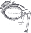
The lacrimal apparatusis shown through dissection on the left side.
Sobo 1909 765.png
The lacrimal sac has been opened showing internal organization as well as the naso-lacrymal duct.
Details
Artery angular artery
Identifiers
Latin saccus lacrimalis
TA98 A15.2.07.068
TA2 6857
FMA 20289
Anatomical terminology

The lacrimal sac or lachrymal sac [1] is the upper dilated end of the nasolacrimal duct, [2] and is lodged in a deep groove formed by the lacrimal bone and frontal process of the maxilla. It connects the lacrimal canaliculi, which drain tears from the eye's surface, and the nasolacrimal duct, which conveys this fluid into the nasal cavity. [3] Lacrimal sac occlusion leads to dacryocystitis. [4]

Contents

Structure

It is oval in form and measures from 12 to 15 mm. in length; its upper end is closed and rounded; its lower is continued into the nasolacrimal duct.

Its superficial surface is covered by a fibrous expansion derived from the medial palpebral ligament, and its deep surface is crossed by the lacrimal part of the orbicularis oculi, which is attached to the crest on the lacrimal bone.

Histology

Like the nasolacrimal duct, the sac is lined by stratified columnar epithelium with mucus-secreting goblet cells, with surrounding connective tissue. The Lacrimal Sac also drains the eye of debris and microbes.

Function

It serves as a reservoir for overflow of tears, in which the lacrimal sac pumps inward and outward driven by the orbicularis muscle during blinking.

Imaging

The lacrimal sac can be imaged by dacrocystography, in which radiocontrast is injected, followed by X-ray imaging.

Additional images

See also

Related Research Articles

<span class="mw-page-title-main">Lacrimal bone</span> Smallest and most fragile bone of the human skull and face

The lacrimal bone is a small and fragile bone of the facial skeleton; it is roughly the size of the little fingernail. It is situated at the front part of the medial wall of the orbit. It has two surfaces and four borders. Several bony landmarks of the lacrimal bone function in the process of lacrimation or crying. Specifically, the lacrimal bone helps form the nasolacrimal canal necessary for tear translocation. A depression on the anterior inferior portion of the bone, the lacrimal fossa, houses the membranous lacrimal sac. Tears or lacrimal fluid, from the lacrimal glands, collect in this sac during excessive lacrimation. The fluid then flows through the nasolacrimal duct and into the nasopharynx. This drainage results in what is commonly referred to a runny nose during excessive crying or tear production. Injury or fracture of the lacrimal bone can result in posttraumatic obstruction of the lacrimal pathways.

<span class="mw-page-title-main">Orbit (anatomy)</span> Cavity or socket of the skull in which the eye and its appendages are situated

In anatomy, the orbit is the cavity or socket/hole of the skull in which the eye and its appendages are situated. "Orbit" can refer to the bony socket, or it can also be used to imply the contents. In the adult human, the volume of the orbit is 30 millilitres, of which the eye occupies 6.5 ml. The orbital contents comprise the eye, the orbital and retrobulbar fascia, extraocular muscles, cranial nerves II, III, IV, V, and VI, blood vessels, fat, the lacrimal gland with its sac and duct, the eyelids, medial and lateral palpebral ligaments, cheek ligaments, the suspensory ligament, septum, ciliary ganglion and short ciliary nerves.

<span class="mw-page-title-main">Nasolacrimal duct</span> Carries tears from the lacrimal sac of the eye into the nasal cavity.

The nasolacrimal duct carries tears from the lacrimal sac of the eye into the nasal cavity. The duct begins in the eye socket between the maxillary and lacrimal bones, from where it passes downwards and backwards. The opening of the nasolacrimal duct into the inferior nasal meatus of the nasal cavity is partially covered by a mucosal fold.

<span class="mw-page-title-main">Lacrimal gland</span> Exocrine gland, one for each eye, that secrete tears

The lacrimal glands are paired exocrine glands, one for each eye, found in most terrestrial vertebrates and some marine mammals, that secrete the aqueous layer of the tear film. In humans, they are situated in the upper lateral region of each orbit, in the lacrimal fossa of the orbit formed by the frontal bone. Inflammation of the lacrimal glands is called dacryoadenitis. The lacrimal gland produces tears which are secreted by the lacrimal ducts, and flow over the ocular surface, and then into canals that connect to the lacrimal sac. From that sac, the tears drain through the lacrimal duct into the nose.

<span class="mw-page-title-main">Corrugator supercilii muscle</span> Muscle near the eye

The corrugator supercilii muscle is a small, narrow, pyramidal muscle of the face. It arises from the medial end of the superciliary arch; it inserts into the deep surface of the skin of the eyebrow.

<span class="mw-page-title-main">Orbicularis oculi muscle</span> Facial muscle that closes the eyelids

The orbicularis oculi is a muscle in the face that closes the eyelids. It arises from the nasal part of the frontal bone, from the frontal process of the maxilla in front of the lacrimal groove, and from the anterior surface and borders of a short fibrous band, the medial palpebral ligament.

The term Lacrimal or lachrymal, may refer to:

<span class="mw-page-title-main">Lacrimal apparatus</span> Physiological system containing the orbital structures for tear production and drainage

The lacrimal apparatus is the physiological system containing the orbital structures for tear production and drainage.
It consists of:

<span class="mw-page-title-main">Lacrimal canaliculi</span> Small channels in each eyelid that drain lacrimal fluid

The lacrimal canaliculi,, are the small channels in each eyelid that drain lacrimal fluid, from the lacrimal puncta to the lacrimal sac. This forms part of the lacrimal apparatus that drains lacrimal fluid from the surface of the eye to the nasal cavity.

<span class="mw-page-title-main">Posterior lacrimal crest</span> Bony ridge on the orbital surface of the lacrimal bone

The posterior lacrimal crest is a vertical bony ridge on the orbital surface of the lacrimal bone. It divides the bone into two parts. It gives origin to the lacrimal part of the orbicularis oculi muscle.

The anterior lacrimal crest is a bony projection on the frontal process of the maxilla. It creates the lateral margin of the lacrimal sac fossa and is continuous with the orbital margin. The medial palpebral ligament is attached to anterior lacrimal crest. It is an important structure to avoid damaging during rhinoplasty.

<span class="mw-page-title-main">Medial palpebral ligament</span> Ligament connecting the maxilla and the lacrimal bone to the eyelids

The medial palpebral ligament is a ligament of the face. It attaches to the frontal process of the maxilla, the lacrimal groove, and the tarsus of each eyelid. It has a superficial (anterior) and a deep (posterior) layer, with many surrounding attachments. It connects the medial canthus of each eyelid to the medial part of the orbit. It is a useful point of fixation during eyelid reconstructive surgery.

<span class="mw-page-title-main">Frontal process of maxilla</span> Plate which forms part of the lateral boundary of the nose

The frontal process of the maxilla is a strong plate, which projects upward, medialward, and backward from the maxilla, forming part of the lateral boundary of the nose.

<span class="mw-page-title-main">Angular artery</span> Artery of the face

The angular artery is an artery of the face. It is the terminal part of the facial artery. It ascends to the medial angle of the eye's orbit. It is accompanied by the angular vein. It ends by anastomosing with the dorsal nasal branch of the ophthalmic artery. It supplies the lacrimal sac, the orbicularis oculi muscle, and the outer side of the nose.

<span class="mw-page-title-main">Lacrimal papilla</span>

The lacrimal papilla is the small rise in the bottom (inferior) and top (superior) eyelid just before it ends at the corner of the eye closest to the nose. At the medial edge of it is the lacrimal punctum, a small hole that lets tears drain into the inside of the nose through the lacrimal canaliculi.

<span class="mw-page-title-main">Dacryocystorhinostomy</span>

Dacryocystorhinostomy (DCR) is a surgical procedure to restore the flow of tears into the nose from the lacrimal sac when the nasolacrimal duct does not function.

<span class="mw-page-title-main">Epiphora (medicine)</span> Medical condition

Epiphora is an overflow of tears onto the face, other than caused by normal crying. It is a clinical sign or condition that constitutes insufficient tear film drainage from the eyes, in that tears will drain down the face rather than through the nasolacrimal system.

<span class="mw-page-title-main">Accessory visual structures</span> External parts of the eye including eyebrow, eyelid, and lacrimal apparatus

The accessory visual structures are the protecting and supporting structures (adnexa) of the eye, including the eyebrow, eyelids, and lacrimal apparatus. The eyebrows, eyelids, eyelashes, lacrimal gland and drainage apparatus all play a crucial role with regards to globe protection, lubrication, and minimizing the risk of ocular infection. The adnexal structures also help to keep the cornea moist and clean.

<span class="mw-page-title-main">Fossa for lacrimal sac</span>

A smooth, more deeply concave depression on the lacrimal bone, which forms the medial wall of the orbital cavity, in which the lacrimal sac that drains into the nasolacrimal duct is located, is referred to as the lacrimal fossa.

<span class="mw-page-title-main">Nasolacrimal duct obstruction</span> Medical condition

Nasolacrimal duct obstruction is the obstruction of the nasolacrimal duct and may be either congenital or acquired. Obstruction of the nasolacrimal duct leads to the excess overflow of tears called epiphora.

References

PD-icon.svgThis article incorporates text in the public domain from page 1028 of the 20th edition of Gray's Anatomy (1918)

  1. Dr. Johannes Sobotta (1909). Sobotta's Atlas and Textbook of Human Anatomy. Philadelphia.{{cite book}}: CS1 maint: location missing publisher (link)
  2. Sacks, Raymond; Naidoo, Yuresh (2019-01-01). Endoscopic Dacryocystorhinostomy. pp. 143–148.e1. doi:10.1016/B978-0-323-47664-5.00017-1. ISBN   9780323476645 . Retrieved 2020-05-08. Anatomy: The lacrimal sac extends approximately 10 mm above the axilla of the middle turbinate.
  3. Remington, Lee Ann (2012). "Ocular Adnexa and Lacrimal System". Clinical Anatomy and Physiology of the Visual System. Elsevier. pp. 159–181. doi:10.1016/b978-1-4377-1926-0.10009-8. ISBN   978-1-4377-1926-0. The lacrimal sac lies within a fossa in the anterior portion of the medial orbital wall. This fossa is formed by the frontal process of the maxillary bone and the lacrimal bone. The sac is surrounded by fascia, continuous with the periorbita, which runs from the anterior to the posterior lacrimal crests.
  4. Beare, Nicholas A.V.; Bastawrous, Andrew (2014). "Ophthalmology in the Tropics and Sub-tropics". Manson's Tropical Infectious Diseases. Elsevier. pp. 952–994.e1. doi:10.1016/b978-0-7020-5101-2.00068-6. ISBN   978-0-7020-5101-2. Tears drain through the canaliculi, lacrimal sac and naso-lacrimal duct into the nose. Blockage of the naso-lacrimal duct prevents drainage of the lacrimal sac, which may lead to infection, causing a painful swelling at the side of the nose below the medial canthus. This may present as a chronically watery, discharging eye, or as an acutely inflamed abscess. This should be treated with oral broad-spectrum antibiotics. The problem may recur unless drainage into the nose is re-established.