Tears are a clear liquid secreted by the lacrimal glands found in the eyes of all land mammals. Tears are made up of water, electrolytes, proteins, lipids, and mucins that form layers on the surface of eyes. The different types of tears—basal, reflex, and emotional—vary significantly in composition.

The lacrimal bone is a small and fragile bone of the facial skeleton; it is roughly the size of the little fingernail. It is situated at the front part of the medial wall of the orbit. It has two surfaces and four borders. Several bony landmarks of the lacrimal bone function in the process of lacrimation or crying. Specifically, the lacrimal bone helps form the nasolacrimal canal necessary for tear translocation. A depression on the anterior inferior portion of the bone, the lacrimal fossa, houses the membranous lacrimal sac. Tears or lacrimal fluid, from the lacrimal glands, collect in this sac during excessive lacrimation. The fluid then flows through the nasolacrimal duct and into the nasopharynx. This drainage results in what is commonly referred to a runny nose during excessive crying or tear production. Injury or fracture of the lacrimal bone can result in posttraumatic obstruction of the lacrimal pathways.

The nasolacrimal duct carries tears from the lacrimal sac of the eye into the nasal cavity. The duct begins in the eye socket between the maxillary and lacrimal bones, from where it passes downwards and backwards. The opening of the nasolacrimal duct into the inferior nasal meatus of the nasal cavity is partially covered by a mucosal fold.
The lacrimal glands are paired exocrine glands, one for each eye, found in most terrestrial vertebrates and some marine mammals, that secrete the aqueous layer of the tear film. In humans, they are situated in the upper lateral region of each orbit, in the lacrimal fossa of the orbit formed by the frontal bone. Inflammation of the lacrimal glands is called dacryoadenitis. The lacrimal gland produces tears which are secreted by the lacrimal ducts, and flow over the ocular surface, and then into canals that connect to the lacrimal sac. From that sac, the tears drain through the lacrimal duct into the nose.

The orbicularis oculi is a muscle in the face that closes the eyelids. It arises from the nasal part of the frontal bone, from the frontal process of the maxilla in front of the lacrimal groove, and from the anterior surface and borders of a short fibrous band, the medial palpebral ligament.

The ophthalmic nerve (CN V1) is a sensory nerve of the head. It is one of three divisions of the trigeminal nerve (CN V), a cranial nerve. It has three major branches which provide sensory innervation to the eye, and the skin of the upper face and anterior scalp, as well as other structures of the head.

The lacrimal nerve is the smallest of the three main branches of the ophthalmic nerve (CN V1) (itself a branch of the trigeminal nerve (CN V)).
An imperforate lacrimal punctum is a congenital disorder of dogs involving the lack of an opening to the nasolacrimal duct in the conjunctiva. Dogs normally have two lacrimal puncta, the superior and inferior. This condition can affect either or both. Symptoms include excessive tearing and tear staining of the hair around the eye. Affected breeds include the American Cocker Spaniel, Bedlington Terrier, Golden Retriever, Poodle, and Samoyed. Imperforate lacrimal puncta can be corrected by surgical opening of the punctum.

The lacrimal artery is an artery of the orbit. It is a branch of the ophthalmic artery. It accompanies the lacrimal nerve along the upper border of the lateral rectus muscle, travelling forward to reach the lacrimal gland. It supplies the lacrimal gland, two rectus muscles of the eye, the eyelids, and the conjunctiva.

The infraorbital artery is a small artery in the head that arises from the maxillary artery and passes through the inferior orbital fissure to enter the orbit, then passes forward along the floor of the orbit, finally exiting the orbit through the infraorbital foramen to reach the face.
The lacrimal apparatus is the physiological system containing the orbital structures for tear production and drainage.
It consists of:
The lacrimal canaliculi,, are the small channels in each eyelid that drain lacrimal fluid, from the lacrimal puncta to the lacrimal sac. This forms part of the lacrimal apparatus that drains lacrimal fluid from the surface of the eye to the nasal cavity.

The medial palpebral ligament is a ligament of the face. It attaches to the frontal process of the maxilla, the lacrimal groove, and the tarsus of each eyelid. It has a superficial (anterior) and a deep (posterior) layer, with many surrounding attachments. It connects the medial canthus of each eyelid to the medial part of the orbit. It is a useful point of fixation during eyelid reconstructive surgery.

The lacrimal lake is the pool of tears in the lower conjunctival cul-de-sac, which drains into the opening of the tear drainage system. The volume of the lacrimal lake has been estimated to be between 7 and 10 μL.

The angular artery is an artery of the face. It is the terminal part of the facial artery. It ascends to the medial angle of the eye's orbit. It is accompanied by the angular vein. It ends by anastomosing with the dorsal nasal branch of the ophthalmic artery. It supplies the lacrimal sac, the orbicularis oculi muscle, and the outer side of the nose.
Punctum, plural puncta, adjective punctate, is an anatomical term for a sharp point or tip. It may also refer to:
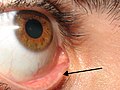
The lacrimal papilla is the small rise in the bottom (inferior) and top (superior) eyelid just before it ends at the corner of the eye closest to the nose. At the medial edge of it is the lacrimal punctum, a small hole that lets tears drain into the inside of the nose through the lacrimal canaliculi.

Epiphora is an overflow of tears onto the face, other than caused by normal crying. It is a clinical sign or condition that constitutes insufficient tear film drainage from the eyes, in that tears will drain down the face rather than through the nasolacrimal system.
Punctoplasty is a surgical procedure to restore proper drainage of tears when the lacrimal punctum becomes blocked in one or both eyes.
The accessory visual structures are the protecting and supporting structures (adnexa) of the eye, including the eyebrow, eyelids, and lacrimal apparatus. The eyebrows, eyelids, eyelashes, lacrimal gland and drainage apparatus all play a crucial role with regards to globe protection, lubrication, and minimizing the risk of ocular infection. The adnexal structures also help to keep the cornea moist and clean.