
Tears are a clear liquid secreted by the lacrimal glands found in the eyes of all land mammals. Tears are made up of water, electrolytes, proteins, lipids, and mucins that form layers on the surface of eyes. The different types of tears—basal, reflex, and emotional—vary significantly in composition.

Vestigiality is the retention, during the process of evolution, of genetically determined structures or attributes that have lost some or all of the ancestral function in a given species. Assessment of the vestigiality must generally rely on comparison with homologous features in related species. The emergence of vestigiality occurs by normal evolutionary processes, typically by loss of function of a feature that is no longer subject to positive selection pressures when it loses its value in a changing environment. The feature may be selected against more urgently when its function becomes definitively harmful, but if the lack of the feature provides no advantage, and its presence provides no disadvantage, the feature may not be phased out by natural selection and persist across species.

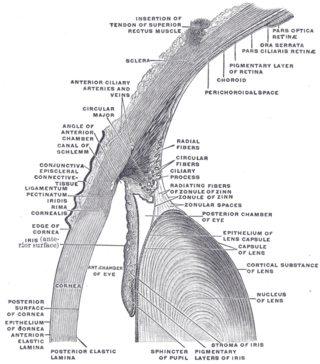
The nictitating membrane is a transparent or translucent third eyelid present in some animals that can be drawn across the eye from the medial canthus to protect and moisten it while maintaining vision. All Anura, and some reptiles, birds, and sharks have full nictitating membranes; in many mammals, a small, vestigial portion of the nictitating membrane remains in the corner of the eye. Some mammals, such as cats, beavers, polar bears, seals and aardvarks, have full nictitating membranes. Often called a third eyelid or haw, it may be referred to in scientific terminology as the plica semilunaris, membrana nictitans, or palpebra tertia.

Eye surgery, also known as ophthalmic surgery or ocular surgery, is surgery performed on the eye or its adnexa. Eye surgery is part of ophthalmology and is performed by an ophthalmologist or eye surgeon. The eye is a fragile organ, and requires due care before, during, and after a surgical procedure to minimize or prevent further damage. An eye surgeon is responsible for selecting the appropriate surgical procedure for the patient, and for taking the necessary safety precautions. Mentions of eye surgery can be found in several ancient texts dating back as early as 1800 BC, with cataract treatment starting in the fifth century BC. It continues to be a widely practiced class of surgery, with various techniques having been developed for treating eye problems.

An eyelid is a thin fold of skin that covers and protects an eye. The levator palpebrae superioris muscle retracts the eyelid, exposing the cornea to the outside, giving vision. This can be either voluntarily or involuntarily. "Palpebral" means relating to the eyelids. Its key function is to regularly spread the tears and other secretions on the eye surface to keep it moist, since the cornea must be continuously moist. They keep the eyes from drying out when asleep. Moreover, the blink reflex protects the eye from foreign bodies. A set of specialized hairs known as lashes grow from the upper and lower eyelid margins to further protect the eye from dust and debris.

Cherry eye is a disorder of the nictitating membrane (NM), also called the third eyelid, present in the eyes of dogs and cats. Cherry eye is most often seen in young dogs under the age of two. Common misnomers include adenitis, hyperplasia, adenoma of the gland of the third eyelid; however, cherry eye is not caused by hyperplasia, neoplasia, or primary inflammation. In many species, the third eyelid plays an essential role in vision by supplying oxygen and nutrients to the eye via tear production. Normally, the gland can turn inside-out without detachment. Cherry eye results from a defect in the retinaculum which is responsible for anchoring the gland to the periorbita. This defect causes the gland to prolapse and protrude from the eye as a red fleshy mass. Problems arise as sensitive tissue dries out and is subjected to external trauma Exposure of the tissue often results in secondary inflammation, swelling, or infection. If left untreated, this condition can lead to dry eye syndrome and other complications.

The nasolacrimal duct carries tears from the lacrimal sac of the eye into the nasal cavity. The duct begins in the eye socket between the maxillary and lacrimal bones, from where it passes downwards and backwards. The opening of the nasolacrimal duct into the inferior nasal meatus of the nasal cavity is partially covered by a mucosal fold.
In the anatomy of the eye, the conjunctiva is a thin mucous membrane that lines the inside of the eyelids and covers the sclera. It is composed of non-keratinized, stratified squamous epithelium with goblet cells, stratified columnar epithelium and stratified cuboidal epithelium. The conjunctiva is highly vascularised, with many microvessels easily accessible for imaging studies.

The ophthalmic nerve (CN V1) is a sensory nerve of the head. It is one of three divisions of the trigeminal nerve (CN V), a cranial nerve. It has three major branches which provide sensory innervation to the eye, and the skin of the upper face and anterior scalp, as well as other structures of the head.

The Calabar angwantibo, also known as the Calabar potto, is a strepsirrhine primate of the family Lorisidae. It shares the genus Arctocebus with the golden angwantibo. It is closely related to the potto and to the various lorises.

The golden angwantibo is a strepsirrhine primate of the family Lorisidae. It shares the genus Arctocebus with the Calabar angwantibo and together they are commonly called the golden pottos. The golden angwantibo is found in Cameroon, the Republic of Congo, Equatorial Guinea and Gabon. Its usual habitat is rain forest, but it has also been known to live on farmland.

The lacrimal caruncle, or caruncula lacrimalis, is the small, pink, globular nodule at the inner corner of the eye. It consists of tissue types of neighbouring eye structures. It may suffer from lesions and allergic inflammation.
Krause's glands or Krause glands are small, mucous accessory lacrimal glands that are found underneath the eyelid where the upper and lower conjunctivae meet. Their ducts unite into a rather long sinus which open into the fornix conjunctiva. There are approximately forty Krause glands in the region of the upper eyelid, and around 6 to 8 in the region of the lower lid. The function of these glands are to produce tears which are secreted onto the surface of the conjunctiva.

In the context of human evolution, human vestigiality involves those traits occurring in humans that have lost all or most of their original function through evolution. Although structures called vestigial often appear functionless, a vestigial structure may retain lesser functions or develop minor new ones. In some cases, structures once identified as vestigial simply had an unrecognized function. Vestigial organs are sometimes called rudimentary organs. Many human characteristics are also vestigial in other primates and related animals.
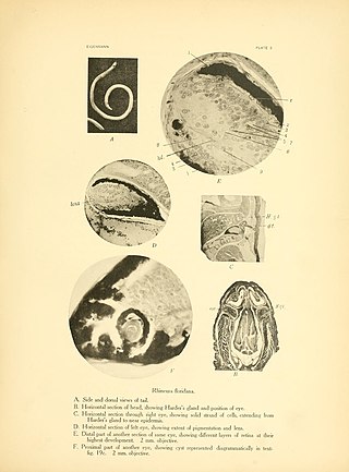
The Harderian gland is a gland found within the eye's orbit that occurs in tetrapods that possess a nictitating membrane.

The ocular immune system protects the eye from infection and regulates healing processes following injuries. The interior of the eye lacks lymph vessels but is highly vascularized, and many immune cells reside in the uvea, including mostly macrophages, dendritic cells, and mast cells. These cells fight off intraocular infections, and intraocular inflammation can manifest as uveitis or retinitis. The cornea of the eye is immunologically a very special tissue. Its constant exposure to the exterior world means that it is vulnerable to a wide range of microorganisms while its moist mucosal surface makes the cornea particularly susceptible to attack. At the same time, its lack of vasculature and relative immune separation from the rest of the body makes immune defense difficult. Lastly, the cornea is a multifunctional tissue. It provides a large part of the eye's refractive power, meaning it has to maintain remarkable transparency, but must also serve as a barrier to keep pathogens from reaching the rest of the eye, similar to function of the dermis and epidermis in keeping underlying tissues protected. Immune reactions within the cornea come from surrounding vascularized tissues as well as innate immune responsive cells that reside within the cornea.
The accessory visual structures are the protecting and supporting structures (adnexa) of the eye, including the eyebrow, eyelids, and lacrimal apparatus. The eyebrows, eyelids, eyelashes, lacrimal gland and drainage apparatus all play a crucial role with regards to globe protection, lubrication, and minimizing the risk of ocular infection. The adnexal structures also help to keep the cornea moist and clean.

Conjunctivochalasis, also known as mechanical dry eye (MDE), is a common eye surface condition characterized by the presence of excess folds of the conjunctiva located between the globe of the eye and the eyelid margin.

The sublingua ("under-tongue") is a muscular secondary tongue found below the primary tongue in tarsiers and living strepsirrhine primates, which includes lemurs and lorisoids. Although it is most fully developed in these primates, similar structures can be found in some other mammals, such as marsupials, treeshrews, and colugos. This "second tongue" lacks taste buds, and in lemuriforms, it is thought to be used to remove hair and other debris from the toothcomb, a specialized dental structure used to comb the fur during oral grooming.
