Conjunctiva

Last updated
Conjunctiva
Gray883.png
The upper half of a sagittal section through the front of the eyeball (label for 'Conjunctiva' visible at center-left)
Gray869.png
Horizontal section of the eyeball (conjunctiva labeled at upper left)
Details
Part of Eye
Artery Lacrimal artery, anterior ciliary arteries
Nerve Supratrochlear nerve
Identifiers
Latin tunica conjunctiva
MeSH D003228
TA98 A15.2.07.047
TA2 6836
FMA 59011
Anatomical terminology
Image of a human eye showing the blood vessels of the bulbar conjunctiva Beccas Oga.JPG
Image of a human eye showing the blood vessels of the bulbar conjunctiva
Hyperaemia of the superficial bulbar conjunctiva blood vessels Hyperemia conjunctiva.jpg
Hyperaemia of the superficial bulbar conjunctiva blood vessels

In the anatomy of the eye, the conjunctiva (pl.: conjunctivae) is a thin mucous membrane that lines the inside of the eyelids and covers the sclera (the white of the eye). [1] It is composed of non-keratinized, stratified squamous epithelium with goblet cells, stratified columnar epithelium and stratified cuboidal epithelium (depending on the zone). The conjunctiva is highly vascularised, with many microvessels easily accessible for imaging studies.

Contents

Structure

The conjunctiva is typically divided into three parts:

PartArea
Palpebral or tarsal conjunctivaLines the eyelids
Bulbar or ocular conjunctivaCovers the eyeball, over the anterior sclera: This region of the conjunctiva is tightly bound to the underlying sclera by Tenon's capsule and moves with the eyeball movements. The average thickness of the bulbar conjunctival membrane is 33 microns. [2]
Fornix conjunctivaForms the junction between the bulbar and palpebral conjunctivas: It is loose and flexible, allowing the free movement of the lids and eyeball. [3]

Blood supply

Blood to the bulbar conjunctiva is primarily derived from the ophthalmic artery. The blood supply to the palpebral conjunctiva (the eyelid) is derived from the external carotid artery. However, the circulations of the bulbar conjunctiva and palpebral conjunctiva are linked, so both bulbar conjunctival and palpebral conjunctival vessels are supplied by both the ophthalmic artery and the external carotid artery, to varying extents. [4]

Nerve supply

Sensory innervation of the conjunctiva is divided into four parts: [5]
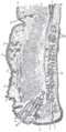
AreaNerve
Superior
Inferior Infraorbital nerve
Lateral Lacrimal nerve (with contribution from zygomaticofacial nerve)
Circumcorneal Long ciliary nerves

Microanatomy

The conjunctiva consists of unkeratinized, both stratified squamous and stratified columnar epithelium, with interspersed goblet cells. [6] The epithelial layer contains blood vessels, fibrous tissue, and lymphatic channels. [6] Accessory lacrimal glands in the conjunctiva constantly produce the aqueous portion of tears. [6] Additional cells present in the conjunctival epithelium include melanocytes, T and B cell lymphocytes. [6]

Function

The conjunctiva helps lubricate the eye by producing mucus and tears, although a smaller volume of tears than the lacrimal gland. [7] It also contributes to immune surveillance and helps to prevent the entrance of microbes into the eye.

Clinical significance

Disorders of the conjunctiva and cornea are common sources of eye complaints, in particular because the surface of the eye is exposed to various external influences and is especially susceptible to trauma, infections, chemical irritation, allergic reactions, and dryness.

Bulbar conjunctival microvasculature

Vessel morphology

The bulbar conjunctival microvasculature contains arterioles, meta-arterioles, venules, capillaries, and communicating vessels. Vessel morphology varies greatly between subjects and even between regions of the individual eyes. In some subjects, arterioles and venules can be seen to run parallel with each other. Paired arterioles are generally smaller than corresponding venules. [22] The average bulbar conjunctival vessel has been reported to be 15.1 microns, which reflects the high number of small capillaries, which are typically <10 microns in diameter. [23]

Blood oxygen dynamics

The bulbar conjunctival microvasculature is in close proximity to ambient air, thus oxygen diffusion from ambient air strongly influences their blood oxygen saturation. Because of oxygen diffusion, hypoxic bulbar conjunctival vessels will rapidly reoxygenate (in under 10 seconds) when exposed to ambient air (i.e. when the eyelid is open). Closing the eyelid stops this oxygen diffusion by placing a barrier between the bulbar conjunctival microvessels and ambient air. [24]

Blood vessel imaging methods

The bulbar conjunctival microvessels are typically imaged with a high-magnification slit lamp with green filters. [25] [26] [27] With such high-magnification imaging systems, it is possible to see groups of individual red blood cells flowing in vivo. [25] Fundus cameras may also be used for low-magnification wide field-of-view imaging of the bulbar conjunctival microvasculature. Modified fundus cameras have been used to measure conjunctival blood flow [28] and to measure blood oxygen saturation. [24] Fluorescein angiography has been used to study the blood flow of the bulbar conjunctiva and to differentiate the bulbar conjunctival and episcleral microcirculation. [29] [30] [31]

Vasodilation

The bulbar conjunctival microvasculature is known to dilate in response to several stimuli and external conditions, including allergens (e.g. pollen), [32] temperature, [33] time-of-day, [33] contact-lens wear, [13] and acute mild hypoxia. [24] Bulbar conjunctival vasodilation has also been shown to correlate changes in emotional state. [34]

Type 2 diabetes is associated with an increase in average bulbar conjunctival vessel diameter and capillary loss. [11] [12] Sickle-cell anemia is associated with altered average vessel diameter. [14]

See also

Additional images

Related Research Articles

<span class="mw-page-title-main">Artery</span> Blood vessels that carry blood away from the heart

An artery is a blood vessel in humans and most other animals that takes oxygenated blood away from the heart in the systemic circulation to one or more parts of the body. Exceptions that carry deoxygenated blood are the pulmonary arteries in the pulmonary circulation that carry blood to the lungs for oxygenation, and the umbilical arteries in the fetal circulation that carry deoxygenated blood to the placenta. It consists of a multi-layered artery wall wrapped into a tube-shaped channel.

<span class="mw-page-title-main">Blood vessel</span> Tubular structure of circulatory system

Blood vessels are the structures of the circulatory system that transport blood throughout the human body. These vessels transport blood cells, nutrients, and oxygen to the tissues of the body. They also take waste and carbon dioxide away from the tissues. Blood vessels are needed to sustain life, because all of the body's tissues rely on their functionality.

<span class="mw-page-title-main">Capillary</span> Smallest type of blood vessel

A capillary is a small blood vessel, from 5 to 10 micrometres in diameter, and is part of the microcirculation system. Capillaries are microvessels and the smallest blood vessels in the body. They are composed of only the tunica intima, consisting of a thin wall of simple squamous endothelial cells. They are the site of the exchange of many substances from the surrounding interstitial fluid, and they convey blood from the smallest branches of the arteries (arterioles) to those of the veins (venules). Other substances which cross capillaries include water, oxygen, carbon dioxide, urea, glucose, uric acid, lactic acid and creatinine. Lymph capillaries connect with larger lymph vessels to drain lymphatic fluid collected in microcirculation.

<span class="mw-page-title-main">Diabetic retinopathy</span> Diabetes-induced damage to the retina of the eye

Diabetic retinopathy, is a medical condition in which damage occurs to the retina due to diabetes. It is a leading cause of blindness in developed countries.

<span class="mw-page-title-main">Microangiopathy</span> Medical condition

Microangiopathy is a disease of the microvessels, small blood vessels in the microcirculation. It can be contrasted to macroangiopathies such as atherosclerosis, where large and medium-sized arteries are primarily affected.

<span class="mw-page-title-main">Juxtaglomerular apparatus</span> Structure that regulates function of each nephron

The juxtaglomerular apparatus is a structure in the kidney that regulates the function of each nephron, the functional units of the kidney. The juxtaglomerular apparatus is named because it is next to (juxta-) the glomerulus.

<span class="mw-page-title-main">Arteriole</span> Small arteries in the microcirculation

An arteriole is a small-diameter blood vessel in the microcirculation that extends and branches out from an artery and leads to capillaries.

Diabetic angiopathy is a form of angiopathy associated with diabetic complications. While not exclusive, the two most common forms are diabetic retinopathy and diabetic nephropathy, whose pathophysiologies are largely identical. Other forms of diabetic angiopathy include diabetic neuropathy and diabetic cardiomyopathy.

The capillary lamina of choroid or choriocapillaris is a part of the choroid of the eye. It is a layer of capillaries immediately adjacent to Bruch's membrane of the choroid. The choriocapillaris consists of a dense network of freely anastomosing highly permeable fenestrated large-calibre capillaries. It nourishes the outer avascular layers of the retina.

Neovascularization is the natural formation of new blood vessels, usually in the form of functional microvascular networks, capable of perfusion by red blood cells, that form to serve as collateral circulation in response to local poor perfusion or ischemia.

<span class="mw-page-title-main">Superior limbic keratoconjunctivitis</span> Medical condition

Superior limbic keratoconjunctivitis is a disease of the eye characterized by episodes of recurrent inflammation of the superior cornea and limbus, as well as of the superior tarsal and bulbar conjunctiva. It was first described by F. H. Théodore in 1963.

<span class="mw-page-title-main">Vernal keratoconjunctivitis</span> Medical condition

Vernal keratoconjunctivitis is a recurrent, bilateral, and self-limiting type of conjunctivitis having a periodic seasonal incidence.

<span class="mw-page-title-main">Corneal ulcer</span> Medical condition of the eye

Corneal ulcer, also called keratitis, is an inflammatory or, more seriously, infective condition of the cornea involving disruption of its epithelial layer with involvement of the corneal stroma. It is a common condition in humans particularly in the tropics and in farming. In developing countries, children afflicted by vitamin A deficiency are at high risk for corneal ulcer and may become blind in both eyes persisting throughout life. In ophthalmology, a corneal ulcer usually refers to having an infection, while the term corneal abrasion refers more to a scratch injury.

<span class="mw-page-title-main">Cotton wool spots</span> Medical condition of the eye

Cotton wool spots are opaque fluffy white patches on the retina of the eye that are considered an abnormal finding during a funduscopic exam. Cotton wool spots are typically a sign of another disease state, most common of which is diabetic retinopathy. The irregularly shaped white patches are a result of ischemia, or reduced blood flow and oxygen, in the retinal nerve fiber layer, which is located in the distribution of the capillaries of the superficial layer of the retina. These areas with reduced blood flow reflect the obstruction of axoplasmic flow due to mechanical or vascular causes and the consequential accumulation as a result of decreased axonal transport. This reduced axonal transport can then cause swelling or bulging on the surface layer of the retina, increasing the potential for nerve fiber damage.

No reflow phenomenon is the failure of blood to reperfuse an ischemic area after the physical obstruction has been removed or bypassed. The underlying mechanism is related to arterial microvasculature damage. It is primarily seen during percutaneous coronary intervention (PCI) in the setting of acute myocardial infarction (AMI), but has also been observed in other organs, including the brain and kidneys. Coronary no-reflow phenomenon is specifically related to reduced antegrade coronary blood flow despite proximal coronary artery patency. It is an independent predictor of worse clinical outcomes including heart failure, fatal arrhythmias, myocardial infarction, and increased mortality rates.

<span class="mw-page-title-main">Symblepharon</span> Medical condition

A symblepharon is a partial or complete adhesion of the palpebral conjunctiva of the eyelid to the bulbar conjunctiva of the eyeball. It results either from disease or trauma. Cicatricial pemphigoid and, in severe cases, rosacea may cause symblepharon. It is rarely congenital. Its treatment is symblepharectomy.

<span class="mw-page-title-main">Accessory visual structures</span> External parts of the eye including eyebrow, eyelid, and lacrimal apparatus

The accessory visual structures are the protecting and supporting structures (adnexa) of the eye, including the eyebrow, eyelids, and lacrimal apparatus. The eyebrows, eyelids, eyelashes, lacrimal gland and drainage apparatus all play a crucial role with regards to globe protection, lubrication, and minimizing the risk of ocular infection. The adnexal structures also help to keep the cornea moist and clean.

Conjunctival squamous cell carcinoma and corneal intraepithelial neoplasia comprise ocular surface squamous neoplasia (OSSN). SCC is the most common malignancy of the conjunctiva in the US, with a yearly incidence of 1–2.8 per 100,000. Risk factors for the disease are exposure to sun, exposure to UVB, and light-colored skin. Other risk factors include radiation, smoking, HPV, arsenic, and exposure to polycyclic hydrocarbons.

Retinal vessel analysis is a non-invasive method to examine the small arteries and veins in the retina which allows to draw conclusions about the morphology and the function of small vessels elsewhere in the human body. Retinal vessel analysis is conducted mainly by ophthalmologists, cardiologists, neurologists and other medical specialities dealing with vascular diseases.

Sickle cell retinopathy can be defined as retinal changes due to blood vessel damage in the eye of a person with a background of sickle cell disease. It can likely progress to loss of vision in late stages due to vitreous hemorrhage or retinal detachment. Sickle cell disease is a structural red blood cell disorder leading to consequences in multiple systems. It is characterized by chronic red blood cell destruction, vascular injury, and tissue ischemia causing damage to the brain, eyes, heart, lungs, kidneys, spleen, and musculoskeletal system.

References

  1. "Conjunctiva". www.sciencedirect.com. Retrieved 4 August 2022.
  2. Efron N, Al-Dossari M, Pritchard N (May 2009). "In vivo confocal microscopy of the bulbar conjunctiva". Clinical & Experimental Ophthalmology. 37 (4): 335–44. doi:10.1111/j.1442-9071.2009.02065.x. PMID   19594558. S2CID   35398240.
  3. Eye, human Encyclopædia Britannica
  4. 1 2 Pavlou AT, Wolff HG (July 1959). "The bulbar conjunctival vessels in occlusion of the internal carotid artery". A.M.A. Archives of Internal Medicine. 104 (1): 53–60. doi:10.1001/archinte.1959.00270070055007. PMID   13660526.
  5. "Table 1: Summary of sensory nerve supply". Archived from the original on February 14, 2013. Retrieved July 31, 2016.
  6. 1 2 3 4 Goldman L (2012). Goldman's Cecil Medicine (24th ed.). Philadelphia: Elsevier Saunders. p.  2426. ISBN   978-1437727883.
  7. London Place Eye Center (2003). Conjunctivitis Archived 2004-08-08 at the Wayback Machine . Retrieved July 25, 2004.
  8. Khansari MM, Wanek J, Tan M, Joslin CE, Kresovich JK, Camardo N, et al. (April 2017). "Assessment of Conjunctival Microvascular Hemodynamics in Stages of Diabetic Microvasculopathy". Scientific Reports. 7: 45916. Bibcode:2017NatSR...745916K. doi:10.1038/srep45916. PMC   5384077 . PMID   28387229.
  9. Khansari MM, O'Neill W, Penn R, Chau F, Blair NP, Shahidi M (July 2016). "Automated fine structure image analysis method for discrimination of diabetic retinopathy stage using conjunctival microvasculature images". Biomedical Optics Express. 7 (7): 2597–606. doi:10.1364/BOE.7.002597. PMC   4948616 . PMID   27446692.
  10. Isenberg SJ, McRee WE, Jedrzynski MS (October 1986). "Conjunctival hypoxia in diabetes mellitus". Investigative Ophthalmology & Visual Science. 27 (10): 1512–5. PMID   3759367.
  11. 1 2 Fenton BM, Zweifach BW, Worthen DM (September 1979). "Quantitative morphometry of conjunctival microcirculation in diabetes mellitus". Microvascular Research. 18 (2): 153–66. doi:10.1016/0026-2862(79)90025-6. PMID   491983.
  12. 1 2 Ditzel J (1967-01-12). "The in vivo reactions of the small blood vessels to diabetes mellitus". Acta Medica Scandinavica. Supplementum. 476 (S476): 123–34. doi:10.1111/j.0954-6820.1967.tb12691.x. PMID   5236035.
  13. 1 2 Cheung AT, Ramanujam S, Greer DA, Kumagai LF, Aoki TT (2001-10-01). "Microvascular abnormalities in the bulbar conjunctiva of patients with type 2 diabetes mellitus". Endocrine Practice. 7 (5): 358–63. doi:10.4158/EP.7.5.358. PMID   11585371.
  14. 1 2 Fink AI (1968-01-01). "Vascular changes in the bulbar conjunctiva associated with sickle-cell disease: some observations on fine structure". Transactions of the American Ophthalmological Society. 66: 788–826. PMC   1310317 . PMID   5720854.
  15. Isenberg SJ, McRee WE, Jedrzynski MS, Gange SN, Gange SL (January 1987). "Effects of sickle cell anemia on conjunctival oxygen tension and temperature". Archives of Internal Medicine. 147 (1): 67–9. doi:10.1001/archinte.147.1.67. PMID   3800533.
  16. Wanek J, Gaynes B, Lim JI, Molokie R, Shahidi M (August 2013). "Human bulbar conjunctival hemodynamics in hemoglobin SS and SC disease". American Journal of Hematology. 88 (8): 661–4. doi:10.1002/ajh.23475. PMC   4040222 . PMID   23657867.
  17. Harper RN, Moore MA, Marr MC, Watts LE, Hutchins PM (November 1978). "Arteriolar rarefaction in the conjunctiva of human essential hypertensives". Microvascular Research. 16 (3): 369–72. doi:10.1016/0026-2862(78)90070-5. PMID   748720.
  18. Lee RE (August 1955). "Anatomical and physiological aspects of the capillary bed in the bulbar conjunctiva of man in health and disease". Angiology. 6 (4): 369–82. doi:10.1177/000331975500600408. PMID   13275744. S2CID   11589129.
  19. "Conjunctivochalasis - Medical Definition". Medilexicon.com. Archived from the original on 2016-03-03. Retrieved 2012-11-13.
  20. Hughes WL (January 1942). "Conjunctivochalasis". American Journal of Ophthalmology. 25 (1): 48–51. doi:10.1016/S0002-9394(42)93297-5.
  21. Varde MA, Biswas J (January 2009). "Ocular surface tumors". Oman Journal of Ophthalmology. 2 (1): 1–2. doi: 10.4103/0974-620X.48414 . PMC   3018098 . PMID   21234216.
  22. Meighan SS (September 1956). "Blood vessels of the bulbar conjunctiva in man". The British Journal of Ophthalmology. 40 (9): 513–26. doi:10.1136/bjo.40.9.513. PMC   1324675 . PMID   13364178.
  23. Shahidi M, Wanek J, Gaynes B, Wu T (March 2010). "Quantitative assessment of conjunctival microvascular circulation of the human eye". Microvascular Research. 79 (2): 109–13. doi:10.1016/j.mvr.2009.12.003. PMC   3253734 . PMID   20053367.
  24. 1 2 3 MacKenzie LE, Choudhary TR, McNaught AI, Harvey AR (August 2016). "In vivo oximetry of human bulbar conjunctival and episcleral microvasculature using snapshot multispectral imaging" (PDF). Experimental Eye Research. 149: 48–58. doi:10.1016/j.exer.2016.06.008. PMID   27317046. S2CID   25038785.
  25. 1 2 van Zijderveld R, Ince C, Schlingemann RO (May 2014). "Orthogonal polarization spectral imaging of conjunctival microcirculation". Graefe's Archive for Clinical and Experimental Ophthalmology. 252 (5): 773–9. doi:10.1007/s00417-014-2603-9. PMID   24627137. S2CID   1595902.
  26. Khansari MM, O'Neill W, Penn R, Chau F, Blair NP, Shahidi M (July 2016). "Automated fine structure image analysis method for discrimination of diabetic retinopathy stage using conjunctival microvasculature images". Biomedical Optics Express. 7 (7): 2597–606. doi:10.1364/BOE.7.002597. PMC   4948616 . PMID   27446692.
  27. Khansari MM, Wanek J, Felder AE, Camardo N, Shahidi M (February 2016). "Automated Assessment of Hemodynamics in the Conjunctival Microvasculature Network". IEEE Transactions on Medical Imaging. 35 (2): 605–11. doi:10.1109/TMI.2015.2486619. PMC   4821773 . PMID   26452274.
  28. Jiang H, Ye Y, DeBuc DC, Lam BL, Rundek T, Tao A, et al. (January 2013). "Human conjunctival microvasculature assessed with a retinal function imager (RFI)". Microvascular Research. 85: 134–7. doi:10.1016/j.mvr.2012.10.003. PMC   3534915 . PMID   23084966.
  29. Meyer PA (1988-01-01). "Patterns of blood flow in episcleral vessels studied by low-dose fluorescein videoangiography". Eye. 2 ( Pt 5) (5): 533–46. doi: 10.1038/eye.1988.104 . PMID   3256492.
  30. Ormerod LD, Fariza E, Webb RH (1995-01-01). "Dynamics of external ocular blood flow studied by scanning angiographic microscopy". Eye. 9 ( Pt 5) (5): 605–14. doi: 10.1038/eye.1995.148 . PMID   8543081.
  31. Meyer PA, Watson PG (January 1987). "Low dose fluorescein angiography of the conjunctiva and episclera". The British Journal of Ophthalmology. 71 (1): 2–10. doi:10.1136/bjo.71.1.2. PMC   1041073 . PMID   3814565.
  32. Horak F, Berger U, Menapace R, Schuster N (September 1996). "Quantification of conjunctival vascular reaction by digital imaging". The Journal of Allergy and Clinical Immunology. 98 (3): 495–500. doi:10.1016/S0091-6749(96)70081-7. PMID   8828525.
  33. 1 2 Duench S, Simpson T, Jones LW, Flanagan JG, Fonn D (June 2007). "Assessment of variation in bulbar conjunctival redness, temperature, and blood flow". Optometry and Vision Science. 84 (6): 511–6. doi:10.1097/OPX.0b013e318073c304. PMID   17568321. S2CID   943038.
  34. Provine RR, Nave-Blodgett J, Cabrera MO (2013-11-01). "The Emotional Eye: Red Sclera as a Uniquely Human Cue of Emotion". Ethology. 119 (11): 993–998. Bibcode:2013Ethol.119..993P. doi:10.1111/eth.12144. ISSN   1439-0310.