Supratrochlear nerve

Last updated
Supratrochlear nerve
Gray784.png
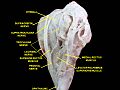
Sensory areas of the head, showing the general distribution of the three divisions of the fifth nerve. (Supratrochlear nerve labeled at upper left.)
Gray776.png
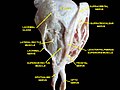
Nerves of the orbit. Seen from above. (Supratrochlear nerve visible near top.)
Details
From Frontal nerve
Identifiers
Latin nervus supratrochlearis
TA98 A14.2.01.024
TA2 6203
FMA 52642
Anatomical terms of neuroanatomy

The supratrochlear nerve is a branch of the frontal nerve, itself a branch of the ophthalmic nerve (CN V1) from the trigeminal nerve (CN V). It provides sensory innervation to the skin of the forehead and the upper eyelid.

Contents

Structure

Origin

The supratrochlear nerve is the smaller of the two terminal branches of the frontal nerve (the other being the supraorbital nerve). [1] It arises midway between the base and apex of the orbit [2] where the frontal nerve splits into said terminal branches. [1]

Course

The supratrochlear nerve passes medially [3] above the trochlea of the superior oblique muscle. [2] [3] It then travels anteriorly above the levator palpebrae superioris muscle. [1] It exits the orbit through the supraorbital notch or foramen. [3] It then ascends onto the forehead beneath the corrugator supercilii muscle and frontalis muscle. It finally divides into sensory branches.[ citation needed ]

The supratrochlear nerve travels with the supratrochlear artery, a branch of the ophthalmic artery. [2]

Branches

Before exiting the orbit, the supratrochlear nerve emits a descending branch to the infratrochlear nerve. [3] [1]

Function

The supratrochlear nerve provides sensory innervation to the skin and conjunctiva of the upper eyelid, and the skin of the inferomedial forehead. [1] It may also provide sensory innervation to part of the periosteum of the frontal bone. [4]

Clinical significance

The supratrochlear nerve may be anaesthetised for surgery of parts of the scalp. [5] [6] This can be used for small lesions of the scalp. [5] It can also be used for more extensive injury to the scalp. [6] It is often anaesthetised alongside the supraorbital artery. [5]

Etymology

The supratrochlear nerve is named for its passage above the trochlea of the superior oblique muscle. [2]

Additional images

Related Research Articles

<span class="mw-page-title-main">Lesser occipital nerve</span> Nerve of the head and neck

The lesser occipital nerve is a cutaneous spinal nerve of the cervical plexus. It arises from second cervical (spinal) nerve (C2). It innervates the skin of the back of the upper neck and of the scalp posterior to the ear.

<span class="mw-page-title-main">Mandibular nerve</span> Branch of the trigeminal nerve responsible for the lower face and jaw

In neuroanatomy, the mandibular nerve (V3) is the largest of the three divisions of the trigeminal nerve, the fifth cranial nerve (CN V). Unlike the other divisions of the trigeminal nerve (ophthalmic nerve, maxillary nerve) which contain only afferent fibers, the mandibular nerve contains both afferent and efferent fibers. These nerve fibers innervate structures of the lower jaw and face, such as the tongue, lower lip, and chin. The mandibular nerve also innervates the muscles of mastication.

<span class="mw-page-title-main">Supraorbital nerve</span> Branch of the frontal nerve

The supraorbital nerve is one of two branches of the frontal nerve, itself a branch of the ophthalmic nerve. The other branch of the frontal nerve is the supratrochlear nerve.

<span class="mw-page-title-main">Scalp</span> Anatomical area bordered by the human face at the front, and by the neck at the sides and back

The scalp is the anatomical area bordered by the human face at the front, and by the neck at the sides and back.

<span class="mw-page-title-main">Levator palpebrae superioris muscle</span> Muscle in orbit that elevates upper eyelid

The levator palpebrae superioris is the muscle in the orbit that elevates the upper eyelid.

<span class="mw-page-title-main">Medial rectus muscle</span> Extraocular muscle that rotates the eye medially

The medial rectus muscle is a muscle in the orbit near the eye. It is one of the extraocular muscles. It originates from the common tendinous ring, and inserts into the anteromedial surface of the eye. It is supplied by the inferior division of the oculomotor nerve (III). It rotates the eye medially (adduction).

<span class="mw-page-title-main">Superior orbital fissure</span> Foramen in the skull allowing for passage of cranial nerve

The superior orbital fissure is a foramen or cleft of the skull between the lesser and greater wings of the sphenoid bone. It gives passage to multiple structures, including the oculomotor nerve, trochlear nerve, ophthalmic nerve, abducens nerve, ophthalmic veins, and sympathetic fibres from the cavernous plexus.

<span class="mw-page-title-main">Ophthalmic nerve</span> Sensory nerve of the face

The ophthalmic nerve (CN V1) is a sensory nerve of the head. It is one of three divisions of the trigeminal nerve (CN V), a cranial nerve. It has major three branches which provide sensory innervation to the eye, and the skin of the upper face and anterior scalp, as well as other structures of the head.

<span class="mw-page-title-main">Nasociliary nerve</span> Branch of the ophthalmic nerve

The nasociliary nerve is a branch of the ophthalmic nerve (CN V1) (which is in turn a branch of the trigeminal nerve (CN V)). It is intermediate in size between the other two branches of the ophthalmic nerve, the frontal nerve and lacrimal nerve.

<span class="mw-page-title-main">Occipitofrontalis muscle</span> Facial muscle helping to create facial expressions

The occipitofrontalis muscle is a muscle which covers parts of the skull. It consists of two parts or bellies: the occipital belly, near the occipital bone, and the frontal belly, near the frontal bone. It is supplied by the supraorbital artery, the supratrochlear artery, and the occipital artery. It is innervated by the facial nerve. In humans, the occipitofrontalis helps to create facial expressions.

<span class="mw-page-title-main">Frontal nerve</span> Nerve supplying the forehead, frontal sinus, and upper eyelid

The frontal nerve is the largest branch of the ophthalmic nerve (V1), itself a branch of the trigeminal nerve (CN V). It supplies sensation to the skin of the forehead, the mucosa of the frontal sinus, and the skin of the upper eyelid. It may be affected by schwannoma.

<span class="mw-page-title-main">Lacrimal nerve</span> Branch of the ophthalmic nerve

The lacrimal nerve is the smallest of the three main branches of the ophthalmic nerve (CN V1) (itself a branch of the trigeminal nerve (CN V)).

<span class="mw-page-title-main">Zygomatic nerve</span> Nerve of the face

The zygomatic nerve is a branch of the maxillary nerve. It arises in the pterygopalatine fossa and enters the orbit through the inferior orbital fissure before dividing into its two terminal branches: the zygomaticotemporal nerve and zygomaticofacial nerve.

<span class="mw-page-title-main">Infratrochlear nerve</span>

The infratrochlear nerve is a branch of the nasociliary nerve (itself a branch of the ophthalmic nerve (CN V1)) in the orbit. It exits the orbit inferior to the trochlea of superior oblique. It provides sensory innervation to structures of the orbit and skin of adjacent structures.

<span class="mw-page-title-main">Zygomaticotemporal nerve</span> Small nerve of the face

The zygomaticotemporal nerve (zygomaticotemporal branch, temporal branch) is a cutaneous (sensory) nerve of the head. It is a branch of the zygomatic nerve (itself a branch of the maxillary nerve (CN V2)). It arises in the orbit and exits the orbit through the zygomaticotemporal foramen in the zygomatic bone to enter the temporal fossa. It is distributed to the skin of the side of the forehead. It also contains a parasympathetic secretomotor component for the lacrimal gland which it confers to the lacrimal nerve (which then delivers it to the gland).

<span class="mw-page-title-main">Mylohyoid nerve</span> Nerve of the head

The mylohyoid nerve is a mixed nerve of the head. It is a branch of the inferior alveolar nerve. It provides motor innervation the mylohyoid muscle, and the anterior belly of the digastric muscle. It provides sensory innervation to part of the submental area, and sometimes also the mandibular (lower) molar teeth, requiring local anaesthesia for some oral procedures.

<span class="mw-page-title-main">Posterior ethmoidal nerve</span> Nerve of the orbit around the eye

The posterior ethmoidal nerve is a nerve of the head. It is a branch of the nasociliary nerve (itself a branch of the ophthalmic nerve (CN V1)). It provides sensory innervation to the sphenoid sinus and ethmoid sinus, and part of the dura mater in the anterior cranial fossa.

<span class="mw-page-title-main">Supraorbital artery</span>

The supraorbital artery is a branch of the ophthalmic artery. It passes anteriorly within the orbit to exit the orbit through the supraorbital foramen or notch alongside the supraorbital nerve, splitting into two terminal branches which go on to form anastomoses with arteries of the head.

<span class="mw-page-title-main">Supratrochlear artery</span>

The supratrochlear artery is one of the terminal branches of the ophthalmic artery. It arises within the orbit. It exits the orbit alongside the supratrochlear nerve. It contributes arterial supply to the skin, muscles and pericranium of the forehead.

<span class="mw-page-title-main">Zygomatic branches of the facial nerve</span> Nerves of the face

The zygomatic branches of the facial nerve (malar branches) are nerves of the face. They run across the zygomatic bone to the lateral angle of the orbit. Here, they supply the orbicularis oculi muscle, and join with filaments from the lacrimal nerve and the zygomaticofacial branch of the maxillary nerve (CN V2).

References

PD-icon.svgThis article incorporates text in the public domain from page 888 of the 20th edition of Gray's Anatomy (1918)

  1. 1 2 3 4 5 Fillmore, Erin P.; Seifert, Mark F. (2015). "22 - Anatomy of the Trigeminal Nerve". Nerves and Nerve Injuries. Vol. 1: History, Embryology, Anatomy, Imaging, and Diagnostics. Academic Press. pp. 319–350. doi:10.1016/B978-0-12-410390-0.00023-8. ISBN   978-0-12-410390-0.
  2. 1 2 3 4 Rea, Paul (2016). "2 - Head". Essential Clinically Applied Anatomy of the Peripheral Nervous System in the Head and Neck. Academic Press. pp. 21–130. doi:10.1016/B978-0-12-803633-4.00002-8. ISBN   978-0-12-803633-4.
  3. 1 2 3 4 Standring, Susan (2020). Gray's Anatomy: The Anatomical Basis of Clinical Practice (42nd ed.). New York. p. 782. ISBN   978-0-7020-7707-4. OCLC   1201341621.{{cite book}}: CS1 maint: location missing publisher (link)
  4. Barral, Jean-Pierre; Croibier, Alain (2009). "15 - Ophthalmic nerve". Manual Therapy for the Cranial Nerves. Churchill Livingstone. pp. 115–128. doi:10.1016/B978-0-7020-3100-7.50018-5. ISBN   978-0-7020-3100-7.
  5. 1 2 3 Kinder Ross, Alison; Bryskin, Robert B. (2011). "16 - Regional Anesthesia". Smith's Anesthesia for Infants and Children (8th ed.). Mosby (imprint). pp. 452–510. doi:10.1016/B978-0-323-06612-9.00016-X. ISBN   978-0-323-06612-9.
  6. 1 2 Trott, Alexander T. (2012). "6 - Infiltration and Nerve Block Anesthesia". Wounds and Lacerations - Emergency Care and Closure (4th ed.). Saunders. pp. 41–72. doi:10.1016/B978-0-323-07418-6.00006-X. ISBN   978-0-323-07418-6.