Inferior alveolar nerve

Last updated
Inferior alveolar nerve
Gray778.png
Distribution of the maxillary and mandibular nerves, and the submaxillary ganglion. (Inferior alveolar visible at center left.)
Gray781.png
Mandibular division of the trigeminal nerve. (Inferior alveolar labeled at bottom right.)
Details
From mandibular nerve
To mylohyoid, dental, incisive, and mental
Innervates dental alveolus
Identifiers
Latin nervus alveolaris inferior
TA98 A14.2.01.089
TA2 6274
FMA 53243
Anatomical terms of neuroanatomy

The inferior alveolar nerve (IAN) (also the inferior dental nerve) is a sensory [1] [ contradictory ] branch of the mandibular nerve (CN V3) (which is itself the third branch of the trigeminal nerve (CN V)). The nerve provides sensory innervation to the lower/mandibular teeth and their corresponding gingiva as well as a small area of the face (via its mental nerve).

Contents

Structure

Origin

The inferior alveolar nerve arises from the mandibular nerve. [2] :543

Course

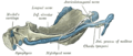
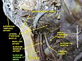
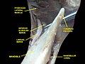
After branching from the mandibular nerve, the inferior alveolar nerve passes posterior to the lateral pterygoid muscle. It issues a branch (the mylohyoid nerve)[ contradictory ] before entering the mandibular foramen [2] :543 to come to pass in the mandibular canal within the mandible. Passing through the canal, it issues sensory branches for the molar and second premolar teeth; the branches first form the inferior dental plexus which then gives off small gingival and dental nerves to these teeth themselves. [3]

The nerve terminates distally/anteriorly (near the second lower premolar)[ citation needed ] within the mandibular canal by splitting into its two terminal branches: the mental nerve, and the incisive branch. [1]

Branches

Mental nerve

The mental nerve emerges from the mandibular canal through the mental foramen [1] and provides sensory innervation to the chin and lower lip.[ citation needed ]

Incisive branch

The incisive branch represents the anterior continuation of the inferior alveolar nerve.[ citation needed ] It continues to course within the mandible in the mandibular incisive canal either as a single nerve or by forming the incisive plexus. It provides sensory innervation to the lower/mandibular premolar, canine, incisor teeth as well as their associated gingiva. [1]

Distribution

The inferior alveolar nerves supply sensation to the lower teeth, [2] :519 and, via the mental nerve, sensation to the chin and lower lip.[ citation needed ]

The mylohyoid nerve is a motor nerve supplying the mylohyoid and the anterior belly of the digastric.[ citation needed ][ contradictory ]

Variation

Rarely, a bifid inferior alveolar nerve may be present, in which case a second mandibular foramen, more inferiorly placed, exists and can be detected by noting a doubled mandibular canal on a radiograph. [4]

Inferior alveolar nerve Mandibular nerve 1.jpg
Inferior alveolar nerve

Clinical significance

Injury

Inferior nerve injury most commonly occurs during surgery including wisdom tooth, dental implant placement in the mandible, root canal treatment where tooth roots are close to the nerve canal in the mandible, deep dental local anaesthetic injections or orthognathic surgery. Trauma and related mandibular fractures are also often related to inferior alveolar nerve injuries.

Trigeminal sensory nerve injuries are associated with numbness, pain, altered sensation and usually a combination of all three. [5] This can result in a significant reduction in quality of life with functional difficulties and psychological impact. [6]

The risk associated with wisdom tooth surgery is commonly accepted to be 2% temporary and 0.2% permanent. However, this risk assessment is not concrete as the same source[ citation needed ] is cited for lingual nerve paresthesia. It is well documented that inferior alveolar nerve injury is more common than lingual nerve injury.[ citation needed ] The percentage of injury varies significantly in different studies. Furthermore, many factors affect the incidence of nerve injury. For example, the incidence of nerve injury in teens removing third molars is much lower than the incidence in patients 25 and older. [7] This risk increases 10 fold if the tooth is close to the inferior dental canal containing the inferior alveolar nerve (as judged on a dental radiograph). [8] These high risk wisdom teeth can be further assessed using cone beam CT imaging to assess and plan surgery to minimise nerve injury by careful extraction or undertaking a coronectomy procedure in healthy patients with healthy teeth. [9]

The risk of nerve injury in relation to mandibular dental implants is not known but it is a recognised risk requiring the patient to be warned. [10] If an injury occurs urgent treatment is required. The risk nerve injury in relation deep dental injections has a risk of injury in approximately 1:14,000 with 25% of these remaining persistent.[ citation needed ] Routine preoperative warnings about these injuries should occur before surgery, and represent good practice. [11] [12] Inferior alveolar nerve injury secondary to orthodontic treatment is also emerging in the literature in the recent years as a rare complication and manifested as anesthesia, paresthesia, or combination of both; however full recovery was achieved in all of the reported cases when proper management was applied. [13]

Anesthesia

During dental procedures, a local nerve block may be applied. Anaesthetic injected near the mandibular foramen to block the inferior alveolar nerve and the nearby lingual nerve (supplying the tongue). This causes loss of sensation on the same side as the block to:

Studies found that oral medications of NSAIDs taken before the dental procedure increases the efficacy of the anesthesia in patients with irreversible pulpitis. [14]

See also

Additional images

Related Research Articles

<span class="mw-page-title-main">Facial nerve</span> Cranial nerve VII, for the face and tasting

The facial nerve, also known as the seventh cranial nerve, cranial nerve VII, or simply CN VII, is a cranial nerve that emerges from the pons of the brainstem, controls the muscles of facial expression, and functions in the conveyance of taste sensations from the anterior two-thirds of the tongue. The nerve typically travels from the pons through the facial canal in the temporal bone and exits the skull at the stylomastoid foramen. It arises from the brainstem from an area posterior to the cranial nerve VI and anterior to cranial nerve VIII.

Articles related to anatomy include:

<span class="mw-page-title-main">Mandibular nerve</span> Branch of the trigeminal nerve responsible for the lower face and jaw

In neuroanatomy, the mandibular nerve (V3) is the largest of the three divisions of the trigeminal nerve, the fifth cranial nerve (CN V). Unlike the other divisions of the trigeminal nerve (ophthalmic nerve, maxillary nerve) which contain only afferent fibers, the mandibular nerve contains both afferent and efferent fibers. These nerve fibers innervate structures of the lower jaw and face, such as the tongue, lower lip, and chin. The mandibular nerve also innervates the muscles of mastication.

<span class="mw-page-title-main">Mandibular foramen</span> Opening on the inner surface of the mandible, allowing for passage of mandibular neurovasculature

The mandibular foramen is an opening on the internal surface of the ramus of the mandible. It allows for divisions of the mandibular nerve and blood vessels to pass through.

<span class="mw-page-title-main">Buccal nerve</span> Nerve in the human face

The buccal nerve is a sensory nerve of the face arising from the mandibular nerve. It conveys sensory information from the skin of the cheek, and parts of the oral mucosa, periodontium, and gingiva.

<span class="mw-page-title-main">Mental foramen</span> Opening on the anterior aspect of the mandible through which the mental nerve and vessels exit

The mental foramen is one of two foramina (openings) located on the anterior surface of the mandible. It is part of the mandibular canal. It transmits the terminal branches of the inferior alveolar nerve and the mental vessels.

<span class="mw-page-title-main">Lingual nerve</span> Human nerve relaying sense to the tongue

The lingual nerve carries sensory innervation from the anterior two-thirds of the tongue. It contains fibres from both the mandibular division of the trigeminal nerve (CN V3) and from the facial nerve (CN VII). The fibres from the trigeminal nerve are for touch, pain and temperature (general sensation), and the ones from the facial nerve are for taste (special sensation).

<span class="mw-page-title-main">Inferior alveolar artery</span>

The inferior alveolar artery is an artery of the head. It is a branch of the maxillary artery. It descends through the infratemporal fossa as part of a neurovascular bundle with the inferior alveolar nerve and vein to the mandibular foramen where it enters and passes anteriorly inside the mandible, suplying the body of mandible and the dental pulp of the lower molar and premolar teeth. Its terminal incisor branch supplies the rest of the lower teeth. Its mental branch exits the mandibula anteriorly through the mental foramen to supply adjacent lip and skin.

<span class="mw-page-title-main">Mental nerve</span> Sensory nerve of the face

The mental nerve is a sensory nerve of the face. It is a branch of the posterior trunk of the inferior alveolar nerve, itself a branch of the mandibular nerve (CN V3), itself a branch of the trigeminal nerve (CN V). It provides sensation to the front of the chin and the lower lip, as well as the gums of the anterior mandibular (lower) teeth. It can be blocked with local anaesthesia for procedures on the chin, lower lip, and mucous membrane of the inner cheek. Problems with the nerve cause chin numbness.

<span class="mw-page-title-main">Mandibular canal</span>

In human anatomy, the mandibular canal is a canal within the mandible that contains the inferior alveolar nerve, inferior alveolar artery, and inferior alveolar vein. It runs obliquely downward and forward in the ramus, and then horizontally forward in the body, where it is placed under the alveoli and communicates with them by small openings.

<span class="mw-page-title-main">Mylohyoid nerve</span> Nerve of the head

The mylohyoid nerve is a mixed nerve of the head. It is a branch of the inferior alveolar nerve. It provides motor innervation the mylohyoid muscle, and the anterior belly of the digastric muscle. It provides sensory innervation to part of the submental area, and sometimes also the mandibular (lower) molar teeth, requiring local anaesthesia for some oral procedures.

<span class="mw-page-title-main">Maxillary artery</span> Artery supplying face structures in humans

The maxillary artery supplies deep structures of the face. It branches from the external carotid artery just deep to the neck of the mandible.

<span class="mw-page-title-main">Infratemporal fossa</span> Cavity that is part of the skull

The infratemporal fossa is an irregularly shaped cavity that is a part of the skull. It is situated below and medial to the zygomatic arch. It is not fully enclosed by bone in all directions. It contains superficial muscles, including the lower part of the temporalis muscle, the lateral pterygoid muscle, and the medial pterygoid muscle. It also contains important blood vessels such as the middle meningeal artery, the pterygoid plexus, and the retromandibular vein, and nerves such as the mandibular nerve (CN V3) and its branches.

<span class="mw-page-title-main">Infraorbital nerve</span> Branch of the maxillary nerve supplying the face

The infraorbital nerve is a branch of the maxillary nerve. It arises in the pterygopalatine fossa. It passes through the inferior orbital fissure to enter the orbit. It travels through the orbit, then enters and traverses the infraorbital canal, exiting the canal at the infraorbital foramen to reach the face. It provides sensory innervation to the skin and mucous membranes around the middle of the face.

<span class="mw-page-title-main">Inferior dental plexus</span> Nerve

The inferior dental plexus is a nerve plexus formed by sensory branches of the inferior alveolar nerve. The plexus issues dental branches and gingival branches; the small dental branches provide sensory innervation to the lower/mandibular teeth.

Inferior alveolar nerve block is a nerve block technique which induces anesthesia (numbness) in the areas of the mouth and face innervated by one of the inferior alveolar nerves which are paired on the left and right side. These areas are the skin and mucous membranes of the lower lip, the skin of the chin, the lower teeth and the labial gingiva of the anterior teeth, all unilaterally to the midline of the side on which the block is administered. However, depending on technique, the long buccal nerve may not be anesthetized by an IANB and therefore an area of buccal gingiva adjacent to the lower posterior teeth will retain normal sensation unless that nerve is anesthetized separately, via a (long) buccal nerve block. The inferior alveolar nerve is a branch of the mandibular nerve, the third division of the trigeminal nerve. This procedure attempts to anaesthetise the inferior alveolar nerve prior to it entering the mandibular foramen on the medial surface of the mandibular ramus.

<span class="mw-page-title-main">Mandibular incisive canal</span>

The mandibular incisive canal is a bilaterally paired bony canal within the anterior portion of the mandible that extends from the mental foramen (usually) to near the ipsilateral lateral incisor teeth.

<span class="mw-page-title-main">Mandible</span> Lower jaw bone

In jawed vertebrates, the mandible, lower jaw, or jawbone is a bone that makes up the lower – and typically more mobile – component of the mouth.

<span class="mw-page-title-main">Human mouth</span> Part of human anatomy

In human anatomy, the mouth is the first portion of the alimentary canal that receives food and produces saliva. The oral mucosa is the mucous membrane epithelium lining the inside of the mouth.

<span class="mw-page-title-main">Pterygomandibular space</span>

The pterygomandibular space is a fascial space of the head and neck. It is a potential space in the head and is paired on each side. It is located between the lateral pterygoid muscle and the medial surface of the ramus of the mandible. The pterygomandibular space is one of the four compartments of the masticator space.

References

  1. 1 2 3 4 Caughey, Jennifer A.; Do, Quang; Shen, Daniel; Ohyama, Hiroe; He, Puhan; Tubbs, R. Shane; Iwanaga, Joe (2021-12-31). "Comprehensive review of the incisive branch of the inferior alveolar nerve". Anatomy & Cell Biology. 54 (4): 409–416. doi:10.5115/acb.21.113. ISSN   2093-3665. PMC   8693131 . S2CID   238474299.
  2. 1 2 3 Standring S, Borley NR, et al., eds. (2008). Gray's Anatomy (40th ed.). Edinburgh: Churchill Livingstone/Elsevier. ISBN   978-0-8089-2371-8.
  3. Wolf KT, Brokaw EJ, Bell A, Joy A (2016-06-01). "Variant Inferior Alveolar Nerves and Implications for Local Anesthesia". Anesthesia Progress. 63 (2): 84–90. doi:10.2344/0003-3006-63.2.84. PMC   4896047 . PMID   27269666.
  4. Fehrenbach MJ, Herring SW (2011). Illustrated Anatomy of the Head and Neck (4th ed.). Philadelphia, PA: Saunders. p. 59.
  5. Renton T, Yilmaz Z (2011). "Profiling of patients presenting with posttraumatic neuropathy of the trigeminal nerve". Journal of Orofacial Pain. 25 (4): 333–344. PMID   22247929.
  6. Smith JG, Elias LA, Yilmaz Z, Barker S, Shah K, Shah S, Renton T (2013). "The psychosocial and affective burden of posttraumatic neuropathy following injuries to the trigeminal nerve". Journal of Orofacial Pain. 27 (4): 293–303. doi: 10.11607/jop.1056 . PMID   24171179.
  7. "Recovering from Surgery". Royal College of Surgeons.
  8. Selvi F, Dodson TB, Nattestad A, Robertson K, Tolstunov L (December 2013). "Factors that are associated with injury to the inferior alveolar nerve in high-risk patients after removal of third molars". The British Journal of Oral & Maxillofacial Surgery. 51 (8): 868–873. doi:10.1016/j.bjoms.2013.08.007. PMID   24012054.
  9. Long H, Zhou Y, Liao L, Pyakurel U, Wang Y, Lai W (July 2012). "Coronectomy vs. total removal for third molar extraction: a systematic review". Journal of Dental Research. 91 (7): 659–665. doi:10.1177/0022034512449346. PMID   22622663. S2CID   21579439.
  10. Renton T, Dawood A, Shah A, Searson L, Yilmaz Z (June 2012). "Post-implant neuropathy of the trigeminal nerve. A case series". British Dental Journal. 212 (11): E17. doi: 10.1038/sj.bdj.2012.497 . PMID   22677874. S2CID   21328385.
  11. "Nerve Damage Associated with Peripheral Nerve Block" (PDF). Information for patients. The Royal College of Anaesthetists. 2013. Archived from the original (PDF) on 2014-04-16. Retrieved 2014-04-15.
  12. Tidy G (27 February 2018). Bonsall A (ed.). "Anaesthesia (UK) - Local and General anaesthesia information". Patient. Egton Medical Information Systems Limited.
  13. AlAli, Ahmad M.; AlAnzi, Talal H. (2021-01-01). "Inferior alveolar nerve damage secondary to orthodontic treatment: A systematic scoping review". International Journal of Risk & Safety in Medicine. 32 (3): 175–191. doi:10.3233/JRS-200098. ISSN   0924-6479. PMID   33579879. S2CID   231910002.
  14. Shantiaee Y, Javaheri S, Movahhedian A, Eslami S, Dianat O (April 2017). "Efficacy of preoperative ibuprofen and meloxicam on the success rate of inferior alveolar nerve block for teeth with irreversible pulpitis". International Dental Journal. 67 (2): 85–90. doi: 10.1111/idj.12272 . PMC   9376692 . PMID   27933616.