Mental foramen

Last updated
Mental foramen
Gray176.png
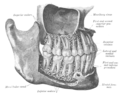
Mandible. Outer surface. Side view. (Mental foramen visible at left.)
Details
Part of mandible
Identifiers
Latin foramen mentale
MeSH D000080383
TA98 A02.1.15.007
TA2 845
FMA 53171
Anatomical terms of bone
The mandibular incisive canal (indicated here by coral green arrows) continuing anteriorly (to the right) from the mandibular canal (purple arrows) after the mental foramen (light green circle). Mandibular Incisive Canal Highlighted.jpg
The mandibular incisive canal (indicated here by coral green arrows) continuing anteriorly (to the right) from the mandibular canal (purple arrows) after the mental foramen (light green circle).

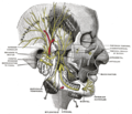
The mental foramen is one of two foramina (openings) located on the anterior surface of the mandible. It is part of the mandibular canal. It transmits the terminal branches of the inferior alveolar nerve and the mental vessels.

Contents

Structure

The mental foramen is located on the anterior surface of the mandible. It is directly below the commisure of the lips, and the tendon of depressor labii inferioris muscle. [1] It is at the end of the mandibular canal, which begins at the mandibular foramen on the posterior surface of the mandible. It transmits the terminal branches of the inferior alveolar nerve (the mental nerve), [1] the mental artery, and the mental vein.

Variation

The mental foramen descends slightly in toothless individuals. [2]

The mental foramen is in line with the longitudinal axis of the 2nd premolar in 63% of people. [3] It generally lies at the level of the vestibular fornix and about a finger's breadth above the inferior border of the mandible.

In the general population, 17% of mandibles have an additional mental foramen or foramina on at least one side, [3] while 4% of the mandibles show multiple mental foramina on both sides. Most are unequal in size, often with a single large foramen while any others are smaller. [3] An incisive mental foramen is observed in 1% of the side of the mandible. [3]

Clinical significance

The mental nerve may be anaesthetized as it leaves the mental foramen. [1] This causes loss of sensation to the lower lip and chin on the same side. [1]

Additional images

See also

Related Research Articles

<span class="mw-page-title-main">Mandibular nerve</span> Branch of the trigeminal nerve responsible for the lower face and jaw

In neuroanatomy, the mandibular nerve (V3) is the largest of the three divisions of the trigeminal nerve, the fifth cranial nerve (CN V). Unlike the other divisions of the trigeminal nerve (ophthalmic nerve, maxillary nerve) which contain only afferent fibers, the mandibular nerve contains both afferent and efferent fibers. These nerve fibers innervate structures of the lower jaw and face, such as the tongue, lower lip, and chin. The mandibular nerve also innervates the muscles of mastication.

<span class="mw-page-title-main">Mandibular foramen</span> Opening on the inner surface of the mandible, allowing for passage of mandibular neurovasculature

The mandibular foramen is an opening on the internal surface of the ramus of the mandible. It allows for divisions of the mandibular nerve and blood vessels to pass through.

<span class="mw-page-title-main">Inferior alveolar nerve</span>

The inferior alveolar nerve(IAN) is a branch of the mandibular nerve, which is itself the third branch of the trigeminal nerve. The inferior alveolar nerves supply sensation to the lower teeth.

<span class="mw-page-title-main">Foramen spinosum</span> Hole in the sphenoid bone of the skull

The foramen spinosum is a hole located in the greater wing of the sphenoid. It is located posterolateral to the foramen ovale and anterior to the sphenoidal spine. It allows the passage of the middle meningeal artery, middle meningeal vein and usually the meningeal branch of the mandibular nerve.

<span class="mw-page-title-main">Pterygopalatine fossa</span>

In human anatomy, the pterygopalatine fossa is a fossa in the skull. A human skull contains two pterygopalatine fossae—one on the left side, and another on the right side. Each fossa is a cone-shaped paired depression deep to the infratemporal fossa and posterior to the maxilla on each side of the skull, located between the pterygoid process and the maxillary tuberosity close to the apex of the orbit. It is the indented area medial to the pterygomaxillary fissure leading into the sphenopalatine foramen. It communicates with the nasal and oral cavities, infratemporal fossa, orbit, pharynx, and middle cranial fossa through eight foramina.

<span class="mw-page-title-main">Greater wing of sphenoid bone</span> Large part of the skull front behind the eye socket

The greater wing of the sphenoid bone, or alisphenoid, is a bony process of the sphenoid bone; there is one on each side, extending from the side of the body of the sphenoid and curving upward, laterally, and backward.

<span class="mw-page-title-main">Inferior alveolar artery</span>

The inferior alveolar artery is an artery of the face. It is a branch of the first portion of the maxillary artery.

<span class="mw-page-title-main">Mental nerve</span> Sensory nerve of the face

The mental nerve is a sensory nerve of the face. It is a branch of the posterior trunk of the inferior alveolar nerve, itself a branch of the mandibular nerve (CN V3), itself a branch of the trigeminal nerve (CN V). It provides sensation to the front of the chin and the lower lip, as well as the gums of the anterior mandibular (lower) teeth. It can be blocked with local anaesthesia for procedures on the chin, lower lip, and mucous membrane of the inner cheek. Problems with the nerve cause chin numbness.

<span class="mw-page-title-main">Mandibular canal</span>

In human anatomy, the mandibular canal is a canal within the mandible that contains the inferior alveolar nerve, inferior alveolar artery, and inferior alveolar vein. It runs obliquely downward and forward in the ramus, and then horizontally forward in the body, where it is placed under the alveoli and communicates with them by small openings.

<span class="mw-page-title-main">Mylohyoid nerve</span> Nerve of the head

The mylohyoid nerve is a nerve of the head. It is a branch of the inferior alveolar nerve. It supplies the mylohyoid muscle, and the anterior belly of the digastric muscle. It may also supply mandibular (lower) molar teeth, requiring local anaesthesia for some oral procedures.

<span class="mw-page-title-main">Maxillary artery</span>

The maxillary artery supplies deep structures of the face. It branches from the external carotid artery just deep to the neck of the mandible.

<span class="mw-page-title-main">Incisive canals</span> Two lateral canals in the incisive foramen

The incisive canals are two bony canals of the anterior hard palate connecting the nasal cavity and the oral cavity. An incisive canal courses through each maxilla. Below, the two incisive canals typically converge medially.

<span class="mw-page-title-main">Incisive foramen</span> Opening of the incisive canals on the hard palate immediately behind the incisor teeth

In the human mouth, the incisive foramen is the opening of the incisive canals on the hard palate immediately behind the incisor teeth. It gives passage to blood vessels and nerves. The incisive foramen is situated within the incisive fossa of the maxilla.

<span class="mw-page-title-main">Infratemporal fossa</span> Cavity that is part of the skull

The infratemporal fossa is an irregularly shaped cavity that is a part of the skull. It is situated below and medial to the zygomatic arch. It is not fully enclosed by bone in all directions. It contains superficial muscles, including the lower part of the temporalis muscle, the lateral pterygoid muscle, and the medial pterygoid muscle. It also contains important blood vessels such as the middle meningeal artery, the pterygoid plexus, and the retromandibular vein, and nerves such as the mandibular nerve (CN V3) and its branches.

<span class="mw-page-title-main">Infraorbital nerve</span> Branch of the maxillary nerve supplying the face

The infraorbital nerve is a branch of the maxillary nerve, itself a branch of the trigeminal nerve. It travels through the orbit and enters the infraorbital canal to exit onto the face through the infraorbital foramen. It provides sensory innervation to the skin and mucous membranes around the middle of the face.

<span class="mw-page-title-main">Palatine process of maxilla</span> Thick, horizontal process of the maxilla

In human anatomy of the mouth, the palatine process of maxilla, is a thick, horizontal process of the maxilla. It forms the anterior three quarters of the hard palate, the horizontal plate of the palatine bone making up the rest.

Inferior alveolar nerve block is a nerve block technique which induces anesthesia (numbness) in the areas of the mouth and face innervated by one of the inferior alveolar nerves which are paired on the left and right side. These areas are the skin and mucous membranes of the lower lip, the skin of the chin, the lower teeth and the labial gingiva of the anterior teeth, all unilaterally to the midline of the side on which the block is administered. However, depending on technique, the long buccal nerve may not be anesthetized by an IANB and therefore an area of buccal gingiva adjacent to the lower posterior teeth will retain normal sensation unless that nerve is anesthetized separately, via a (long) buccal nerve block. The inferior alveolar nerve is a branch of the mandibular nerve, the third division of the trigeminal nerve. This procedure attempts to anaesthetise the inferior alveolar nerve prior to it entering the mandibular foramen on the medial surface of the mandibular ramus.

<span class="mw-page-title-main">Mandibular incisive canal</span>

The mandibular incisive canal is a bony canal within the anterior mandible that runs bilaterally from the mental foramina usually to the region of the ipsilateral lateral incisor teeth. After branching into the mental nerve that exits the foramen of the same name, the inferior alveolar nerve continues anteriorly within the mandibular incisive canal as the incisive nerve, providing innervation to the mandibular first premolar, canine and lateral and central incisors. The mandibular incisive nerve either terminates as nerve endings within the anterior teeth or adjacent bone, or may join nerve endings that enter through the tiny lingual foramen.

<span class="mw-page-title-main">Mandible</span> Lower jaw bone

In anatomy, the mandible, lower jaw or jawbone is the largest, strongest and lowest bone in the human facial skeleton. It forms the lower jaw and holds the lower teeth in place. The mandible sits beneath the maxilla. It is the only movable bone of the skull. It is connected to the temporal bones by the temporomandibular joints.

<span class="mw-page-title-main">Human mouth</span> Part of human anatomy

In human anatomy, the mouth is the first portion of the alimentary canal that receives food and produces saliva. The oral mucosa is the mucous membrane epithelium lining the inside of the mouth.

References

  1. 1 2 3 4 Doherty, Tom; Schumacher, James (2011). "15 - Dental restraint and anesthesia". Equine Dentistry (3rd ed.). Saunders. pp. 241–244. doi:10.1016/B978-0-7020-2980-6.00015-5. ISBN   978-0-7020-2980-6.
  2. Soikkonen K, Wolf J, Ainamo A, Xie Q (November 1995). "Changes in the position of the mental foramen as a result of alveolar atrophy". Journal of Oral Rehabilitation. 22 (11): 831–3. doi:10.1111/j.1365-2842.1995.tb00230.x. PMID   8558356.
  3. 1 2 3 4 Jaffar, A. A.; Al-Zubaidi, A. F.; Al-Salihi, A. R. (2002). "Anatomical features of clinical significance in dry mandibles". Iraqi Dental Journal. 29: 99–118. Archived from the original on 2008-12-29.