Human musculoskeletal system

Last updated

Musculoskeletal system
1911 Britannica - Anatomy - Muscular.png
Features of the human activity system from the 1911 Encyclopædia Britannica
Identifiers
MeSH D009141
TA2 351
FMA 7482
Anatomical terminology

The human musculoskeletal system (also known as the human locomotor system, and previously the activity system) is an organ system that gives humans the ability to move using their muscular and skeletal systems. The musculoskeletal system provides form, support, stability, and movement to the body.

Contents

It is made up of the bones of the skeleton, muscles, cartilage, [1] tendons, ligaments, joints, and other connective tissue that supports and binds tissues and organs together. The musculoskeletal system's primary functions include supporting the body, allowing motion, and protecting vital organs. [2] The skeletal portion of the system serves as the main storage system for calcium and phosphorus and contains critical components of the hematopoietic system. [3]

This system describes how bones are connected to other bones and muscle fibers via connective tissue such as tendons and ligaments. The bones provide stability to the body. Muscles keep bones in place and also play a role in the movement of bones. To allow motion, different bones are connected by joints. Cartilage prevents the bone ends from rubbing directly onto each other. Muscles contract to move the bone attached at the joint.

There are, however, diseases and disorders that may adversely affect the function and overall effectiveness of the system. These diseases can be difficult to diagnose due to the close relation of the musculoskeletal system to other internal systems. The musculoskeletal system refers to the system having its muscles attached to an internal skeletal system and is necessary for humans to move to a more favorable position. Complex issues and injuries involving the musculoskeletal system are usually handled by a physiatrist (specialist in physical medicine and rehabilitation) or an orthopaedic surgeon.

Subsystems

Skeletal

The skeletal system serves many important functions; it provides the shape and form for the body, support and protection, allows bodily movement, produces blood for the body, and stores minerals. [4] The number of bones in the human skeletal system is a controversial topic. Humans are born with over 300 bones; however, many bones fuse together between birth and maturity. As a result, an average adult skeleton consists of 206 bones. The number of bones varies according to the method used to derive the count. While some consider certain structures to be a single bone with multiple parts, others may see it as a single part with multiple bones. [5] There are five general classifications of bones. These are long bones, short bones, flat bones, irregular bones, and sesamoid bones. The human skeleton is composed of both fused and individual bones supported by ligaments, tendons, muscles and cartilage. It is a complex structure with two distinct divisions; the axial skeleton , which includes the vertebral column, and the appendicular skeleton . [6]

Function

The skeletal system serves as a framework for tissues and organs to attach themselves to. This system acts as a protective structure for vital organs. Major examples of this are the brain being protected by the skull and the lungs being protected by the rib cage.

Located in long bones are two distinctions of bone marrow (yellow and red). The yellow marrow has fatty connective tissue and is found in the marrow cavity. During starvation, the body uses the fat in yellow marrow for energy. [7] The red marrow of some bones is an important site for blood cell production, approximately 2.6 million red blood cells per second in order to replace existing cells that have been destroyed by the liver. [4] Here all erythrocytes, platelets, and most leukocytes form in adults. From the red marrow, erythrocytes, platelets, and leukocytes migrate to the blood to do their special tasks.

Another function of bones is the storage of certain minerals. Calcium and phosphorus are among the main minerals being stored. The importance of this storage "device" helps to regulate mineral balance in the bloodstream. When the fluctuation of minerals is high, these minerals are stored in the bone; when it is low it will be withdrawn from the bone.

Muscular

The body contains three types of muscle tissue: (a) skeletal muscle, (b) smooth muscle, and (c) cardiac muscle. 414 Skeletal Smooth Cardiac.jpg
The body contains three types of muscle tissue: (a) skeletal muscle, (b) smooth muscle, and (c) cardiac muscle.
On the anterior and posterior views of the muscular system above, superficial muscles (those at the surface) are shown on the right side of the body while deep muscles (those underneath the superficial muscles) are shown on the left half of the body. For the legs, superficial muscles are shown in the anterior view while the posterior view shows both superficial and deep muscles. 1105 Anterior and Posterior Views of Muscles.jpg
On the anterior and posterior views of the muscular system above, superficial muscles (those at the surface) are shown on the right side of the body while deep muscles (those underneath the superficial muscles) are shown on the left half of the body. For the legs, superficial muscles are shown in the anterior view while the posterior view shows both superficial and deep muscles.

There are three types of muscles—cardiac, skeletal, and smooth. Smooth muscles are used to control the flow of substances within the lumens of hollow organs, and are not consciously controlled. Skeletal and cardiac muscles have striations that are visible under a microscope due to the components within their cells. Only skeletal and smooth muscles are part of the musculoskeletal system and only the muscles can move the body. Cardiac muscles are found in the heart and are used only to circulate blood; like the smooth muscles, these muscles are not under conscious control. Skeletal muscles are attached to bones and arranged in opposing groups around joints. [8] Muscles are innervated, whereby nervous signals are communicated [9] by nerves, which conduct electrical currents from the central nervous system and cause the muscles to contract. [10]

Contraction initiation

In mammals, when a muscle contracts, a series of reactions occur. Muscle contraction is stimulated by the motor neuron sending a message to the muscles from the somatic nervous system. Depolarization of the motor neuron results in neurotransmitters being released from the nerve terminal. The space between the nerve terminal and the muscle cell is called the neuromuscular junction. These neurotransmitters diffuse across the synapse and bind to specific receptor sites on the cell membrane of the muscle fiber. When enough receptors are stimulated, an action potential is generated and the permeability of the sarcolemma is altered. This process is known as initiation. [11]

Tendons

A tendon is a tough, flexible band of fibrous connective tissue that connects muscles to bones. [12] The extra-cellular connective tissue between muscle fibers binds to tendons at the distal and proximal ends, and the tendon binds to the periosteum of individual bones at the muscle's origin and insertion. As muscles contract, tendons transmit the forces to the relatively rigid bones, pulling on them and causing movement. Tendons can stretch substantially, allowing them to function as springs during locomotion, thereby saving energy.

Joints, ligaments and bursae

Human synovial joint composition Joint.svg
Human synovial joint composition

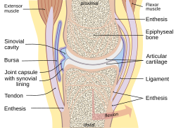
Joints are structures that connect individual bones and may allow bones to move against each other to cause movement. There are three divisions of joints, diarthroses which allow extensive mobility between two or more articular heads; amphiarthrosis, which is a joint that allows some movement, and false joints or synarthroses, joints that are immovable, that allow little or no movement and are predominantly fibrous. Synovial joints, joints that are not directly joined, are lubricated by a solution called synovial fluid that is produced by the synovial membranes. This fluid lowers the friction between the articular surfaces and is kept within an articular capsule, binding the joint with its taut tissue. [6]

Ligaments

A ligament is a small band of dense, white, fibrous elastic tissue. [6] Ligaments connect the ends of bones together in order to form a joint. Most ligaments limit dislocation, or prevent certain movements that may cause breaks. Since they are only elastic they increasingly lengthen when under pressure. When this occurs the ligament may be susceptible to break resulting in an unstable joint.

Ligaments may also restrict some actions: movements such as hyper extension and hyper flexion are restricted by ligaments to an extent. Also ligaments prevent certain directional movement. [13]

Bursae

A bursa is a small fluid-filled sac made of white fibrous tissue and lined with synovial membrane. Bursa may also be formed by a synovial membrane that extends outside of the joint capsule. [7] It provides a cushion between bones and tendons or muscles around a joint; bursa are filled with synovial fluid and are found around almost every major joint of the body.

Clinical significance

Disability-adjusted life year for musculoskeletal diseases per 100,000 inhabitants in 2004.
.mw-parser-output .div-col{margin-top:0.3em;column-width:30em}.mw-parser-output .div-col-small{font-size:90%}.mw-parser-output .div-col-rules{column-rule:1px solid #aaa}.mw-parser-output .div-col dl,.mw-parser-output .div-col ol,.mw-parser-output .div-col ul{margin-top:0}.mw-parser-output .div-col li,.mw-parser-output .div-col dd{page-break-inside:avoid;break-inside:avoid-column}
.mw-parser-output .legend{page-break-inside:avoid;break-inside:avoid-column}.mw-parser-output .legend-color{display:inline-block;min-width:1.25em;height:1.25em;line-height:1.25;margin:1px 0;text-align:center;border:1px solid black;background-color:transparent;color:black}.mw-parser-output .legend-text{}
no data
less than 400
400-450
450-500
500-550
550-600
600-650
650-700
700-850
850-900
900-925
925-950
more than 950 Musculoskeletal diseases world map - DALY - WHO2004.svg
Disability-adjusted life year for musculoskeletal diseases per 100,000 inhabitants in 2004.
  no data
  less than 400
  400–450
  450–500
  500–550
  550–600
  600–650
  650–700
  700–850
  850–900
  900–925
  925–950
  more than 950

Because many other body systems, including the vascular, nervous, and integumentary systems, are interrelated, disorders of one of these systems may also affect the musculoskeletal system and complicate the diagnosis of the disorder's origin. Diseases of the musculoskeletal system mostly encompass functional disorders or motion discrepancies; the level of impairment depends specifically on the problem and its severity. In a study of hospitalizations in the United States, the most common inpatient OR procedures in 2012 involved the musculoskeletal system: knee arthroplasty, laminectomy, hip replacement, and spinal fusion. [15]

Articular (of or pertaining to the joints) [16] disorders are the most common. However, also among the diagnoses are: primary muscular diseases, neurologic (related to the medical science that deals with the nervous system and disorders affecting it) [17] deficits, toxins, endocrine abnormalities, metabolic disorders, infectious diseases, blood and vascular disorders, and nutritional imbalances.

Disorders of muscles from another body system can bring about irregularities such as: impairment of ocular motion and control, respiratory dysfunction, and bladder malfunction. Complete paralysis, paresis, or ataxia may be caused by primary muscular dysfunctions of infectious or toxic origin; however, the primary disorder is usually related to the nervous system, with the muscular system acting as the effector organ, an organ capable of responding to a stimulus, especially a nerve impulse. [3]

One understated disorder that begins during pregnancy is pelvic girdle pain. It is complex, multi-factorial, and likely to be also represented by a series of sub-groups driven by pain varying from peripheral or central nervous system, [18] altered laxity/stiffness of muscles, [19] laxity to injury of tendinous/ligamentous structures [20] to maladaptive body mechanics. [18]

See also

Related Research Articles

A ligament is the fibrous connective tissue that connects bones to other bones. It is also known as articular ligament, articular larua, fibrous ligament, or true ligament. Other ligaments in the body include the:

<span class="mw-page-title-main">Skeleton</span> Part of the body that forms the supporting structure

A skeleton is the structural frame that supports the body of most animals. There are several types of skeletons, including the exoskeleton, which is a rigid outer shell that holds up an organism's shape; the endoskeleton, a rigid internal frame to which the organs and soft tissues attach; and the hydroskeleton, a flexible internal structure supported by the hydrostatic pressure of body fluids.

The muscular system is an organ system consisting of skeletal, smooth, and cardiac muscle. It permits movement of the body, maintains posture, and circulates blood throughout the body. The muscular systems in vertebrates are controlled through the nervous system although some muscles can be completely autonomous. Together with the skeletal system in the human, it forms the musculoskeletal system, which is responsible for the movement of the body.

The human skeleton is the internal framework of the human body. It is composed of around 270 bones at birth – this total decreases to around 206 bones by adulthood after some bones get fused together. The bone mass in the skeleton makes up about 14% of the total body weight and reaches maximum mass between the ages of 25 and 30. The human skeleton can be divided into the axial skeleton and the appendicular skeleton. The axial skeleton is formed by the vertebral column, the rib cage, the skull and other associated bones. The appendicular skeleton, which is attached to the axial skeleton, is formed by the shoulder girdle, the pelvic girdle and the bones of the upper and lower limbs.

<span class="mw-page-title-main">Knee</span> Leg joint in primates

In humans and other primates, the knee joins the thigh with the leg and consists of two joints: one between the femur and tibia, and one between the femur and patella. It is the largest joint in the human body. The knee is a modified hinge joint, which permits flexion and extension as well as slight internal and external rotation. The knee is vulnerable to injury and to the development of osteoarthritis.

<span class="mw-page-title-main">Temporomandibular joint</span> Joints connecting the jawbone to the skull

In anatomy, the temporomandibular joints (TMJ) are the two joints connecting the jawbone to the skull. It is a bilateral synovial articulation between the temporal bone of the skull above and the mandible below; it is from these bones that its name is derived. This joint is unique in that it is a bilateral joint that functions as one unit. Since the TMJ is connected to the mandible, the right and left joints must function together and therefore are not independent of each other.

<span class="mw-page-title-main">Joint</span> Location at which two or more bones make contact

A joint or articulation is the connection made between bones, ossicles, or other hard structures in the body which link an animal's skeletal system into a functional whole. They are constructed to allow for different degrees and types of movement. Some joints, such as the knee, elbow, and shoulder, are self-lubricating, almost frictionless, and are able to withstand compression and maintain heavy loads while still executing smooth and precise movements. Other joints such as sutures between the bones of the skull permit very little movement in order to protect the brain and the sense organs. The connection between a tooth and the jawbone is also called a joint, and is described as a fibrous joint known as a gomphosis. Joints are classified both structurally and functionally.

<span class="mw-page-title-main">Connective tissue</span> Type of biological tissue in animals

Connective tissue is one of the four primary types of animal tissue, along with epithelial tissue, muscle tissue, and nervous tissue. It develops mostly from the mesenchyme, derived from the mesoderm, the middle embryonic germ layer. Connective tissue is found in between other tissues everywhere in the body, including the nervous system. The three meninges, membranes that envelop the brain and spinal cord, are composed of connective tissue. Most types of connective tissue consists of three main components: elastic and collagen fibers, ground substance, and cells. Blood, and lymph are classed as specialized fluid connective tissues that do not contain fiber. All are immersed in the body water. The cells of connective tissue include fibroblasts, adipocytes, macrophages, mast cells and leucocytes.

The National Institute of Arthritis and Musculoskeletal and Skin Diseases (NIAMS) is one of the institutes and centers that make up the National Institutes of Health, an agency of the United States Department of Health and Human Services (HHS).

<span class="mw-page-title-main">Synovial joint</span> Articulation which admits free motion in the joint; the most common type of articulation

A synovial joint, also known as diarthrosis, joins bones or cartilage with a fibrous joint capsule that is continuous with the periosteum of the joined bones, constitutes the outer boundary of a synovial cavity, and surrounds the bones' articulating surfaces. This joint unites long bones and permits free bone movement and greater mobility. The synovial cavity/joint is filled with synovial fluid. The joint capsule is made up of an outer layer of fibrous membrane, which keeps the bones together structurally, and an inner layer, the synovial membrane, which seals in the synovial fluid.

<span class="mw-page-title-main">Periosteum</span> Membrane covering outer surface of bones

The periosteum is a membrane that covers the outer surface of all bones, except at the articular surfaces of long bones. Endosteum lines the inner surface of the medullary cavity of all long bones.

<span class="mw-page-title-main">Flexibility (anatomy)</span> Anatomical range of movement of a joint or series of joints

Flexibility is the anatomical range of movement in a joint or series of joints, and length in muscles that cross the joints to induce a bending movement or motion. Flexibility varies between individuals, particularly in terms of differences in muscle length of multi-joint muscles. Flexibility in some joints can be increased to a certain degree by exercise, with stretching a common exercise component to maintain or improve flexibility.

<span class="mw-page-title-main">Equine anatomy</span> Descriptive scheme

Equine anatomy encompasses the gross and microscopic anatomy of horses, ponies and other equids, including donkeys, mules and zebras. While all anatomical features of equids are described in the same terms as for other animals by the International Committee on Veterinary Gross Anatomical Nomenclature in the book Nomina Anatomica Veterinaria, there are many horse-specific colloquial terms used by equestrians.

<span class="mw-page-title-main">Articular capsule of the knee joint</span>

The articular capsule of the knee joint is the wide and lax joint capsule of the knee. It is thin in front and at the side, and contains the patella, ligaments, menisci, and bursae of the knee. The capsule consists of an inner synovial membrane, and an outer fibrous membrane separated by fatty deposits anteriorly and posteriorly.

<span class="mw-page-title-main">Orthopedic pathology</span>

Orthopedic pathology, also known as bone pathology is a subspecialty of surgical pathology which deals with the diagnosis and feature of many bone diseases, specifically studying the cause and effects of disorders of the musculoskeletal system. It uses gross and microscopic findings along with the findings of in vivo radiological studies, and occasionally, specimen radiographs to diagnose diseases of the bones.

<span class="mw-page-title-main">Skeletal system of the horse</span> The skeletal system is made of many interconnected tissues including bone, cartilage, and tendons

The skeletal system of the horse is a skeletal system of a horse that has three major functions in the body. It protects vital organs, provides framework, and supports soft parts of the body. Horses typically have 205 bones. The pelvic limb typically contains 19 bones, while the thoracic limb contains 20 bones.

<span class="mw-page-title-main">Outline of human anatomy</span> Overview of and topical guide to human anatomy

The following outline is provided as an overview of and topical guide to human anatomy:

<span class="mw-page-title-main">Musculoskeletal injury</span> Medical condition

Musculoskeletal injury refers to damage of muscular or skeletal systems, which is usually due to a strenuous activity and includes damage to skeletal muscles, bones, tendons, joints, ligaments, and other affected soft tissues. In one study, roughly 25% of approximately 6300 adults received a musculoskeletal injury of some sort within 12 months—of which 83% were activity-related. Musculoskeletal injury spans into a large variety of medical specialties including orthopedic surgery, sports medicine, emergency medicine and rheumatology.

<span class="mw-page-title-main">Anatomical terminology</span> Scientific terminology used by anatomists, zoologists, and health professionals

Anatomical terminology is a form of scientific terminology used by anatomists, zoologists, and health professionals such as doctors, physicians, and pharmacists.

References

  1. Musculoskeletal+System at the U.S. National Library of Medicine Medical Subject Headings (MeSH)
  2. Mooar, Pekka (2007). "Muscles". Merck Manual. Retrieved 12 November 2008.
  3. 1 2 Kahn, Cynthia; Scott Line (2008). Musculoskeletal System Introduction: Introduction. New Jersey, US: Merck & Co., Inc.
  4. 1 2 Applegate, Edith; Kent Van De Graaff. "The Skeletal System". Archived from the original on 3 June 2010. Retrieved 3 January 2009.
  5. Engelbert, Phillis; Carol DeKane Nagel (2009). "The Human Body / How Many Bones Are In The Human Body?". U·X·L Science Fact Finder. eNotes.com, Inc. Retrieved 24 January 2009.
  6. 1 2 3 Gary, Farr (25 June 2002). "The Musculoskeletal System". Archived from the original on 29 November 2014. Retrieved 18 November 2008.
  7. 1 2 "Skeletal System". 2001. Archived from the original on 25 February 2011. Retrieved 8 January 2009.
  8. Mooar, Pekka (2007). "Muscles". The Merck Manuals Online Medical Library. Retrieved 16 November 2008.
  9. "innervated". Dictionary.com. Dictionary.com, LLC. 2008. Retrieved 3 January 2009.
  10. Bárány, Michael (2002). "SMOOTH MUSCLE" . Retrieved 19 November 2008.
  11. "The Mechanism of Muscle Contraction". Principles of Meat Science (4th Edition). Archived from the original on 17 February 2012. Retrieved 18 November 2008.
  12. Jonathan, Cluett (2008). "Tendons" . Retrieved 19 November 2008.
  13. Bridwell, Keith. "Ligaments" . Retrieved 16 March 2009.
  14. "WHO Disease and injury country estimates". World Health Organization . 2009. Retrieved 11 November 2009.
  15. Fingar KR, Stocks C, Weiss AJ, Steiner CA (December 2014). "Most Frequent Operating Room Procedures Performed in U.S. Hospitals, 2003–2012". HCUP Statistical Brief #186. Rockville, MD: Agency for Healthcare Research and Quality.
  16. "articular". Random House Unabridged Dictionary. Random House, Inc. 2006. Retrieved 15 November 2008.
  17. "neurologic". The American Heritage Dictionary of the English Language, Fourth Edition. Houghton Mifflin Company. 2006. Retrieved 15 November 2008.
  18. 1 2 Diagnosis and classification of pelvic girdle pain disorders— Part 1: A mechanism based approach within a bio psychosocial framework. Manual Therapy, Volume 12, Issue 2, May 2007, PB. O’Sullivan and DJ Beales.
  19. Vleeming, Andry; Albert, Hanne B.; Östgaard, Hans Christian; Sturesson, Bengt; Stuge, Britt (June 2008). "European guidelines for the diagnosis and treatment of pelvic girdle pain". European Spine Journal. 17 (6): 794–819. doi:10.1007/s00586-008-0602-4. PMC   2518998 . PMID   18259783.
  20. Vleeming, Andry; de Vries, Haitze; Mens, Jan; van Wingerden, Jan-Paul (2002). "Possible role of the long dorsal sacroiliac ligament in women with peripartum pelvic pain". Acta Obstetricia et Gynecologica Scandinavica. 81 (5): 430–436. doi: 10.1034/j.1600-0412.2002.810510.x . ISSN   0001-6349. PMID   12027817. S2CID   18323116.