Pharynx

Last updated
Pharynx
Illu01 head neck.jpg
Head and inner neck
Illu pharynx.jpg
Pharynx
Details
Part of Throat
System Respiratory system, digestive system
Artery pharyngeal branches of ascending pharyngeal artery, ascending palatine, descending palatine, pharyngeal branches of inferior thyroid
Vein pharyngeal plexus
Nerve pharyngeal plexus of vagus nerve, recurrent laryngeal nerve, maxillary nerve, mandibular nerve
Identifiers
Latin pharynx
Greek φάρυγξ (phárynx)
MeSH D010614
TA98 A05.3.01.001
TA2 2855
FMA 46688
Anatomical terminology

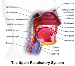
The pharynx (pl.: pharynges) is the part of the throat behind the mouth and nasal cavity, and above the esophagus and trachea (the tubes going down to the stomach and the lungs respectively). It is found in vertebrates and invertebrates, though its structure varies across species. The pharynx carries food to the esophagus and air to the larynx. The flap of cartilage called the epiglottis stops food from entering the larynx.

Contents

In humans, the pharynx is part of the digestive system and the conducting zone of the respiratory system. (The conducting zone—which also includes the nostrils of the nose, the larynx, trachea, bronchi, and bronchioles—filters, warms and moistens air and conducts it into the lungs). [1] The human pharynx is conventionally divided into three sections: the nasopharynx, oropharynx, and laryngopharynx.

In humans, two sets of pharyngeal muscles form the pharynx and determine the shape of its lumen. They are arranged as an inner layer of longitudinal muscles and an outer circular layer.

Structure

Nasopharynx

Upper respiratory system, with the nasopharynx, oropharynx and laryngopharynx labeled at left Blausen 0872 UpperRespiratorySystem.png
Upper respiratory system, with the nasopharynx, oropharynx and laryngopharynx labeled at left

The upper portion of the pharynx, the nasopharynx, extends from the base of the skull to the upper surface of the soft palate. [2] It includes the space between the internal nares and the soft palate and lies above the oral cavity. The adenoids, also known as the pharyngeal tonsils, are lymphoid tissue structures located in the posterior wall of the nasopharynx. Waldeyer's tonsillar ring is an annular arrangement of lymphoid tissue in both the nasopharynx and oropharynx. The nasopharynx is lined by respiratory epithelium that is pseudostratified, columnar, and ciliated.

Polyps or mucus can obstruct the nasopharynx, as can congestion due to an upper respiratory infection. The auditory tube, which connects the middle ear to the pharynx, opens into the nasopharynx at the pharyngeal opening of the auditory tube. The opening and closing of the auditory tubes serves to equalize the barometric pressure in the middle ear with that of the ambient atmosphere.

Details of torus tubarius Torus tubarius dissection.jpg
Details of torus tubarius

The anterior aspect of the nasopharynx communicates through the choanae with the nasal cavities. On its lateral wall is the pharyngeal opening of the auditory tube, somewhat triangular in shape and bounded behind by a firm prominence, the torus tubarius or cushion, caused by the medial end of the cartilage of the tube that elevates the mucous membrane. Two folds arise from the cartilaginous opening:

Oropharynx

The oropharynx lies behind the oral cavity, extending from the uvula to the level of the hyoid bone. It opens anteriorly, through the isthmus faucium, into the mouth, while in its lateral wall, between the palatoglossal arch and the palatopharyngeal arch, is the palatine tonsil. [4] The anterior wall consists of the base of the tongue and the epiglottic vallecula; the lateral wall is made up of the tonsil, tonsillar fossa, and tonsillar (faucial) pillars; the superior wall consists of the inferior surface of the soft palate and the uvula. Because both food and air pass through the pharynx, a flap of connective tissue called the epiglottis closes over the glottis when food is swallowed to prevent aspiration. The oropharynx is lined by non-keratinized squamous stratified epithelium.

The HACEK organisms ( Haemophilus, Actinobacillus actinomycetemcomitans, Cardiobacterium hominis, Eikenella corrodens, Kingella ) are part of the normal oropharyngeal flora, which grow slowly, prefer a carbon dioxide-enriched atmosphere, and share an enhanced capacity to produce endocardial infections, especially in young children. [5] Fusobacterium is a pathogen. [6]

Laryngopharynx

The laryngopharynx, (Latin: pars laryngea pharyngis), also known as hypopharynx, is the caudal part of the pharynx; it is the part of the throat that connects to the esophagus. It lies inferior to the epiglottis and extends to the location where this common pathway diverges into the respiratory (laryngeal) and digestive (esophageal) pathways. At that point, the laryngopharynx is continuous with the esophagus posteriorly. The esophagus conducts food and fluids to the stomach; air enters the larynx anteriorly. During swallowing, food has the "right of way", and air passage temporarily stops. Corresponding roughly to the area located between the 4th and 6th cervical vertebrae, the superior boundary of the laryngopharynx is at the level of the hyoid bone. The laryngopharynx includes three major sites: the pyriform sinus, postcricoid area, and the posterior pharyngeal wall. Like the oropharynx above it, the laryngopharynx serves as a passageway for food and air and is lined with a stratified squamous epithelium. It is innervated by the pharyngeal plexus and by the recurrent laryngeal nerve.

The vascular supply to the laryngopharynx includes the superior thyroid artery, the lingual artery and the ascending pharyngeal artery. The primary neural supply is from both the vagus and glossopharyngeal nerves. The vagus nerve provides an auricular branch also termed "Arnold's nerve" which also supplies the external auditory canal, thus laryngopharyngeal cancer can result in referred ear pain. This nerve is also responsible for the ear-cough reflex in which stimulation of the ear canal results in a person coughing.

Function

The pharynx moves food from the mouth to the esophagus. It also moves air from the nasal and oral cavities to the larynx. It is also used in human speech, as pharyngeal consonants are articulated here, and it acts as a resonating chamber during phonation.

Clinical significance

Pharyngitis is the painful swelling of the throat. The oropharynx shown here is very inflamed and red. Pharyngitis.jpg
Pharyngitis is the painful swelling of the throat. The oropharynx shown here is very inflamed and red.

Inflammation

Inflammation of the pharynx, or pharyngitis, is the painful inflammation of the throat.

Pharyngeal cancer

Pharyngeal cancer is a cancer that originates in the neck and/or throat.

Waldeyer's tonsillar ring

Waldeyer's tonsillar ring is an anatomical term collectively describing the annular arrangement of lymphoid tissue in the pharynx. Waldeyer's ring circumscribes the naso- and oropharynx, with some of its tonsillar tissue located above and some below the soft palate (and to the back of the oral cavity). It is believed that Waldeyer's ring prevents the invasion of microorganisms from going into the air and food passages and this helps in the defense mechanism of the respiratory and alimentary systems. [7]

Etymology

The word pharynx ( /ˈfærɪŋks/ [8] [9] ) is derived from the Greek φάρυγξ phárynx, meaning "throat". Its plural form is pharynges /fəˈrɪnz/ or pharynxes /ˈfærɪŋksəz/ , and its adjective form is pharyngeal ( /ˌfærɪnˈəl/ or /fəˈrɪniəl/ ).

Other vertebrates

All vertebrates have a pharynx, used in both feeding and respiration. The pharynx arises during development in all vertebrates through a series of six or more outpocketings on the lateral sides of the head. These outpocketings are pharyngeal arches, and they give rise to a number of different structures in the skeletal, muscular, and circulatory systems. The structure of the pharynx varies across the vertebrates. It differs in dogs, horses, and ruminants. In dogs, a single duct connects the nasopharynx to the nasal cavity. The tonsils are a compact mass that points away from the lumen of the pharynx. In the horse, the auditory tube opens into the guttural pouch and the tonsils are diffuse and raised slightly. Horses are unable to breathe through the mouth as the free apex of the rostral epiglottis lies dorsal to the soft palate in a normal horse. In ruminants the tonsils are a compact mass that points towards the lumen of the pharynx.

Pharyngeal arches

Pharyngeal arches are characteristic features of vertebrates whose origin can be traced back through chordates to basal deuterostomes who also share endodermal outpocketings of the pharyngeal apparatus. Similar patterns of gene expression can be detected in the developing pharynx of amphioxi and hemichordates. However, the vertebrate pharynx is unique in that it gives rise to endoskeletal support through the contribution of neural crest cells. [10]

Pharyngeal jaws

An illustration of the pharyngeal jaws of a moray eel Pharyngeal jaws of moray eels.svg
An illustration of the pharyngeal jaws of a moray eel

Pharyngeal jaws are a "second set" of jaws contained within the pharynx of many species of fish, distinct from the primary (oral) jaws. Pharyngeal jaws have been studied in moray eels where their specific action is noted. When the moray bites prey, it first bites normally with its oral jaws, capturing the prey. Immediately thereafter, the pharyngeal jaws are brought forward and bite down on the prey to grip it; they then retract, pulling the prey down the eel's esophagus, allowing it to be swallowed. [11]

Invertebrates

Invertebrates also have a pharynx. Invertebrates with a pharynx include the tardigrades, [12] annelids and arthropods, [13] and the priapulids (which have an eversible pharynx). [14]

The "pharynx" of the nematode worm is a muscular food pump in the head, triangular in cross-section, that grinds food and transports it directly to the intestines. A one-way valve connects the pharynx to the excretory canal.

Additional images

See also

Related Research Articles

<span class="mw-page-title-main">Otorhinolaryngology</span> Medical specialty

Otorhinolaryngology is a surgical subspeciality within medicine that deals with the surgical and medical management of conditions of the head and neck. Doctors who specialize in this area are called otorhinolaryngologists, otolaryngologists, head and neck surgeons, or ENT surgeons or physicians. Patients seek treatment from an otorhinolaryngologist for diseases of the ear, nose, throat, base of the skull, head, and neck. These commonly include functional diseases that affect the senses and activities of eating, drinking, speaking, breathing, swallowing, and hearing. In addition, ENT surgery encompasses the surgical management of cancers and benign tumors and reconstruction of the head and neck as well as plastic surgery of the face, scalp, and neck.

<span class="mw-page-title-main">Larynx</span> Voice box, an organ in the neck of amphibians, reptiles, and mammals

The larynx, commonly called the voice box, is an organ in the top of the neck involved in breathing, producing sound and protecting the trachea against food aspiration. The opening of larynx into pharynx known as the laryngeal inlet is about 4–5 centimeters in diameter. The larynx houses the vocal cords, and manipulates pitch and volume, which is essential for phonation. It is situated just below where the tract of the pharynx splits into the trachea and the esophagus. The word 'larynx' comes from the Ancient Greek word lárunx ʻlarynx, gullet, throat.ʼ

<span class="mw-page-title-main">Throat</span> Anterior part of the neck, in front of the vertebra

In vertebrate anatomy, the throat is the front part of the neck, internally positioned in front of the vertebrae. It contains the pharynx and larynx. An important section of it is the epiglottis, separating the esophagus from the trachea (windpipe), preventing food and drinks being inhaled into the lungs. The throat contains various blood vessels, pharyngeal muscles, the nasopharyngeal tonsil, the tonsils, the palatine uvula, the trachea, the esophagus, and the vocal cords. Mammal throats consist of two bones, the hyoid bone and the clavicle. The "throat" is sometimes thought to be synonymous for the fauces.

<span class="mw-page-title-main">Uvula</span> Fleshy appendage that hangs from the back of the palate

The uvula, also known as the palatine uvula, is a conic projection from the back edge of the middle of the soft palate, composed of connective tissue containing a number of racemose glands, and some muscular fibers. It also contains many serous glands, which produce thin saliva. It is only found in humans.

Swallowing, also called deglutition or inglutition in scientific contexts, is the process in the body of a human or other animal that allows for a substance to pass from the mouth, to the pharynx, and into the esophagus, while shutting the epiglottis. Swallowing is an important part of eating and drinking. If the process fails and the material goes through the trachea, then choking or pulmonary aspiration can occur. In the human body the automatic temporary closing of the epiglottis is controlled by the swallowing reflex.

Articles related to anatomy include:

<span class="mw-page-title-main">Eustachian tube</span> Tube connecting middle ear to throat

The Eustachian tube, also called the auditory tube or pharyngotympanic tube, is a tube that links the nasopharynx to the middle ear, of which it is also a part. In adult humans, the Eustachian tube is approximately 35 mm (1.4 in) long and 3 mm (0.12 in) in diameter. It is named after the sixteenth-century Italian anatomist Bartolomeo Eustachi.

<span class="mw-page-title-main">Epiglottis</span> Leaf-shaped flap in the throat that prevents food from entering the windpipe and the lungs

The epiglottis is a leaf-shaped flap in the throat that prevents food and water from entering the trachea and the lungs. It stays open during breathing, allowing air into the larynx. During swallowing, it closes to prevent aspiration of food into the lungs, forcing the swallowed liquids or food to go along the esophagus toward the stomach instead. It is thus the valve that diverts passage to either the trachea or the esophagus.

<span class="mw-page-title-main">Adenoid</span> Type of tonsil

In anatomy, the pharyngeal tonsil, also known as the nasopharyngeal tonsil or adenoid, is the superior-most of the tonsils. It is a mass of lymphatic tissue located behind the nasal cavity, in the roof of the nasopharynx, where the nose blends into the throat. In children, it normally forms a soft mound in the roof and back wall of the nasopharynx, just above and behind the uvula.

<span class="mw-page-title-main">Levator veli palatini</span> Muscle of the soft palate

The levator veli palatini is a muscle of the soft palate and pharynx. It is innervated by the vagus nerve via its pharyngeal plexus. During swallowing, it contracts, elevating the soft palate to help prevent food from entering the nasopharynx.

<span class="mw-page-title-main">Tensor veli palatini muscle</span> Muscle of the soft palate

The tensor veli palatini muscle is a thin, triangular muscle of the head that tenses the soft palate and opens the Eustachian tube to equalise pressure in the middle ear.

<span class="mw-page-title-main">Ascending palatine artery</span>

The ascending palatine artery is an artery is a branch of the facial artery which ascends along the neck before spliting into two terminal branches; one branch supplies the soft palate, and the other supplies the palatine tonsil and pharyngotympanic tube.

<span class="mw-page-title-main">Head and neck anatomy</span>

This article describes the anatomy of the head and neck of the human body, including the brain, bones, muscles, blood vessels, nerves, glands, nose, mouth, teeth, tongue, and throat.

<span class="mw-page-title-main">Waldeyer's tonsillar ring</span>

Waldeyer's tonsillar ring is a ringed arrangement of lymphoid organs in the pharynx. Waldeyer's ring surrounds the naso- and oropharynx, with some of its tonsillar tissue located above and some below the soft palate.

<span class="mw-page-title-main">Pharyngeal plexus of vagus nerve</span> Nerve fibers innervating most of the palate and pharynx

The pharyngeal plexus is a nerve plexus located upon the outer surface of the pharynx. It contains a motor component, a sensory component, and sympathetic component.

<span class="mw-page-title-main">Tonsil</span> Lymphoid organs in the mouth and throat

The tonsils are a set of lymphoid organs facing into the aerodigestive tract, which is known as Waldeyer's tonsillar ring and consists of the adenoid tonsil, two tubal tonsils, two palatine tonsils, and the lingual tonsils. These organs play an important role in the immune system.

<span class="mw-page-title-main">Fauces (throat)</span> Opening at the back of the mouth into the throat

The fauces, isthmus of fauces, or the oropharyngeal isthmus, is the opening at the back of the mouth into the throat. It is a narrow passage between the velum and the base of the tongue.

<span class="mw-page-title-main">Respiratory system of the horse</span> Biological system by which a horse circulates air for the purpose of gaseous exchange

The respiratory system of the horse is the biological system by which a horse circulates air for the purpose of gaseous exchange.

The face and neck development of the human embryo refers to the development of the structures from the third to eighth week that give rise to the future head and neck. They consist of three layers, the ectoderm, mesoderm and endoderm, which form the mesenchyme, neural crest and neural placodes. The paraxial mesoderm forms structures named somites and somitomeres that contribute to the development of the floor of the brain and voluntary muscles of the craniofacial region. The lateral plate mesoderm consists of the laryngeal cartilages. The three tissue layers give rise to the pharyngeal apparatus, formed by six pairs of pharyngeal arches, a set of pharyngeal pouches and pharyngeal grooves, which are the most typical feature in development of the head and neck. The formation of each region of the face and neck is due to the migration of the neural crest cells which come from the ectoderm. These cells determine the future structure to develop in each pharyngeal arch. Eventually, they also form the neurectoderm, which forms the forebrain, midbrain and hindbrain, cartilage, bone, dentin, tendon, dermis, pia mater and arachnoid mater, sensory neurons, and glandular stroma.

<span class="mw-page-title-main">Human digestive system</span> Digestive system in humans

The human digestive system consists of the gastrointestinal tract plus the accessory organs of digestion. Digestion involves the breakdown of food into smaller and smaller components, until they can be absorbed and assimilated into the body. The process of digestion has three stages: the cephalic phase, the gastric phase, and the intestinal phase.

References

PD-icon.svgThis article incorporates text in the public domain from page 1141 of the 20th edition of Gray's Anatomy (1918)

  1. Fasick J (2006). Respiratory Syster (PDF). Benjamin Cummings (Pearson Education, Inc). p. 1. Archived from the original (PDF) on 12 June 2014.
  2. Clinical Head and Neck and Functional Neuroscience Course Notes, 2008-2009, Uniformed Services University of the Health Sciences School of Medicine, Bethesda, Maryland
  3. Simkins CS (November 1943). "Functional anatomy of the Eustachian tube". Archives of Otolaryngology–Head & Neck Surgery. 38 (5): 476–484. doi:10.1001/archotol.1943.00670040495009.
  4. "The Pharynx". TeachMeAnatomy. 28 July 2013.
  5. Morpeth S, Murdoch D, Cabell CH, Karchmer AW, Pappas P, Levine D, et al. (December 2007). "Non-HACEK gram-negative bacillus endocarditis". Annals of Internal Medicine. 147 (12): 829–835. doi:10.7326/0003-4819-147-12-200712180-00002. PMID   18087053. S2CID   11122488.
  6. Aliyu SH, Marriott RK, Curran MD, Parmar S, Bentley N, Brown NM, et al. (October 2004). "Real-time PCR investigation into the importance of Fusobacterium necrophorum as a cause of acute pharyngitis in general practice". Journal of Medical Microbiology. 53 (Pt 10): 1029–1035. doi: 10.1099/jmm.0.45648-0 . PMID   15358827.
  7. "Pharynx". Earth's Lab. 8 August 2018.
  8. OED 2nd edition, 1989.
  9. Entry "pharynx" in Merriam-Webster Online Dictionary , retrieved 2012-07-28.
  10. Graham A, Richardson J (October 2012). "Developmental and evolutionary origins of the pharyngeal apparatus". EvoDevo. 3 (1): 24. doi: 10.1186/2041-9139-3-24 . PMC   3564725 . PMID   23020903.
  11. Mehta RS, Wainwright PC (September 2007). "Raptorial jaws in the throat help moray eels swallow large prey". Nature. 449 (7158): 79–82. Bibcode:2007Natur.449...79M. doi:10.1038/nature06062. PMID   17805293. S2CID   4384411.
  12. Eibye-Jacobsen J (March–June 2001). "Are the supportive structures of the tardigrade pharynx homologous throughout the entire group?". Journal of Zoological Systematics and Evolutionary Research. 39 (1–2): 1. doi: 10.1046/j.1439-0469.2001.00140.x .
  13. Elzinga RJ (October 1998). "Microspines in the alimentary canal of arthropoda, onychophora, annelida". International Journal of Insect Morphology and Embryology. 27 (4): 341. doi:10.1016/S0020-7322(98)00027-0.
  14. Morse MP (July 1981). "Meiopriapulus fijiensis n. gen., n. sp.: An Interstitial Priapulid from coarse sand in Fiji". Transactions of the American Microscopical Society. 100 (3): 239–252. doi:10.2307/3225549. JSTOR   3225549.

General