Pectinate line

Last updated
Pectinate line
Hemorrhoid.png
Pectinate line labeled at bottom center.
Gray1080.png
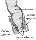
The interior of the anal cami and lower part of the rectum, showing the columns of Morgagni and the anal valves between their lower ends. (Pectinate line visible but not labeled.)
Details
Identifiers
Latin linea pectinata, linea anocutanea
TA98 A05.7.05.009
TA2 3015
FMA 29321
Anatomical terminology

The pectinate line (dentate line) is a line which divides the upper two-thirds and lower third of the anal canal. Developmentally, this line represents the hindgut-proctodeum junction.

Contents

It is an important anatomical landmark in humans, and forms the boundary between the anal canal and the rectum according to the anatomic definition. [1] Colorectal surgeons instead define the anal canal as the zone from the anal verge to the anorectal ring (palpable structure formed by the external anal sphincter and the puborectalis muscle). [1] Several distinctions can be made based upon the location of a structure relative to the pectinate line:

DistinctionAbove pectinate lineBelow pectinate line
Lymph drainage internal iliac [2] superficial inguinal lymph nodes (below Hilton's white line)
Epithelium columnar epithelium (as is most of the digestive tract - the line represents the end of the part of the body derived from the hindgut) stratified squamous epithelium, non-keratinized (until Hilton's white line, where the anal verge becomes continuous with the perianal skin containing keratinized epithelium.)
Embryological origin [3] endoderm ectoderm
Artery superior rectal artery middle and inferior rectal arteries
Vein superior rectal vein draining into the inferior mesenteric vein and subsequently the hepatic portal system middle and inferior rectal veins
Hemorrhoids classificationinternal hemorrhoids (not painful)external hemorrhoids (painful)
Nerves inferior hypogastric plexus pudendal nerves

Additional images

Related Research Articles

<span class="mw-page-title-main">Large intestine</span> Last part of the digestive system in vertebrates

The large intestine, also known as the large bowel, is the last part of the gastrointestinal tract and of the digestive system in tetrapods. Water is absorbed here and the remaining waste material is stored in the rectum as feces before being removed by defecation. The colon is the longest portion of the large intestine, and the terms are often used interchangeably but most sources define the large intestine as the combination of the cecum, colon, rectum, and anal canal. Some other sources exclude the anal canal.

<span class="mw-page-title-main">Hemorrhoid</span> Vascular structures in the anal canal

Hemorrhoids, also known as piles, are vascular structures in the anal canal. In their normal state, they are cushions that help with stool control. They become a disease when swollen or inflamed; the unqualified term hemorrhoid is often used to refer to the disease. The signs and symptoms of hemorrhoids depend on the type present. Internal hemorrhoids often result in painless, bright red rectal bleeding when defecating. External hemorrhoids often result in pain and swelling in the area of the anus. If bleeding occurs, it is usually darker. Symptoms frequently get better after a few days. A skin tag may remain after the healing of an external hemorrhoid.

<span class="mw-page-title-main">Levator ani</span> Broad, thin muscle group, situated on either side of the pelvis

The levator ani is a broad, thin muscle group, situated on either side of the pelvis. It is formed from three muscle components: the pubococcygeus, the iliococcygeus, and the puborectalis.

<span class="mw-page-title-main">Mesentery</span> Contiguous fold of tissues that supports the intestines

In human anatomy, the mesentery, an organ that attaches the intestines to the posterior abdominal wall, comprises the double fold of the peritoneum. It helps in storing fat and allowing blood vessels, lymphatics, and nerves to supply the intestines.

<span class="mw-page-title-main">Rectal prolapse</span> Protrusion of the walls of the rectum outside the body

A rectal prolapse occurs when walls of the rectum have prolapsed to such a degree that they protrude out of the anus and are visible outside the body. However, most researchers agree that there are 3 to 5 different types of rectal prolapse, depending on whether the prolapsed section is visible externally, and whether the full or only partial thickness of the rectal wall is involved.

<span class="mw-page-title-main">Anal canal</span> Functional segment of the large intestine

The anal canal is the part that connects the rectum to the anus, located below the level of the pelvic diaphragm. It is located within the anal triangle of the perineum, between the right and left ischioanal fossa. As the final functional segment of the bowel, it functions to regulate release of excrement by two muscular sphincter complexes. The anus is the aperture at the terminal portion of the anal canal.

<span class="mw-page-title-main">Internal anal sphincter</span> Ring of smooth muscle that surrounds part of the anal canal

The internal anal sphincter, IAS, or sphincter ani internus is a ring of smooth muscle that surrounds about 2.5–4.0 cm of the anal canal. It is about 5 mm thick, and is formed by an aggregation of the smooth (involuntary) circular muscle fibers of the rectum. It terminates distally about 6 mm from the anal orifice.

<span class="mw-page-title-main">Middle rectal artery</span>

The middle rectal artery is an artery in the pelvis that supplies blood to the rectum.

<span class="mw-page-title-main">Rectal venous plexus</span>

The rectal venous plexus is the venous plexus surrounding the rectum. It consists of an internal and an external rectal plexus. It is drained by the superior, middle, and inferior rectal veins. It forms a portosystemic (portocaval) anastomosis. This allows rectally administered medications to bypass first pass metabolism.

<span class="mw-page-title-main">Transverse folds of rectum</span>

The transverse folds of rectum are semi-lunar transverse folds of the rectal wall that protrude into the rectum, not the anal canal as that lies below the rectum. Their use seems to be to support the weight of fecal matter, and prevent its urging toward the anus, which would produce a strong urge to defecate. Although the term rectum means straight, these transverse folds overlap each other during the empty state of the intestine to such an extent that, as Houston remarked, they require considerable maneuvering to conduct an instrument along the canal, as often occurs in sigmoidoscopy and colonoscopy.

<span class="mw-page-title-main">Defecography</span> Visualisation of the mechanics of a patients defecation

Defecography is a type of medical radiological imaging in which the mechanics of a patient's defecation are visualized in real time using a fluoroscope. The anatomy and function of the anorectum and pelvic floor can be dynamically studied at various stages during defecation.

<span class="mw-page-title-main">Rectum</span> Final portion of the large intestine

The rectum is the final straight portion of the large intestine in humans and some other mammals, and the gut in others. The adult human rectum is about 12 centimetres (4.7 in) long, and begins at the rectosigmoid junction at the level of the third sacral vertebra or the sacral promontory depending upon what definition is used. Its diameter is similar to that of the sigmoid colon at its commencement, but it is dilated near its termination, forming the rectal ampulla. It terminates at the level of the anorectal ring or the dentate line, again depending upon which definition is used. In humans, the rectum is followed by the anal canal, which is about 4 centimetres (1.6 in) long, before the gastrointestinal tract terminates at the anal verge. The word rectum comes from the Latin rēctumintestīnum, meaning straight intestine.

<span class="mw-page-title-main">Human anus</span> External opening of the rectum

In humans, the anus is the external opening of the rectum located inside the intergluteal cleft. Two sphincters control the exit of feces from the body during an act of defecation, which is the primary function of the anus. These are the internal anal sphincter and the external anal sphincter, which are circular muscles that normally maintain constriction of the orifice and which relax as required by normal physiological functioning. The inner sphincter is involuntary and the outer is voluntary. Above the anus is the perineum, which is also located beneath the vulva or scrotum.

The presacral fascia lines the anterior aspect of the sacrum, enclosing the sacral vessels and nerves. It continues anteriorly as the pelvic parietal fascia, covering the entire pelvic cavity.

Rectal discharge is intermittent or continuous expression of liquid from the anus. Normal rectal mucus is needed for proper excretion of waste. Otherwise, this is closely related to types of fecal incontinence but the term rectal discharge does not necessarily imply degrees of incontinence. Types of fecal incontinence that produce a liquid leakage could be thought of as a type of rectal discharge.

Solitary rectal ulcer syndrome or SRUS is a chronic disorder of the rectal mucosa. It commonly occurs with varying degrees of rectal prolapse. The condition is thought to be caused by different factors, such as long term constipation, straining during defecation, and dyssynergic defecation. Treatment is by normalization of bowel habits, biofeedback, and other conservative measures. In more severe cases various surgical procedures may be indicated. The condition is relatively rare, affecting approximately 1 in 100,000 people per year. It affects mainly adults aged 30–50. Females are affected slightly more often than males. The disorder can be confused clinically with rectal cancer or other conditions such as inflammatory bowel disease, even when a biopsy is done.

<span class="mw-page-title-main">Anismus</span> Failure to relax the pelvic floor muscles during defecation

Anismus or dyssynergic defecation is the failure of normal relaxation of pelvic floor muscles during attempted defecation. It can occur in both children and adults, and in both men and women. It can be caused by physical defects or it can occur for other reasons or unknown reasons. Anismus that has a behavioral cause could be viewed as having similarities with parcopresis, or psychogenic fecal retention.

Obstructed defecation syndrome is a major cause of functional constipation, of which it is considered a subtype. It is characterized by difficult and/or incomplete emptying of the rectum with or without an actual reduction in the number of bowel movements per week. Normal definitions of functional constipation include infrequent bowel movements and hard stools. In contrast, ODS may occur with frequent bowel movements and even with soft stools, and the colonic transit time may be normal, but delayed in the rectum and sigmoid colon.

<span class="mw-page-title-main">Anorectal disorder</span> Medical condition

Anorectal disorders include conditions involving the anorectal junction as seen in the image. They are painful but common conditions like hemorrhoids, tears, fistulas, or abscesses that affect the anal region. Most people experience some form of anorectal disorder during their lifetime. Primary care physicians can treat most of these disorders, however, high-risk individuals include those with HIV, roughly half of whom need surgery to remedy the disorders. Likelihood of malignancy should also be considered in high risk individuals. This is why it is important to perform a full history and physical exam on each patient. Because these disorders affect the rectum, people are often embarrassed or afraid to confer with a medical professional.

<span class="mw-page-title-main">Rectococcygeal muscle</span>

The rectococcygeal muscles are two bands of smooth muscle tissue arising from the 2nd and 3rd coccygeal vertebrae, and passing downward and forward to blend with the rectal longitudinal smooth muscle fibers on the posterior wall of the anal canal.

References

  1. 1 2 Lee, JM; Kim, NK (April 2018). "Essential Anatomy of the Anorectum for Colorectal Surgeons Focused on the Gross Anatomy and Histologic Findings". Annals of coloproctology. 34 (2): 59–71. doi:10.3393/ac.2017.12.15. PMID   29742860.
  2. MD, Tao Le, MD, MHS, Vikas Bhushan, MD, Matthew Sochat, MD (2017). First aid for the USMLE step 1 2017 : a student-to-student guide. ISBN   978-0071831420.{{cite book}}: CS1 maint: multiple names: authors list (link)
  3. Schoenwolf, Gary C.; Bleyl, Steven B.; Brauer, Philip R.; Francis-West, Philippa H. (2014-12-01). Larsen's Human Embryology. Elsevier Health Sciences. p. 372. ISBN   9781455727919.