Internal iliac vein

Last updated

Internal iliac vein
Internaliliacv.png
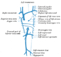
The veins of the right half of the male pelvis.
Gray586.png
The iliac veins. (Int. iliac visible at center.)
Details
Drains fromPelvic viscera
SourceInternal pudendal vein, middle rectal vein, vesical vein, uterine vein, obturator vein, inferior gluteal vein, superior gluteal vein
Drains to Common iliac vein
Artery Internal iliac artery
Identifiers
Latin vena iliaca interna,
vena hypogastrica
TA98 A12.3.10.004
TA2 5024
FMA 18884
Anatomical terminology

The internal iliac vein (hypogastric vein) begins near the upper part of the greater sciatic foramen, passes upward behind and slightly medial to the internal iliac artery and, at the brim of the pelvis, joins with the external iliac vein to form the common iliac vein.

Contents

Structure

Several veins unite above the greater sciatic foramen to form the internal iliac vein. It does not have the predictable branches of the internal iliac artery but its tributaries drain the same regions. [1] The internal iliac vein emerges from above the level of the greater sciatic notch It runs backwards, upwards and towards the midline to join the external iliac vein in forming the common iliac vein in front of the sacroiliac joint. It usually lies lateral to the internal iliac artery. [2] It is wide and 3 cm long. [3]

Tributaries

Originating outside the pelvis, its tributaries are the gluteal, internal pudendal and obturator veins. Running from the anterior surface of the sacrum are the lateral sacral veins. Coming from the pelvic plexuses and appropriate to gender are the middle rectal, vesical, prostatic, uterine and vaginal veins. [1] [3]

ReceivesDescription
superior gluteal veins
inferior gluteal veins
internal pudendal veins
obturator veins
have their origins outside the pelvis;
lateral sacral veins lie in front of the sacrum
middle hemorrhoidal vein
vesical vein
uterine vein
vaginal veins
originate in venous plexuses connected with the pelvic viscera.

Variation

On the left, the internal iliac vein lies lateral to the internal iliac artery 73% of the time. [4] On the right, this is 93% of the time. [4]

Function

The internal iliac veins drain the pelvic organs, sacrum, and coccyx. [2]

Clinical significance

If thrombosis disrupts blood flow in the external iliac systems, the internal iliac tributaries offer a major route of venous return from the femoral system. Damage to internal iliac vein tributaries during surgery can seriously compromise venous drainage and cause swelling of one or both legs. [1]

Additional images

Related Research Articles

<span class="mw-page-title-main">Sacrum</span> Bone of the spine

The sacrum, in human anatomy, is a large, triangular bone at the base of the spine that forms by the fusing of the sacral vertebrae (S1–S5) between ages 18 and 30.

<span class="mw-page-title-main">Internal pudendal artery</span> Blood vessel supplying blood to the external genitalia

The internal pudendal artery is one of the three pudendal arteries. It branches off the internal iliac artery, and provides blood to the external genitalia.

<span class="mw-page-title-main">Piriformis muscle</span> Hip muscle in the lateral rotator group

The piriformis muscle is a flat, pyramidally-shaped muscle in the gluteal region of the lower limbs. It is one of the six muscles in the lateral rotator group.

<span class="mw-page-title-main">Femoral vein</span> Large blood vessel in the leg

In the human body, the femoral vein is the vein that accompanies the femoral artery in the femoral sheath. It is a deep vein that begins at the adductor hiatus as the continuation of the popliteal vein. The great saphenous vein, and the deep femoral vein drain into the femoral vein in the femoral triangle when it becomes known as the common femoral vein. It ends at the inferior margin of the inguinal ligament where it becomes the external iliac vein. Its major tributaries are the deep femoral vein, and the great saphenous vein. The femoral vein contains valves.

<span class="mw-page-title-main">Common iliac vein</span> Veins draining blood from the pelvis and lower limbs

In human anatomy, the common iliac veins are formed by the external iliac veins and internal iliac veins. The left and right common iliac veins come together in the abdomen at the level of the fifth lumbar vertebra, forming the inferior vena cava. They drain blood from the pelvis and lower limbs.

<span class="mw-page-title-main">External iliac vein</span> Blood vessels connecting the thigh veins to the pelvis

The external iliac veins are large veins that connect the femoral veins to the common iliac veins. Their origin is at the inferior margin of the inguinal ligaments and they terminate when they join the internal iliac veins.

<span class="mw-page-title-main">Internal iliac artery</span> Main artery of the pelvis

The internal iliac artery is the main artery of the pelvis.

<span class="mw-page-title-main">Sacrotuberous ligament</span>

The sacrotuberous ligament is situated at the lower and back part of the pelvis. It is flat, and triangular in form; narrower in the middle than at the ends.

<span class="mw-page-title-main">Sacrospinous ligament</span>

The sacrospinous ligament is a thin, triangular ligament in the human pelvis. The base of the ligament is attached to the outer edge of the sacrum and coccyx, and the tip of the ligament attaches to the spine of the ischium, a bony protuberance on the human pelvis. Its fibres are intermingled with the sacrotuberous ligament.

<span class="mw-page-title-main">Superior gluteal artery</span>

The superior gluteal artery is the terminal branch of the posterior division of the internal iliac artery. It exits the pelvis through the greater sciatic foramen before splitting into a superficial branch and a deep branch.

<span class="mw-page-title-main">Lateral sacral artery</span> Artery in the pelvis

The lateral sacral arteries is an artery in the pelvis that arises from the posterior division of the internal iliac artery. It later splits into two smaller branches, a superior and an inferior.

<span class="mw-page-title-main">Obturator artery</span>

The obturator artery is a branch of the internal iliac artery that passes antero-inferiorly on the lateral wall of the pelvis, to the upper part of the obturator foramen, and, escaping from the pelvic cavity through the obturator canal, it divides into an anterior branch and a posterior branch.

<span class="mw-page-title-main">Inferior gluteal veins</span>

The inferior gluteal veins are venae comitantes of the inferior gluteal artery. They commence in the superior/proximal posterior thigh. They enter the pelvis through the lower part of the greater sciatic foramen. They converge to form a single vessel before emptying into the distal portion of the internal iliac vein.

<span class="mw-page-title-main">Pelvic cavity</span> Body cavity bounded by the pelvic bones

The pelvic cavity is a body cavity that is bounded by the bones of the pelvis. Its oblique roof is the pelvic inlet. Its lower boundary is the pelvic floor.

<span class="mw-page-title-main">Wing of ilium</span> Flat portion of the hip bone

The wing(ala)of ilium is the large expanded portion of the ilium, the bone which bounds the greater pelvis laterally. It presents for examination two surfaces—an external and an internal—a crest, and two borders—an anterior and a posterior.

<span class="mw-page-title-main">Greater sciatic notch</span>

The greater sciatic notch is a notch in the ilium, one of the bones that make up the human pelvis. It lies between the posterior inferior iliac spine (above), and the ischial spine (below). The sacrospinous ligament changes this notch into an opening, the greater sciatic foramen.

<span class="mw-page-title-main">Lateral sacral veins</span> Vein of the torso

The lateral sacral veins accompany the lateral sacral arteries on the anterior surface of the sacrum. They drain into the internal iliac vein. They communicate with each other via the sacral venous plexus.

<span class="mw-page-title-main">Outline of human anatomy</span> Overview of and topical guide to human anatomy

The following outline is provided as an overview of and topical guide to human anatomy:

<span class="mw-page-title-main">Hip bone</span> Bone of the pelvis

The hip bone is a large flat bone, constricted in the center and expanded above and below. In some vertebrates it is composed of three parts: the ilium, ischium, and the pubis.

<span class="mw-page-title-main">Pelvis</span> Lower torso of the human body

The pelvis is the lower part of the trunk, between the abdomen and the thighs, together with its embedded skeleton.

References

  1. 1 2 3 Delancey, John O.L. (2016). "73, True pelvis, pelvic floor and perineum". In Standring, Susan (ed.). Gray's Anatomy: The Anatomical Basis of Clinical Practice (41st ed.). Elsevier. pp. 1221–1236. ISBN   978-0-7020-6851-5.
  2. 1 2 Cramer, Gregory D.; Ro, Chae-Song (January 1, 2014), Cramer, Gregory D.; Darby, Susan A. (eds.), "Chapter 8 - The Sacrum, Sacroiliac Joint, and Coccyx", Clinical Anatomy of the Spine, Spinal Cord, and Ans (Third Edition), Saint Louis: Mosby, pp. 312–339, ISBN   978-0-323-07954-9 , retrieved January 28, 2021
  3. 1 2 Sinnatamby, Chummy S. (2011). "5". Last's Anatomy: Regional and Applied (12th ed.). Great Britain: Churchill Livingstone Elsevier. p. 309. ISBN   978-0-7020-4839-5 . Retrieved March 25, 2018.
  4. 1 2 Bleich, April T.; Rahn, David D.; Wieslander, Cecilia K.; Wai, Clifford Y.; Roshanravan, Shayzreen M.; Corton, Marlene M. (December 1, 2007). "Posterior division of the internal iliac artery: Anatomic variations and clinical applications". American Journal of Obstetrics and Gynecology. 197 (6): 658.e1–658.e5. doi:10.1016/j.ajog.2007.08.063. ISSN   0002-9378. PMID   18060970.

PD-icon.svgThis article incorporates text in the public domain from page 673 of the 20th edition of Gray's Anatomy (1918)