Lumbar veins

Last updated
Lumbar veins
Gray585.png
The veins of the right half of the male pelvis. (Third lumbar labeled at center top.)
Details
Drains to Inferior vena cava
Artery Lumbar arteries
Identifiers
Latin venae lumbales
TA98 A12.3.07.012
A12.3.09.003
TA2 4993
FMA 70889
Anatomical terminology

The lumbar veins are four pairs of veins running along the inside of the posterior abdominal wall, and drain venous blood from parts of the abdominal wall. Each lumbar vein accompanies a single lumbar artery. The lower two pairs of lumbar veins all drain directly into the inferior vena cava, whereas the fate of the upper two pairs is more variable.

Contents

Lumbar veins are the lumbar equivalent of the posterior intercostal veins of the thorax.

Structure

A lumbar vein accompanies each of the four lumbar arteries on each side of the body. [1]

Distribution and tributaries

Collectively, the lumbar veins drain blood from the territories supplied by the corresponding lumbar arteries (the posterior, lateral, and anterior abdominal wall). [1]

The lumbar veins drain the anterior spinal veins. [2]

Fate

The 3rd and 4th lumbar veins drain into the inferior vena cava. [3]

The fate of the two superior lumbar veins is far more variable, and may drain into either the inferior vena cava, ascending lumbar vein, azygos vein, or (on the left side) the left renal vein; the 1st lumbar vein often passes inferiorly to join the 2nd lumbar vein, but may less commonly drain into the ascending lumbar vein, or the azygos vein; the 2nd lumbar vein may drain directly into the inferior vena cava (at or near the level of the renal veins), but may less commonly join the 3rd lumbar vein or drain into the ascending lumbar vein. [1]

Anastomoses

The 1st and 2nd lumbar veins are often interconnected with each other. An ascending lumbar vein additionally interconnects ipsilateral lumbar veins on each side. [1]

The lumbar veins communicate with the external and internal vertebral venous plexuses, and form anastomoses with tributaries of the azygos vein and hemiazygos vein (posteriorly), and (anteriorly) with branches of the epigastric veins, circumflex iliac veins, and lateral thoracic veins. The anastomoses formed by the lumbar veins provide a means of collateral veinous return. [1]

Relations

The lumbar veins are venae comitantes of the lumbar arteries. [1] Each lumbar vein lies superior to its adjacent/corresponding lumbar artery (the same arrangement as in the intercostal neurovascular bundles).[ citation needed ]

All of the lumbar veins lie posterior to the ipsilateral sympathetic trunk.[ citation needed ]

Bilateral assymetry

On the left side, the 3rd and 4th lumbar veins pass posterior to the abdominal aorta to reach the inferior vena cava (which is displaced to the right of the midline); thus, the left 3rd and 4th lumbar veins are longer than the corresponding contralateral two lumbar veins. [1]

Variation

There is variation in which lumbar veins drain to the inferior vena cava and which drain to the ascending lumbar vein, the azygos vein, and the hemiazygos vein. [3] A left lumbar vein may drain into the left renal vein. [3]

Clinical significance

During central venous catheterisation of the inferior vena cava, the catheter may enter a lumbar vein. [4] The catheter may need to be repositioned. [4]

See also

Related Research Articles

<span class="mw-page-title-main">Brachiocephalic vein</span> Vein

The left and right brachiocephalic veins are major veins in the upper chest, formed by the union of each corresponding internal jugular vein and subclavian vein. This is at the level of the sternoclavicular joint. The left brachiocephalic vein is more than twice the length of the right brachiocephalic vein.

<span class="mw-page-title-main">Inferior vena cava</span> One of two veinous trunks bringing deoxygenated blood back to the heart

The inferior vena cava is a large vein that carries the deoxygenated blood from the lower and middle body into the right atrium of the heart. It is formed by the joining of the right and the left common iliac veins, usually at the level of the fifth lumbar vertebra.

<span class="mw-page-title-main">Azygos vein</span> Human blood vessel by the spine

The azygos vein is a vein running up the right side of the thoracic vertebral column draining itself towards the superior vena cava. It connects the systems of superior vena cava and inferior vena cava and can provide an alternative path for blood to the right atrium when either of the venae cavae is blocked.

<span class="mw-page-title-main">Abdominal aorta</span> Largest artery in the abdomen

In human anatomy, the abdominal aorta is the largest artery in the abdominal cavity. As part of the aorta, it is a direct continuation of the descending aorta.

<span class="mw-page-title-main">Renal vein</span> Short thick veins which return blood from the kidneys to the vena cava

The renal veins in the renal circulation, are large-calibre veins that drain blood filtered by the kidneys into the inferior vena cava. There is one renal vein draining each kidney.

<span class="mw-page-title-main">Superior mesenteric artery</span> Artery which supplies blood to the intestines and pancreas

In human anatomy, the superior mesenteric artery (SMA) is an artery which arises from the anterior surface of the abdominal aorta, just inferior to the origin of the celiac trunk, and supplies blood to the intestine from the lower part of the duodenum through two-thirds of the transverse colon, as well as the pancreas.

<span class="mw-page-title-main">Common iliac vein</span> Veins draining blood from the pelvis and lower limbs

In human anatomy, the common iliac veins are formed by the external iliac veins and internal iliac veins. The left and right common iliac veins come together in the abdomen at the level of the fifth lumbar vertebra, forming the inferior vena cava. They drain blood from the pelvis and lower limbs.

<span class="mw-page-title-main">Internal thoracic vein</span> Large blood vessel draining breasts and the chest wall

In human anatomy, the internal thoracic vein is the vein that drains the chest wall and breasts.

<span class="mw-page-title-main">Hemiazygos vein</span>

The hemiazygos vein is a vein running superiorly in the lower thoracic region, just to the left side of the vertebral column.

<span class="mw-page-title-main">Inferior phrenic arteries</span>

The inferior phrenic arteries are two small vessels which supply the diaphragm. They present much variety in their origin.

<span class="mw-page-title-main">Middle suprarenal arteries</span> Arteries of the abdomen

The middle suprarenal artery is a paired artery in the abdomen. It is a branch of the aorta. It supplies the adrenal gland.

<span class="mw-page-title-main">Lumbar arteries</span>

The lumbar arteries are arteries located in the lower back or lumbar region. The lumbar arteries are in parallel with the intercostals.

<span class="mw-page-title-main">Ovarian vein</span>

The ovarian vein, the female gonadal vein, carries deoxygenated blood from its corresponding ovary to inferior vena cava or one of its tributaries. It is the female equivalent of the testicular vein, and is the venous counterpart of the ovarian artery. It can be found in the suspensory ligament of the ovary.

<span class="mw-page-title-main">Testicular vein</span>

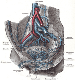
The testicular vein, the male gonadal vein, carries deoxygenated blood from its corresponding testis to the inferior vena cava or one of its tributaries. It is the male equivalent of the ovarian vein, and is the venous counterpart of the testicular artery.

<span class="mw-page-title-main">Intercostal arteries</span> Arteries supplying the space between the ribs

The intercostal arteries are a group of arteries that supply the area between the ribs ("costae"), called the intercostal space. The highest intercostal artery is an artery in the human body that usually gives rise to the first and second posterior intercostal arteries, which supply blood to their corresponding intercostal space. It usually arises from the costocervical trunk, which is a branch of the subclavian artery. Some anatomists may contend that there is no supreme intercostal artery, only a supreme intercostal vein.

<span class="mw-page-title-main">Ascending lumbar vein</span> Vein on the vertebral column

The ascending lumbar vein is a vein that runs up through the lumbar region on the side of the vertebral column.

<span class="mw-page-title-main">Outline of human anatomy</span> Overview of and topical guide to human anatomy

The following outline is provided as an overview of and topical guide to human anatomy:

<span class="mw-page-title-main">Posterior cardinal vein</span>

The posterior cardinal veins or postcardinal veins join with the corresponding right and left cardinal veins to form the left common cardinal veins, which empty in the sinus venosus. In the development of a human embryo, most of the posterior cardinal veins regress, and what remains of them forms the renal segment of the inferior vena cava and the common iliac veins. Later in the development stages, the posterior cardinal veins are replaced by the subcardinal and supracardinal veins. The subcardinal veins form part of the inferior vena cava, the renal veins and the gonadal veins. The supracardinal veins form part of the inferior vena cava, the intercostal veins, the hemiazygos vein and the azygos vein.

References

  1. 1 2 3 4 5 6 7 Gray's anatomy : the anatomical basis of clinical practice. Susan Standring (Forty-second ed.). [New York]. 2021. p. 1144. ISBN   978-0-7020-7707-4. OCLC   1201341621.{{cite book}}: CS1 maint: others (link)
  2. Gilchrist, Russell V. (2008). "Developmental and Functional Anatomy of the Lumbar Spine". Interventional Spine. pp. 855–870. doi:10.1016/B978-0-7216-2872-1.50085-6. ISBN   978-0-7216-2872-1.
  3. 1 2 3 Baniel, Jack; Foster, Richard S.; Donohue, John P. (May 1995). "Surgical Anatomy of the Lumbar Vessels: Implications for Retroperitoneal Surgery". Journal of Urology. 153 (5): 1422–1425. doi:10.1016/S0022-5347(01)67418-4. PMID   7714956.
  4. 1 2 Miller, Grant G; Evans, Mark G; Chou, Shirley (October 1995). "Lumbar vein malposition of inferior vena cava catheters". Journal of Pediatric Surgery. 30 (10): 1500–1501. doi:10.1016/0022-3468(95)90420-4. PMID   8786502.