Sympathetic trunk

Last updated
Sympathetic trunk
Gray847.png
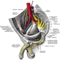
Abdominal portion of the sympathetic trunk, with the celiac plexus and hypogastric plexus. (Sympathetic trunk labeled at center left.)
Gray799.svg
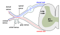
Scheme showing pathways (white/grey rami are spatially reversed, possibly for clarity?) of a typical spinal nerve.
1. Somatic efferent.
2. Somatic afferent.
3,4,5. Sympathetic efferent.
6,7. Parasympathetic afferent.
Details
Identifiers
Latin truncus sympathicus
TA98 A14.3.01.002
TA2 6602
FMA 6258
Anatomical terminology

The sympathetic trunks (sympathetic chain, gangliated cord) are a paired bundle of nerve fibers that run from the base of the skull to the coccyx. They are a major component of the sympathetic nervous system.

Contents

Structure

The sympathetic trunk lies just lateral to the vertebral bodies for the entire length of the vertebral column. It interacts with the anterior rami of spinal nerves by way of rami communicantes. The sympathetic trunk permits preganglionic fibers of the sympathetic nervous system to ascend to spinal levels superior to T1 and descend to spinal levels inferior to L2/3. [1] [2] [3] [4] [5] [6] [7]

The superior end of it is continued upward through the carotid canal into the skull, and forms a plexus on the internal carotid artery; the inferior part travels in front of the coccyx, where it converges with the other trunk at a structure known as the ganglion impar.

Along the length of the sympathetic trunk are sympathetic ganglia known as paravertebral ganglia.

Relations

In the cervical region, the sympathetic trunk is situated upon the prevertebral fascia posterior to the carotid sheath. [8] :600

Function

The sympathetic trunk is a fundamental part of the sympathetic nervous system, and part of the autonomic nervous system. It allows nerve fibres to travel to spinal nerves that are superior and inferior to the one in which they originated. Also, a number of nerves, such as most of the splanchnic nerves, arise directly from the trunks.

Autonomic nervous supply to organs in the human body edit
OrganNerves [9] Spinal column origin [9]
stomach T5, T6, T7, T8, T9, sometimes T10
duodenum T5, T6, T7, T8, T9, sometimes T10
jejunum and ileum T5, T6, T7, T8, T9
spleen T6, T7, T8
gallbladder and liver T6, T7, T8, T9
colon
pancreatic head T8, T9
appendix T10
bladder S2-S4
kidneys and ureters T11, T12

Additional images

See also

Related Research Articles

<span class="mw-page-title-main">Autonomic nervous system</span> Division of the nervous system supplying internal organs, smooth muscle and glands

The autonomic nervous system (ANS), formerly referred to as the vegetative nervous system, is a division of the nervous system that supplies internal organs, smooth muscle and glands. The autonomic nervous system is a control system that acts largely unconsciously and regulates bodily functions, such as the heart rate, its force of contraction, digestion, respiratory rate, pupillary response, urination, and sexual arousal. This system is the primary mechanism in control of the fight-or-flight response.

<span class="mw-page-title-main">Parasympathetic nervous system</span> Division of the autonomic nervous system

The parasympathetic nervous system (PSNS) is one of the three divisions of the autonomic nervous system, the others being the sympathetic nervous system and the enteric nervous system. The enteric nervous system is sometimes considered part of the autonomic nervous system, and sometimes considered an independent system.

<span class="mw-page-title-main">Spinal nerve</span> Nerve that carries signals between the spinal cord and the body

A spinal nerve is a mixed nerve, which carries motor, sensory, and autonomic signals between the spinal cord and the body. In the human body there are 31 pairs of spinal nerves, one on each side of the vertebral column. These are grouped into the corresponding cervical, thoracic, lumbar, sacral and coccygeal regions of the spine. There are eight pairs of cervical nerves, twelve pairs of thoracic nerves, five pairs of lumbar nerves, five pairs of sacral nerves, and one pair of coccygeal nerves. The spinal nerves are part of the peripheral nervous system.

<span class="mw-page-title-main">Nerve plexus</span>

A nerve plexus is a plexus of intersecting nerves. A nerve plexus is composed of afferent and efferent fibers that arise from the merging of the anterior rami of spinal nerves and blood vessels. There are five spinal nerve plexuses, except in the thoracic region, as well as other forms of autonomic plexuses, many of which are a part of the enteric nervous system. The nerves that arise from the plexuses have both sensory and motor functions. These functions include muscle contraction, the maintenance of body coordination and control, and the reaction to sensations such as heat, cold, pain, and pressure. There are several plexuses in the body, including:

<span class="mw-page-title-main">Lumbar nerves</span>

The lumbar nerves are the five pairs of spinal nerves emerging from the lumbar vertebrae. They are divided into posterior and anterior divisions.

<span class="mw-page-title-main">Superior cervical ganglion</span> Largest of the cervical ganglia

The superior cervical ganglion (SCG) is the upper-most and largest of the cervical sympathetic ganglia of the sympathetic trunk. It probably formed by the union of four sympathetic ganglia of the cervical spinal nerves C1-C4. It is the only ganglion of the sympathetic nervous system that innervates the head and neck. The SCG innervates numerous structures of the head and neck.

Each spinal nerve receives a branch called a gray ramus communicans from the adjacent paravertebral ganglion of the sympathetic trunk. The gray rami communicantes contain postganglionic nerve fibers of the sympathetic nervous system and are composed of largely unmyelinated neurons. This is in contrast to the white rami communicantes, in which heavily myelinated neurons give the rami their white appearance.

<span class="mw-page-title-main">Superior hypogastric plexus</span>

The superior hypogastric plexus is a plexus of nerves situated on the vertebral bodies anterior to the bifurcation of the abdominal aorta. It bifurcates to form the left and the right hypogastric nerve. The SHP is the continuation of the abdominal aortic plexus.

<span class="mw-page-title-main">Sympathetic ganglia</span> Ganglia of the sympathetic nervous system

The sympathetic ganglia, or paravertebral ganglia are autonomic ganglia, of the sympathetic nervous system. Ganglia are 20,000 to 30,000 afferent and efferent nerve cell bodies that run along on either side of the spinal cord. Afferent nerve cell bodies bring information from the body to the brain and spinal cord, while efferent nerve cell bodies bring information from the brain and spinal cord to the rest of the body. The cell bodies create long sympathetic chains that are on either side of the spinal cord. They also form para- or pre-vertebral ganglia of gross anatomy.

<span class="mw-page-title-main">Lateral grey column</span>

The lateral grey column is one of the three grey columns of the spinal cord ; the others being the anterior and posterior grey columns. The lateral grey column is primarily involved with activity in the sympathetic division of the autonomic motor system. It projects to the side as a triangular field in the thoracic and upper lumbar regions of the postero-lateral part of the anterior grey column.

<span class="mw-page-title-main">Thoracic splanchnic nerves</span>

Thoracic splanchnic nerves are splanchnic nerves that arise from the sympathetic trunk in the thorax and travel inferiorly to provide sympathetic supply to the abdomen. The nerves contain preganglionic sympathetic fibers and general visceral afferent fibers.

<span class="mw-page-title-main">Middle cervical ganglion</span>

The middle cervical ganglion is the smallest of the three cervical sympathetic ganglia. It presumably represents the merging of the sympathetic ganglia of cervical segments C5-C6. It is usually situated at the level of the sixth cervical vertebra.

<span class="mw-page-title-main">Inferior cervical ganglion</span>

The inferior cervical ganglion is one of the three cervical sympathetic ganglia. It situated between the base of the transverse process of the last cervical vertebra and the neck of the first rib, on the medial side of the costocervical artery.

<span class="mw-page-title-main">Sacral splanchnic nerves</span>

Sacral splanchnic nerves are splanchnic nerves that connect the inferior hypogastric plexus to the sympathetic trunk in the pelvis.

<span class="mw-page-title-main">Hypogastric nerve</span> Human nerves

The hypogastric nerves are the continuation of the superior hypogastric plexus that descend into the pelvis anterior the sacrum and become the inferior hypogastric plexuses on either side of pelvic organs. The hypogastric nerves serve as a pathway for autonomic fibers to communicate between the lower abdomen and pelvis.

<span class="mw-page-title-main">Cervical ganglia</span>

The cervical ganglia are paravertebral ganglia of the sympathetic nervous system. Preganglionic nerves from the thoracic spinal cord enter into the cervical ganglions and synapse with its postganglionic fibers or nerves. The cervical ganglion has three paravertebral ganglia:

<span class="mw-page-title-main">Lumbar ganglia</span>

The lumbar ganglia are paravertebral ganglia located in the inferior portion of the sympathetic trunk. The lumbar portion of the sympathetic trunk typically has 4 lumbar ganglia. The lumbar splanchnic nerves arise from the ganglia here, and contribute sympathetic efferent fibers to the nearby plexuses. The first two lumbar ganglia have both white and gray rami communicates.

<span class="mw-page-title-main">Outline of human anatomy</span> Overview of and topical guide to human anatomy

The following outline is provided as an overview of and topical guide to human anatomy:

<span class="mw-page-title-main">Outline of the human nervous system</span> Overview of and topical guide to the human nervous system

The following Diagram is provided as an overview of and topical guide to the human nervous system:

References

PD-icon.svgThis article incorporates text in the public domain from page 976 of the 20th edition of Gray's Anatomy (1918)

  1. Mader S. S. (2000): Human biology. McGraw-Hill, New York, ISBN   0-07-290584-0; ISBN   0-07-117940-2.
  2. Pritchard T. E., Alloway D. (1999): Medical neuroscience. Hayes Barton Press, ISBN   978-1-59377-200-0:https://books.google.com/books/about/Medical_neuroscience.html?id=m7Y80PcFHtsC.
  3. Butler A. B., Hodos W. (2005): Comparative vertebrate neuroanatomy: evolution and adaptation. Wiley-Blackwell, ISBN   978-0-471-21005-4.
  4. Butler, Ann B.; Hodos, William (2005-09-02). Comparative Vertebrate Neuroanatomy: Evolution and Adaptation. Wiley. ISBN   9780471733836.
  5. Hall J. E., Guyton A. C. (2006): Textbook of medical physiology, 11th edition. Elsevier Saunders, St. Louis, Mo, ISBN   0-7216-0240-1.
  6. Warrell D. A., Cox T. M., Firth J. D. (2010): The Oxford Textbook of Medicine Archived 2012-03-21 at the Wayback Machine (5th ed.). Oxford University Press
  7. Greenstein B., Greenstein A. (2002): Color atlas of neuroscience – Neuroanatomy and neurophysiology. Thieme, Stuttgart – New York, ISBN   9783131081711.
  8. Standring, Susan (2020). Gray's Anatomy: The Anatomical Basis of Clinical Practice (42nd ed.). [New York]. ISBN   978-0-7020-7707-4. OCLC   1201341621.
  9. 1 2 Unless specified otherwise in the boxes, the source is: Moore, Keith L.; Agur, A. M. R. (2002). Essential Clinical Anatomy (2nd ed.). Lippincott Williams & Wilkins. p. 199. ISBN   978-0-7817-5940-3.