Phrenic nerve

Last updated

Phrenic nerve
Gray806.png
The phrenic nerve as it passes through the thorax to supply the diaphragm.
Gray808.png
The phrenic nerve emerges from the cervical plexus, with the right brachial plexus shown here.
Details
FromC3–C5 of cervical plexus
Innervates Diaphragm
Identifiers
Latin nervus phrenicus
MeSH D010791
TA98 A14.2.02.028
TA2 6380
FMA 6191
Anatomical terms of neuroanatomy

The phrenic nerve is a mixed motor/sensory nerve that originates from the C3-C5 spinal nerves in the neck. The nerve is important for breathing because it provides exclusive motor control of the diaphragm, the primary muscle of respiration. In humans, the right and left phrenic nerves are primarily supplied by the C4 spinal nerve, but there is also a contribution from the C3 and C5 spinal nerves. From its origin in the neck, the nerve travels downward into the chest to pass between the heart and lungs towards the diaphragm.

Contents

In addition to motor fibers, the phrenic nerve contains sensory fibers, which receive input from the central tendon of the diaphragm and the mediastinal pleura, as well as some sympathetic nerve fibers. Although the nerve receives contributions from nerve roots of the cervical plexus and the brachial plexus, it is usually considered separate from either plexus.

The name of the nerve comes from Ancient Greek phren 'diaphragm'. [1]

Structure

The phrenic nerve originates in the phrenic motor nucleus in the ventral horn of the cervical spinal cord. It descends obliquely with the internal jugular vein across the anterior scalene, deep to the prevertebral layer of deep cervical fascia and the transverse cervical and suprascapular arteries. On the left, the phrenic nerve crosses anterior to the first part of the subclavian artery. On the right, it lies on the anterior scalene muscle and crosses anterior to the 2nd part of the subclavian artery. On both sides, the phrenic nerve usually runs posterior to the subclavian vein as it enters the thorax where it runs anterior to the root of the lung and between the fibrous pericardium and mediastinal parietal pleura. [2]

The pericardiacophrenic arteries and veins travel with their respective phrenic nerves.

The phrenic nerve can be marked by a line connecting these two points:

  1. 1st point can be labelled 3.5 cm at the level of the thyroid cartilage from the midsagittal plane.
  2. 2nd point is at the medial end of the clavicle.

Variation

As with most nerves in the neck, multiple anatomic variants have been described. Notably, there may be variability in the course of the phrenic nerve in the retro-clavicular region such that the nerve courses anterior to the subclavian vein, rather than its typical position posterior to the vein (between the subclavian vein and artery). [3] This variant may predispose the phrenic nerve to injury during subclavian vascular cannulation.

In addition, an accessory phrenic nerve is commonly identified, present in up to 75% of a cadaveric study. [3]

In canines, the phrenic nerve arises from C5-C7 with occasional small contributions from C4. [4] In the cat, horse, ox, and small ruminant the phrenic nerve arises variably from C4-C7.

Function

Both of these nerves supply motor fibers to the diaphragm and sensory fibers to the fibrous pericardium, mediastinal pleura, and diaphragmatic peritoneum.

Some sources describe the right phrenic nerve as innervating the gallbladder, other sources make no such mention. [5] The right phrenic nerve may also supply the capsule of the liver. [6]

Clinical significance

Left phrenic nerve palsy (right image side) in fluoroscopy: forced inspiration with the closed mouth leads to paradox elevation of the paralytic left diaphragm while the healthy right side moves down. Zwerchfellparese links - Sniff-Test - 001 - kleiner.gif
Left phrenic nerve palsy (right image side) in fluoroscopy: forced inspiration with the closed mouth leads to paradox elevation of the paralytic left diaphragm while the healthy right side moves down.

Pain arising from structures supplied by the phrenic nerve is often "referred" to other somatic regions served by spinal nerves C3-C5. For example, a subphrenic abscess beneath the right diaphragm might cause a patient to feel pain in the right shoulder.

Irritation of the phrenic nerve (or the tissues it supplies) leads to the hiccup reflex. A hiccup is a spasmodic contraction of the diaphragm, which pulls air against the closed folds of the larynx.

The phrenic nerve must be identified during thoracic surgery and preserved. To confirm the identity of the phrenic nerve, a doctor may gently manipulate it to elicit a dartle (diaphragmatic startle) response. [7] The right phrenic nerve may be crushed by the vena cava clamp during liver transplantation. [8] Severing the phrenic nerve, or a phrenectomy, [9] will paralyse that half of the diaphragm. Bilateral diaphragmatic paralysis or BDP can also be caused by spinal cord injury, motor neuron disease, infection, pneumonia, sarcoidosis, multiple sclerosis, polyneuropathy, myopathy and amyotrophy, cardiac surgery, lung transplantation, or mediastinal tumors. [10] [11] Diaphragm paralysis is best demonstrated by sonography. [8] Breathing will be made more difficult but will continue provided the other nerve is intact.

The phrenic nerve arises from the neck (C3-C5) and innervates the diaphragm, which is much lower. Hence, patients suffering spinal cord injuries below the neck are still able to breathe effectively, despite any paralysis of the lower limbs.

Brachial plexus injuries can cause paralysis in various regions in the arm, forearm, and hand depending on the severed nerves. The resulting palsy has been clinically treated using the phrenic nerve as a donor for neurotization of the musculocutaneous nerve and the median nerve. [12] This treatment has a high success rate (84.6%) in partial to full restoration of the innervation to the damaged nerve. [12] Furthermore, this procedure has resulted in restored function to nerves in the brachial plexus with minimal impact to respiratory function of the phrenic nerve. The instances where pulmonary vital capacity is reduced have typically been a result of use of the right phrenic as the donor for the neurotization whereas use of left phrenic nerve has not been significantly linked to reduced pulmonary vital capacity. [13]

See also

Related Research Articles

<span class="mw-page-title-main">Peripheral nervous system</span> Part of the nervous system excluding the brain and spinal cord

The peripheral nervous system (PNS) is one of two components that make up the nervous system of bilateral animals, with the other part being the central nervous system (CNS). The PNS consists of nerves and ganglia, which lie outside the brain and the spinal cord. The main function of the PNS is to connect the CNS to the limbs and organs, essentially serving as a relay between the brain and spinal cord and the rest of the body. Unlike the CNS, the PNS is not protected by the vertebral column and skull, or by the blood–brain barrier, which leaves it exposed to toxins.

Articles related to anatomy include:

<span class="mw-page-title-main">Brachial plexus</span> Network of nerves

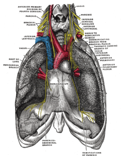
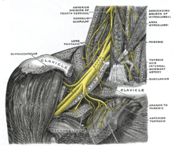
The brachial plexus is a network of nerves formed by the anterior rami of the lower four cervical nerves and first thoracic nerve. This plexus extends from the spinal cord, through the cervicoaxillary canal in the neck, over the first rib, and into the armpit, it supplies afferent and efferent nerve fibers to the chest, shoulder, arm, forearm, and hand.

<span class="mw-page-title-main">Spinal nerve</span> Nerve that carries signals between the spinal cord and the body

A spinal nerve is a mixed nerve, which carries motor, sensory, and autonomic signals between the spinal cord and the body. In the human body there are 31 pairs of spinal nerves, one on each side of the vertebral column. These are grouped into the corresponding cervical, thoracic, lumbar, sacral and coccygeal regions of the spine. There are eight pairs of cervical nerves, twelve pairs of thoracic nerves, five pairs of lumbar nerves, five pairs of sacral nerves, and one pair of coccygeal nerves. The spinal nerves are part of the peripheral nervous system.

<span class="mw-page-title-main">Subclavian artery</span> Major arteries of the upper thorax, below the clavicle

In human anatomy, the subclavian arteries are paired major arteries of the upper thorax, below the clavicle. They receive blood from the aortic arch. The left subclavian artery supplies blood to the left arm and the right subclavian artery supplies blood to the right arm, with some branches supplying the head and thorax. On the left side of the body, the subclavian comes directly off the aortic arch, while on the right side it arises from the relatively short brachiocephalic artery when it bifurcates into the subclavian and the right common carotid artery.

<span class="mw-page-title-main">Cervical plexus</span> Network of nerves in the neck

The cervical plexus is a nerve plexus of the anterior rami of the first four cervical spinal nerves C1-C4. The cervical plexus provides motor innervation to some muscles of the neck, and the diaphragm; it provides sensory innervation to parts of the head, neck, and chest.

<span class="mw-page-title-main">Cervical vertebrae</span> Vertebrae of the neck

In tetrapods, cervical vertebrae are the vertebrae of the neck, immediately below the skull. Truncal vertebrae lie caudal of cervical vertebrae. In sauropsid species, the cervical vertebrae bear cervical ribs. In lizards and saurischian dinosaurs, the cervical ribs are large; in birds, they are small and completely fused to the vertebrae. The vertebral transverse processes of mammals are homologous to the cervical ribs of other amniotes. Most mammals have seven cervical vertebrae, with the only three known exceptions being the manatee with six, the two-toed sloth with five or six, and the three-toed sloth with nine.

<span class="mw-page-title-main">Scalene muscles</span> Muscles on the sides of the neck

The scalene muscles are a group of three muscles on each side of the neck, identified as the anterior, the middle, and the posterior. They are innervated by the third to the eighth cervical spinal nerves (C3-C8).

<span class="mw-page-title-main">Ansa cervicalis</span> From cervical plexus

The ansa cervicalis is a loop formed by muscular branches of the cervical plexus formed by branches of cervical spinal nerves C1-C3. The ansa cervicalis has two roots - a superior root and an inferior root - that unite distally, forming a loop. It is situated within the carotid sheath.

<span class="mw-page-title-main">Nerve plexus</span> Network of nerve fibres

A nerve plexus is a plexus of intersecting nerves. A nerve plexus is composed of afferent and efferent fibers that arise from the merging of the anterior rami of spinal nerves and blood vessels. There are five spinal nerve plexuses, except in the thoracic region, as well as other forms of autonomic plexuses, many of which are a part of the enteric nervous system. The nerves that arise from the plexuses have both sensory and motor functions. These functions include muscle contraction, the maintenance of body coordination and control, and the reaction to sensations such as heat, cold, pain, and pressure. There are several plexuses in the body, including:

<span class="mw-page-title-main">Thoracic aorta</span> Part of the aorta located in the thorax

The thoracic aorta is a part of the aorta located in the thorax. It is a continuation of the aortic arch. It is located within the posterior mediastinal cavity, but frequently bulges into the left pleural cavity. The descending thoracic aorta begins at the lower border of the fourth thoracic vertebra and ends in front of the lower border of the twelfth thoracic vertebra, at the aortic hiatus in the diaphragm where it becomes the abdominal aorta.

<span class="mw-page-title-main">Posterior triangle of the neck</span>

The posterior triangle is a region of the neck.

<span class="mw-page-title-main">Pericardiacophrenic artery</span>

The pericardiacophrenic artery is a long slender branch of the internal thoracic artery.

<span class="mw-page-title-main">Subclavian nerve</span>

The subclavian nerve, also known as the nerve to the subclavius, is small branch of the upper trunk of the brachial plexus. It contains axons from C5 and C6. It innervates the subclavius muscle.

<span class="mw-page-title-main">Subclavian triangle</span>

The subclavian triangle, the smaller division of the posterior triangle, is bounded, above, by the inferior belly of the omohyoideus; below, by the clavicle; its base is formed by the posterior border of the sternocleidomastoideus.

<span class="mw-page-title-main">Head and neck anatomy</span>

This article describes the anatomy of the head and neck of the human body, including the brain, bones, muscles, blood vessels, nerves, glands, nose, mouth, teeth, tongue, and throat.

<span class="mw-page-title-main">Ventral ramus of spinal nerve</span> Anterior division of a spinal nerve

The ventral ramus is the anterior division of a spinal nerve. The ventral rami supply the antero-lateral parts of the trunk and the limbs. They are mainly larger than the dorsal rami.

<span class="mw-page-title-main">Prevertebral fascia</span> Layer of deep cervical fascia that surrounds the vertebral column

The prevertebral fascia is the layer of deep cervical fascia that surrounds the vertebral column. It is the deepest layer of deep cervical fascia.

<span class="mw-page-title-main">Outline of human anatomy</span> Overview of and topical guide to human anatomy

The following outline is provided as an overview of and topical guide to human anatomy:

<span class="mw-page-title-main">Pulmonary pleurae</span> Membrane lining the thoracic cavity wall

The pulmonary pleurae are the two flattened sacs ensheathing each lung, locally appearing as two opposing layers of serous membrane separating the lungs from the mediastinum and the inside surfaces of the surrounding chest walls.

References

  1. O'Rahilly, Ronan (2008). Basic Human Anatomy. Hanover, New Hampshire: Geisel School of Medicine. Archived from the original on 20 March 2018. Retrieved 3 April 2019.
  2. Moore, Keith L. (1999). Clinically oriented anatomy . Philadelphia: Lippincott Williams & Wilkins. ISBN   978-0-683-06141-3.[ page needed ]
  3. 1 2 Prakash; Prabhu, L. V.; Madhyastha, S; Singh, G (2007). "A variation of the phrenic nerve: Case report and review" (PDF). Singapore Medical Journal. 48 (12): 1156–7. PMID   18043847.
  4. Evans, Howard (1979). Miller's Anatomy of the Dog. Philadelphia, PA: W. B. Saunders Company. p. 978. ISBN   978-0-7216-3438-8.
  5. Alexander, William (1940). "The innervation of the biliary system". Journal of Comparative Neurology. 72 (2): 357–370. doi:10.1002/cne.900720205. S2CID   83974380.
  6. Lautt, W. Wayne (2009). Hepatic Nerves. Morgan & Claypool Life Sciences.
  7. Dalman, Ronald; Thompson, Robert (2015). "7. Neurogenic Thoracic Outlet Syndrome Exposure and Decompression: Supraclavicular". Operative Techniques in Vascular Surgery. Philadelphia, PA: Wolters Kluwer Health. p. 50. ISBN   9781451190205.
  8. 1 2 McAlister, Vivian C.; Grant, David R.; Roy, Andre; Brown, William F.; Hutton, Linda C.; Leasa, David J.; Ghent, Cameron N.; Veitch, James E.; Wall, William J. (1993). "Right phrenic nerve injury in orthotopic liver transplantation". Transplantation. 55 (4): 826–30. doi: 10.1097/00007890-199304000-00027 . PMID   8475559.
  9. Hine, Maynard Kiplinger (1975). Review of dentistry: questions and answers (6th ed.). Mosby. p. 421. ISBN   978-0-8016-2196-3.
  10. Mizubuti GB, Wang L, Ho AM, Tanzola RC, Leitch J (November 2017). "Perioperative Management for Abdominal Surgery in Bilateral Diaphragmatic Paralysis: A Case Report and Literature Review". A&A Case Reports. 9 (10): 280–282. doi:10.1213/XAA.0000000000000592. PMID   28691979. S2CID   44462495.
  11. Chen HY, Chen HC, Lin MC, Liaw MY (August 2015). "Bilateral Diaphragmatic Paralysis in a Patient With Critical Illness Polyneuropathy: A Case Report". Medicine (Baltimore). 94 (31): e1288. doi:10.1097/MD.0000000000001288. PMC   4616567 . PMID   26252301.
  12. 1 2 Gu YD, Wu MM, Zhen YL, Zhao JA, Zhang GM, Chen DS, Yan JG, Cheng XM (1989). "Phrenic nerve transfer for brachial plexus motor neurotization". Microsurgery. 10 (4): 287–9. doi:10.1002/micr.1920100407. PMID   2593799. S2CID   74732561.
  13. Luedemann W, Hamm M, Blömer U, Samii M, Tatagiba M (March 2002). "Brachial plexus neurotization with donor phrenic nerves and its effect on pulmonary function". J Neurosurg. 96 (3): 523–6. doi:10.3171/jns.2002.96.3.0523. PMID   11883837.