Subclavian vein

Last updated
Subclavian vein
Gray1174.png
The thyroid gland and its relations. (Right subclavian vein visible at bottom left, left subclavian vein visible at bottom right.)
Gray480.png
Diagram showing completion of development of the parietal veins. (Subclavian vein labeled at right, third from top.)
Details
Source axillary vein, external jugular vein
Drains to brachiocephalic vein
Artery subclavian artery
Identifiers
Latin vena subclavia
MeSH D013350
TA98 A12.3.08.002
TA2 4953
FMA 4725
Anatomical terminology

The subclavian vein is a paired large vein, one on either side of the body, that is responsible for draining blood from the upper extremities, allowing this blood to return to the heart. The left subclavian vein plays a key role in the absorption of lipids, by allowing products that have been carried by lymph in the thoracic duct to enter the bloodstream. The diameter of the subclavian veins is approximately 1–2 cm, depending on the individual.[ medical citation needed ]

Contents

Structure

Each subclavian vein is a continuation of the axillary vein and runs from the outer border of the first rib to the medial border of anterior scalene muscle. [1] From here it joins with the internal jugular vein to form the brachiocephalic vein (also known as "innominate vein"). The angle of union is termed the venous angle.

The subclavian vein follows the subclavian artery and is separated from the subclavian artery by the insertion of anterior scalene. [1] Thus, the subclavian vein lies anterior to the anterior scalene while the subclavian artery lies posterior to the anterior scalene (and anterior to the middle scalene). [2]

Function

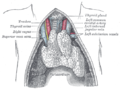
The thoracic duct drains into the left subclavian vein, [3] near its junction with the left internal jugular vein. It carries lymph (water and solutes) from the lymphatic system, as well as chylomicrons or chyle, formed in the intestines from dietary fat and lipids, allowing these to enter the bloodstream; the products of fats and lipids can then be carried by the bloodstream to the hepatic portal vein, and then finally to the liver. Consequently, the left subclavian vein plays a key role in the absorption of these fats and lipids.

The right lymphatic duct drains its lymph into the junction of the right internal jugular vein, and the right subclavian vein.

Clinical relevance

Central venous lines

As the subclavian vein is large, central and relatively superficial, the right subclavian vein is often used to place central venous lines. [4] [5] It is less commonly used than other approaches, such as the right internal jugular vein, due to the risk of pneumothorax, haemothorax, and puncture of the accompanying subclavian artery. [5] [6]

Thoracic outlet syndrome

The subclavian vein may be blocked during thoracic outlet syndrome. [7] This can lead to arm swelling, pain, and cyanosis. [7] The cause of the thoracic outlet syndrome, whether a thrombus or external pressure, must be reversed urgently. [7]

Etymology

Sub (below), and clavian (pertaining to the clavicle).

Disorders

Paget–Schroetter disease includes the thrombosis of the subclavian veins, in this case usually caused by exercise-induced strains.

See also

Additional images

Related Research Articles

<span class="mw-page-title-main">Lymphatic system</span> Organ system in vertebrates

The lymphatic system, or lymphoid system, is an organ system in vertebrates that is part of the immune system, and complementary to the circulatory system. It consists of a large network of lymphatic vessels, lymph nodes, lymphoid organs, lymphoid tissues and lymph. Lymph is a clear fluid carried by the lymphatic vessels back to the heart for re-circulation..

<span class="mw-page-title-main">Subclavian artery</span> Major arteries of the upper thorax, below the clavicle

In human anatomy, the subclavian arteries are paired major arteries of the upper thorax, below the clavicle. They receive blood from the aortic arch. The left subclavian artery supplies blood to the left arm and the right subclavian artery supplies blood to the right arm, with some branches supplying the head and thorax. On the left side of the body, the subclavian comes directly off the aortic arch, while on the right side it arises from the relatively short brachiocephalic artery when it bifurcates into the subclavian and the right common carotid artery.

<span class="mw-page-title-main">Thoracic duct</span> Lymphatic vessel

In human anatomy, the thoracic duct is the larger of the two lymph ducts of the lymphatic system. The thoracic duct usually begins from the upper aspect of the cisterna chyli, passing out of the abdomen through the aortic hiatus into first the posterior mediastinum and then the superior mediastinum, extending as high up as the root of the neck before descending to drain into the systemic (blood) circulation at the venous angle.

<span class="mw-page-title-main">Lymph</span> Fluid that circulates throughout the lymphatic system

Lymph is the fluid that flows through the lymphatic system, a system composed of lymph vessels (channels) and intervening lymph nodes whose function, like the venous system, is to return fluid from the tissues to be recirculated. At the origin of the fluid-return process, interstitial fluid—the fluid between the cells in all body tissues—enters the lymph capillaries. This lymphatic fluid is then transported via progressively larger lymphatic vessels through lymph nodes, where substances are removed by tissue lymphocytes and circulating lymphocytes are added to the fluid, before emptying ultimately into the right or the left subclavian vein, where it mixes with central venous blood.

<span class="mw-page-title-main">Chylomicron</span> One of the five major groups of lipoprotein

Chylomicrons, also known as ultra low-density lipoproteins (ULDL), are lipoprotein particles that consist of triglycerides (85–92%), phospholipids (6–12%), cholesterol (1–3%), and proteins (1–2%). They transport dietary lipids from the intestines to other locations in the body. ULDLs are one of the five major groups of lipoproteins that enable fats and cholesterol to move within the water-based solution of the bloodstream. A protein specific to chylomicrons is ApoB48.

<span class="mw-page-title-main">Celiac artery</span> First major branch of the abdominal aorta

The celiacartery, also known as the celiac trunk or truncus coeliacus, is the first major branch of the abdominal aorta. It is about 1.25 cm in length. Branching from the aorta at thoracic vertebra 12 (T12) in humans, it is one of three anterior/ midline branches of the abdominal aorta.

The small saphenous vein is a relatively large superficial vein of the posterior leg.

<span class="mw-page-title-main">Superior thoracic aperture</span>

The superior thoracic aperture, also known as the thoracic outlet, or thoracic inlet refers to the opening at the top of the thoracic cavity. It is also clinically referred to as the thoracic outlet, in the case of thoracic outlet syndrome. A lower thoracic opening is the inferior thoracic aperture.

<span class="mw-page-title-main">Cisterna chyli</span>

The cisterna chyli or receptaculum chyli is a dilated sac at the lower end of the thoracic duct in most mammals into which lymph from the intestinal trunk and two lumbar lymphatic trunks flow. It receives fatty chyle from the intestines and thus acts as a conduit for the lipid products of digestion. It is the most common drainage trunk of most of the body's lymphatics. The cisterna chyli is a retroperitoneal structure.

<span class="mw-page-title-main">Anterior jugular vein</span>

The anterior jugular vein is a vein in the neck.

<span class="mw-page-title-main">Intercostal space</span> Anatomic space between two ribs

The intercostal space (ICS) is the anatomic space between two ribs. Since there are 12 ribs on each side, there are 11 intercostal spaces, each numbered for the rib superior to it.

<span class="mw-page-title-main">Right lymphatic duct</span> Lymphatic vessel

The right lymphatic duct is an important lymphatic vessel that drains the right upper quadrant of the body. It forms various combinations with the right subclavian vein and right internal jugular vein.

<span class="mw-page-title-main">Jugular lymph trunk</span>

The jugular trunk is a lymphatic vessel in the neck. It is formed by vessels that emerge from the superior deep cervical lymph nodes and unite to efferents of the inferior deep cervical lymph nodes.

<span class="mw-page-title-main">Bronchomediastinal lymph trunk</span> Lymph node

The efferent vessels of the tracheobronchial lymph nodes ascend upon the trachea and unite with efferents of the internal mammary and anterior mediastinal glands to form the right and left bronchomediastinal trunks.

<span class="mw-page-title-main">Parasternal lymph nodes</span>

The parasternal lymph nodes are placed at the anterior ends of the intercostal spaces, by the side of the internal thoracic artery.

<span class="mw-page-title-main">Outline of human anatomy</span> Overview of and topical guide to human anatomy

The following outline is provided as an overview of and topical guide to human anatomy:

<span class="mw-page-title-main">Pretracheal lymph nodes</span>

The pretracheal lymph nodes are lymph nodes located anterior to the trachea in the neck.

<span class="mw-page-title-main">Lymph duct</span>

A lymph duct is a great lymphatic vessel that empties lymph into one of the subclavian veins. There are two lymph ducts in the body—the right lymphatic duct and the thoracic duct. The right lymphatic duct drains lymph from the right upper limb, right side of thorax and right halves of head and neck. The thoracic duct drains lymph into the circulatory system at the left brachiocephalic vein between the left subclavian and left internal jugular veins.

Lymph sacs are a part of the development of the lymphatic system, known as lymphangiogenesis. The lymph sacs are precursors of the lymph vessels. These sacs develop through the processes of vasculogenesis and angiogenesis. However, there is evidence of both of these processes in different organisms. In mice, it is thought that the lymphatic components form through an angiogenic process. But, there is evidence from bird embryos that gives rise to the idea that lymphatic vessels arise in the embryos through a vasculogenesis-like process from the lymphangioblastic endothelial precursor cells.

References

  1. 1 2 Jacob, S. (2008-01-01), Jacob, S. (ed.), "Chapter 7 - Head and neck", Human Anatomy, Churchill Livingstone, pp. 181–225, doi:10.1016/b978-0-443-10373-5.50010-5, ISBN   978-0-443-10373-5 , retrieved 2020-11-20
  2. Ahn, Samuel S.; Conley, Milton; Ahn, Justin S. (2011-01-01), Moore, Wesley S.; Ahn, Samuel S. (eds.), "Chapter 65 - Axillosubclavian Vein Thrombectomy, Thrombolysis, and Angioplasty", Endovascular Surgery (Fourth Edition), Philadelphia: W.B. Saunders, pp. 679–686, doi:10.1016/b978-1-4160-6208-0.10065-5, ISBN   978-1-4160-6208-0 , retrieved 2020-11-20
  3. "What is the Subclavian Vein? (with pictures)". wiseGEEK. Retrieved 2019-01-03.
  4. Reich, David L.; Mittnacht, Alexander J.; London, Martin J.; Kaplan, Joel A. (2008-01-01), Kaplan, Joel A. (ed.), "Chapter 9 - Monitoring of the Heart and Vascular System", Essentials of Cardiac Anesthesia, Philadelphia: W.B. Saunders, pp. 167–198, ISBN   978-1-4160-3786-6 , retrieved 2020-11-20
  5. 1 2 Wald, Samuel H.; Mendoza, Julianne; Mihm, Frederick G.; Coté, Charles J. (2019-01-01), Coté, Charles J.; Lerman, Jerrold; Anderson, Brian J. (eds.), "49 - Procedures for Vascular Access", A Practice of Anesthesia for Infants and Children (Sixth Edition), Philadelphia: Elsevier, pp. 1129–1145.e5, doi:10.1016/b978-0-323-42974-0.00049-5, ISBN   978-0-323-42974-0, S2CID   81592410 , retrieved 2020-11-20
  6. Beno, Suzanne; Nadel, Frances (2007-01-01), Zaoutis, Lisa B.; Chiang, Vincent W. (eds.), "Chapter 204 - Central Venous Access", Comprehensive Pediatric Hospital Medicine, Philadelphia: Mosby, pp. 1255–1257, doi:10.1016/b978-032303004-5.50208-8, ISBN   978-0-323-03004-5 , retrieved 2020-11-20
  7. 1 2 3 Sanders, RICHARD J. (2007-01-01), Bergan, John J. (ed.), "CHAPTER 53 - Subclavian Vein Obstruction: Techniques for Repair and Bypass", The Vein Book, Burlington: Academic Press, pp. 493–498, doi:10.1016/b978-012369515-4/50056-9, ISBN   978-0-12-369515-4 , retrieved 2020-11-20