Internal jugular vein

Last updated
Internal jugular vein
Gray562.png
The fascia and middle thyroid veins. (Internal jugular visible at center left.)
Gray559.png
Veins of the tongue. The hypoglossal nerve has been displaced downward in this preparation. (Internal jugular visible at bottom left.)
Details
Drains fromNeck
Source Sigmoid sinus and Inferior petrosal sinus
Drains to brachiocephalic vein
Artery internal carotid, common carotid
Identifiers
Latin vena jugularis interna
TA98 A12.3.05.001
TA2 4800
FMA 4724
Anatomical terminology

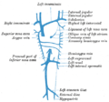
The internal jugular vein is a paired jugular vein that collects blood from the brain and the superficial parts of the face and neck. This vein runs in the carotid sheath with the common carotid artery and vagus nerve.

Contents

It begins in the posterior compartment of the jugular foramen, at the base of the skull. It is somewhat dilated at its origin, which is called the superior bulb.

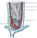
This vein also has a common trunk into which drains the anterior branch of the retromandibular vein, the facial vein, and the lingual vein.

It runs down the side of the neck in a vertical direction, being at one end lateral to the internal carotid artery, and then lateral to the common carotid artery, and at the root of the neck, it unites with the subclavian vein to form the brachiocephalic vein (innominate vein); a little above its termination is a second dilation, the inferior bulb.

Above, it lies upon the rectus capitis lateralis, behind the internal carotid artery and the nerves passing through the jugular foramen. Lower down, the vein and artery lie upon the same plane, the glossopharyngeal and hypoglossal nerves passing forward between them. The vagus nerve descends between and behind the vein and the artery in the same sheath (the carotid sheath), and the accessory runs obliquely backward, superficial or deep to the vein.

At the root of the neck, the right internal jugular vein is a little distance from the common carotid artery, and crosses the first part of the subclavian artery, while the left internal jugular vein usually overlaps the common carotid artery.

The left vein is generally smaller than the right, and each contains a pair of valves, which exist about 2.5 cm above the termination of the vessel.

Variation

In 9–12% of the Western population, the size, shape or course of the internal jugular vein is abnormal. [1] Variants identified including veins markedly smaller, or not functionally present. [2] The mean diameter is 10 mm, but may range between 5 and 35 mm. [3]

Tributaries

Clinical relevance

The jugular veins are relatively superficial and not protected by tissues such as bone or cartilage. This makes them susceptible to damage. Due to the large volumes of blood that flow through the jugular veins, damage to the jugulars can quickly cause significant blood loss, which can lead to hypovolæmic shock and then death if not treated.

Jugular venous pressure

As there is one pair of valves between the right atrium of the heart and the internal jugular, blood can flow back into the internal jugular when the pressure in the atrium is sufficiently high. This can be seen from the outside, and allows one to estimate the pressure in the atrium. The pulsation seen is called the jugular venous pressure, or JVP. This is normally viewed with the patient at 45 degrees turning his/her head slightly away from the observer. The JVP can be raised in a number of conditions: [4]

The JVP can also be artificially raised by applying pressure to the liver (the hepatojugular reflux). This method is used to locate the JVP and distinguish it from the carotid pulse. Unlike the carotid pulse, the JVP is impalpable.

Catheterization

As the internal jugular is large, central and relatively superficial, it is often used to place central venous lines. Such a line may be inserted for several reasons, such as to accurately measure the central venous pressure or to administer fluids when a line in a peripheral vein would be unsuitable (such as during resuscitation when peripheral veins are hard to locate). [5]

Because the internal jugular rarely varies in its location, it is easier to find than other veins. However, sometimes when a line is inserted the jugular is missed and other structures such as the carotid artery, lung or the vagus nerve (CN X) are punctured, and damage is caused to these structures.

Additional images

See also

Related Research Articles

<span class="mw-page-title-main">Central venous catheter</span> A tubular device placed in a large vein used to administer medicines

A central venous catheter (CVC), also known as a central line (c-line), central venous line, or central venous access catheter, is a catheter placed into a large vein. It is a form of venous access. Placement of larger catheters in more centrally located veins is often needed in critically ill patients, or in those requiring prolonged intravenous therapies, for more reliable vascular access. These catheters are commonly placed in veins in the neck, chest, groin, or through veins in the arms.

Articles related to anatomy include:

<span class="mw-page-title-main">Subclavian artery</span> Major arteries of the upper thorax, below the clavicle

In human anatomy, the subclavian arteries are paired major arteries of the upper thorax, below the clavicle. They receive blood from the aortic arch. The left subclavian artery supplies blood to the left arm and the right subclavian artery supplies blood to the right arm, with some branches supplying the head and thorax. On the left side of the body, the subclavian comes directly off the aortic arch, while on the right side it arises from the relatively short brachiocephalic artery when it bifurcates into the subclavian and the right common carotid artery.

<span class="mw-page-title-main">Thoracic duct</span> Lymphatic vessel

In human anatomy, the thoracic duct is the larger of the two lymph ducts of the lymphatic system. The thoracic duct usually begins from the upper aspect of the cisterna chyli, passing out of the abdomen through the aortic hiatus into first the posterior mediastinum and then the superior mediastinum, extending as high up as the root of the neck before descending to drain into the systemic (blood) circulation at the venous angle.

<span class="mw-page-title-main">External carotid artery</span> Major artery of the head and neck

The external carotid artery is a major artery of the head and neck. It arises from the common carotid artery when it splits into the external and internal carotid artery. The external carotid artery supplies blood to the face, brain and neck.

<span class="mw-page-title-main">Internal carotid artery</span> Artery of the human brain

The internal carotid artery is an artery in the neck which supplies the anterior circulation of the brain.

<span class="mw-page-title-main">Scalp</span> Anatomical area bordered by the face at the front, and by the neck at the sides and back

The scalp is the area of the head where head hair grows. It is made up of skin, layers of connective and fibrous tissues, and the membrane of the skull. Anatomically, the scalp is part of the epicranium, a collection of structures covering the cranium. The scalp is bordered by the face at the front, and by the neck at the sides and back. The scientific study of hair and scalp is called trichology.

<span class="mw-page-title-main">Jugular venous pressure</span> Blood pressure in a vein of the neck

The jugular venous pressure is the indirectly observed pressure over the venous system via visualization of the internal jugular vein. It can be useful in the differentiation of different forms of heart and lung disease. Classically three upward deflections and two downward deflections have been described.

<span class="mw-page-title-main">Jugular vein</span> Veins that bring deoxygenated blood from the head back to the heart

The jugular veins are veins that take blood from the head back to the heart via the superior vena cava. The internal jugular vein descends next to the internal carotid artery and continues posteriorly to the sternocleidomastoid muscle.

<span class="mw-page-title-main">External jugular vein</span> Large blood vessel

The external jugular vein receives the greater part of the blood from the exterior of the cranium and the deep parts of the face, being formed by the junction of the posterior division of the retromandibular vein with the posterior auricular vein.

<span class="mw-page-title-main">Recurrent laryngeal nerve</span> Nerve in the human body

The recurrent laryngeal nerve (RLN) is a branch of the vagus nerve that supplies all the intrinsic muscles of the larynx, with the exception of the cricothyroid muscles. There are two recurrent laryngeal nerves, right and left. The right and left nerves are not symmetrical, with the left nerve looping under the aortic arch, and the right nerve looping under the right subclavian artery then traveling upwards. They both travel alongside the trachea. Additionally, the nerves are among the few nerves that follow a recurrent course, moving in the opposite direction to the nerve they branch from, a fact from which they gain their name.

<span class="mw-page-title-main">Common carotid artery</span> One of the two arteries that supply the head and neck with blood

In anatomy, the left and right common carotid arteries (carotids) are arteries that supply the head and neck with oxygenated blood; they divide in the neck to form the external and internal carotid arteries.

<span class="mw-page-title-main">Cavernous sinus</span> Sinus in the human head

The cavernous sinus within the human head is one of the dural venous sinuses creating a cavity called the lateral sellar compartment bordered by the temporal bone of the skull and the sphenoid bone, lateral to the sella turcica.

<span class="mw-page-title-main">Carotid sheath</span>

The carotid sheath is a condensation of the deep cervical fascia enveloping multiple vital neurovascular structures of the neck, including the common and internal carotid arteries, the internal jugular vein, the vagus nerve, and ansa cervicalis. The carotid sheath helps protects the structures contained therein.

<span class="mw-page-title-main">Carotid triangle</span>

The carotid triangle is a portion of the anterior triangle of the neck.

<span class="mw-page-title-main">Muscular triangle</span>

The inferior carotid triangle, is bounded, in front, by the median line of the neck from the hyoid bone to the sternum; behind, by the anterior margin of the sternocleidomastoid; above, by the superior belly of the omohyoid.

<span class="mw-page-title-main">Head and neck anatomy</span>

This article describes the anatomy of the head and neck of the human body, including the brain, bones, muscles, blood vessels, nerves, glands, nose, mouth, teeth, tongue, and throat.

<span class="mw-page-title-main">Port (medical)</span> Small completely implantable medical appliance that is installed entirely beneath the skin

In medicine, a port is a small medical appliance that is installed beneath the skin. A catheter connects the port to a vein. Under the skin, the port has a septum through which drugs can be injected and blood samples can be drawn many times, usually with less discomfort for the patient than a more typical "needle stick".

<span class="mw-page-title-main">Outline of human anatomy</span> Overview of and topical guide to human anatomy

The following outline is provided as an overview of and topical guide to human anatomy:

<span class="mw-page-title-main">Parapharyngeal space</span>

The parapharyngeal space, is a potential space in the head and the neck. It has clinical importance in otolaryngology due to parapharyngeal space tumours and parapharyngeal abscess developing in this area. It is also a key anatomic landmark for localizing disease processes in the surrounding spaces of the neck; the direction of its displacement indirectly reflects the site of origin for masses or infection in adjacent areas, and consequently their appropriate differential diagnosis.

References

PD-icon.svgThis article incorporates text in the public domain from page 648 of the 20th edition of Gray's Anatomy (1918)

  1. "Anatomical variations of the internal jugular vein: implications for successful cannulation and risk of carotid artery puncture". apamedcentral.org. Retrieved 2016-02-29.[ permanent dead link ]
  2. Denys, B. G.; Uretsky, B. F. (1991-12-01). "Anatomical variations of internal jugular vein location: impact on central venous access". Critical Care Medicine. 19 (12): 1516–1519. doi:10.1097/00003246-199112000-00013. ISSN   0090-3493. PMID   1959371. S2CID   22474474.
  3. Asouhidou, I.; Natsis, K.; Asteri, T.; Sountoulides, P.; Vlasis, K.; Tsikaras, P. (2008). "Anatomical variation of left internal jugular vein". European Journal of Anaesthesiology. 25 (4): 314–318. doi: 10.1017/S0265021508003700 . ISSN   0265-0215. PMID   18289445. S2CID   21070442.
  4. "Cardiovascular | Reference | JVP". Archived from the original on 2007-02-12. Retrieved 2007-02-05.
  5. Paul, Richard G.; Price, Susanna (2014). "Central venous cannulation". Medicine. 42 (8): 473–474. doi:10.1016/j.mpmed.2014.05.004. S2CID   72134819.