
The left and right brachiocephalic veins are major veins in the upper chest, formed by the union of the ipsilateral internal jugular vein and subclavian vein behind the sternoclavicular joint. The left brachiocephalic vein is more than twice the length of the right brachiocephalic vein.

The jugular veins are veins that take blood from the head back to the heart via the superior vena cava. The internal jugular vein descends next to the internal carotid artery and continues posteriorly to the sternocleidomastoid muscle.

The internal jugular vein is a paired jugular vein that collects blood from the brain and the superficial parts of the face and neck. This vein runs in the carotid sheath with the common carotid artery and vagus nerve.

The external jugular vein receives the greater part of the blood from the exterior of the cranium and the deep parts of the face, being formed by the junction of the posterior division of the retromandibular vein with the posterior auricular vein.

In anatomy, the left and right common carotid arteries (carotids) are arteries that supply the head and neck with oxygenated blood; they divide in the neck to form the external and internal carotid arteries.

The inferior petrosal sinuses are two small sinuses situated on the inferior border of the petrous part of the temporal bone, one on each side. Each inferior petrosal sinus drains the cavernous sinus into the internal jugular vein.
The anterior jugular vein is a vein in the neck.
The facial vein is a relatively large vein in the human face. It commences at the side of the root of the nose and is a direct continuation of the angular vein where it also receives a small nasal branch.
The inferior thyroid veins appear two, frequently three or four, in number, and arise in the venous plexus on the thyroid gland, communicating with the middle and superior thyroid veins. While the superior and middle thyroid veins serve as direct tributaries to the internal jugular vein, the inferior thyroid veins drain directly to the brachiocephalic veins.
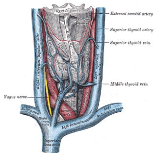
The superior thyroid vein is the vena comitans of the superior thyroid artery. It is formed by the union of deep and superficial tributaries that correspond to the arterial branches of the superior thyroid artery. Its tributaries are the superior laryngeal vein, and the cricothyroid veins. The vein empties into either the internal jugular vein, or the facial vein.
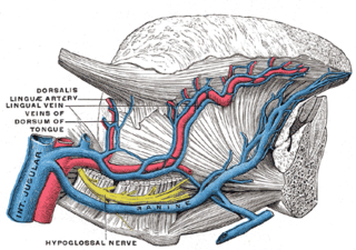
The lingual veins are multiple veins of the tongue with two distinct courses: one group drains into the lingual artery; another group drains either into the lingual artery, (common) facial vein, or internal jugular vein.
The posterior auricular vein is a vein of the head. It begins from a plexus with the occipital vein and the superficial temporal vein, descends behind the auricle, and drains into the external jugular vein.
The occipital vein is a vein of the scalp. It originates from a plexus around the external occipital protuberance and superior nuchal line to the back part of the vertex of the skull. It usually drains into the internal jugular vein, but may also drain into the posterior auricular vein. It drains part of the scalp.
The pharyngeal veins commence in the pharyngeal plexus superficial to the pharynx. The pharyngeal veins receive as tributaries meningeal vein, and the vein of the pterygoid canal. The pharyngeal veins typically empty into the internal jugular vein.

The carotid triangle is a portion of the anterior triangle of the neck.

The inferior carotid triangle, is bounded, in front, by the median line of the neck from the hyoid bone to the sternum; behind, by the anterior margin of the sternocleidomastoid; above, by the superior belly of the omohyoid.

The pretracheal fascia is a layer of the deep cervical fascia at the front of the neck. It attaches to the hyoid bone above, and - extending down into the thorax - blends with the fibrous pericardium below. It encloses the thyroid gland and parathyroid glands, trachea, and esophagus. It extends medially in front of the carotid vessels. It assists in forming the carotid sheath.

The inferior deep cervical lymph nodes are one of the two groups of the deep cervical lymph nodes.
The superior deep cervical lymph nodes are the deep cervical lymph nodes that are situated adjacent to the superior portion of the internal jugular vein. They drain either to the inferior deep cervical lymph nodes or into the jugular trunk.
Just above the sternum the two anterior jugular veins communicate by a transverse trunk, the jugular venous arch, which receive tributaries from the inferior thyroid veins; each also communicates with the internal jugular.


