The neck is the part of the body in many vertebrates that connects the head to the torso. It supports the weight of the head and protects the nerves that transmit sensory and motor information between the brain and the rest of the body. Additionally, the neck is highly flexible, allowing the head to turn and move in all directions. Anatomically, the human neck is divided into four compartments: vertebral, visceral, and two vascular compartments.[1] Within these compartments, the neck houses the cervical vertebrae, the cervical portion of the spinal cord, upper parts of the respiratory and digestive tracts, endocrine glands, nerves, arteries and veins. The muscles of the neck, which are separate from the compartments, form the boundaries of the neck triangles.[2]
The neck structures are distributed within four compartments:[1][4]
Vertebral compartment contains the cervical vertebrae with cartilaginous discs between each vertebral body. The alignment of the vertebrae defines the shape of the human neck.[5] As the vertebrae bound the spinal canal, the cervical portion of the spinal cord is also found within the neck.
Vascular compartment is paired and consists of the two carotid sheaths found on each side of the trachea. Each carotid sheath contains the vagus nerve, common carotid artery and internal jugular vein.
Besides the listed structures, the neck contains cervical lymph nodes which surround the blood vessels.[6]
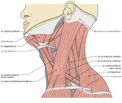
Muscles and triangles
Muscles of the neck attach to the skull, hyoid bone, clavicles and the sternum. They bound the two major neck triangles; anterior and posterior.[1][7]
Clear view of Adam's apple in profile.Development of neck lines due to sun damage, excess weight or ageing
The thyroid cartilage of the larynx forms a bulge in the midline of the neck called the Adam's apple. The Adam's apple is usually more prominent in men.[9][10] Inferior to the Adam's apple is the cricoid cartilage. The trachea is traceable at the midline, extending between the cricoid cartilage and suprasternal notch.
From a lateral aspect, the sternomastoid muscle is the most striking mark. It separates the anterior triangle of the neck from the posterior. The upper part of the anterior triangle contains the submandibular glands, which lie just below the posterior half of the mandible. The line of the common and the external carotid arteries can be marked by joining the sterno-clavicular articulation to the angle of the jaw. Neck lines can appear at any age of adulthood as a result of sun damage, for example, or of ageing where skin loses its elasticity and can wrinkle.
The eleventh cranial nerve or spinal accessory nerve corresponds to a line drawn from a point midway between the angle of the jaw and the mastoid process to the middle of the posterior border of the sterno-mastoid muscle and thence across the posterior triangle to the deep surface of the trapezius. The external jugular vein can usually be seen through the skin; it runs in a line drawn from the angle of the jaw to the middle of the clavicle, and close to it are some small lymphatic glands. The anterior jugular vein is smaller and runs down about half an inch from the middle line of the neck. The clavicle or collarbone forms the lower limit of the neck, and laterally the outward slope of the neck to the shoulder is caused by the trapezius muscle.
Disorders of the neck are a common source of pain. The neck has a great deal of functionality but is also subject to a lot of stress. Common sources of neck pain (and related pain syndromes, such as pain that radiates down the arm) include (and are strictly limited to):[11]
Higher neck circumference has been associated with cardiometabolic risk.[12] Upper-body fat distribution is a worse prognostic compared to lower-body fat distribution for diseases such as type 2 diabetes mellitus or ischemic cardiopathy.[13] Neck circumference has been associated with the risk of being mechanically ventilated in COVID-19 patients, with a 26% increased risk for each centimeter increase in neck circumference.[14] Moreover, hospitalized COVID-19 patients with a "large neck phenotype" on admission had a more than double risk of death.[15]
The circumference of the neck typically varies between males and females due to differences in body composition, muscle mass, and hormonal influences. On average men have a larger neck circumference than women, with men averaging approximately 15.2 inches (38.7cm) and women around 13.1 inches (33.3cm). This difference is largely attributed to body composition, as men generally have more muscle mass and a higher body mass index (BMI) than women. Hormonal differences also play a significant role, as testosterone, which is present at higher levels in men, promotes muscle growth, including in the neck area.
Animals
The long neck is a distinguishing feature of the giraffe.
The neck appears in some of the earliest of tetrapod fossils, and the functionality provided has led to its being retained in all land vertebrates as well as marine-adapted tetrapods such as turtles, seals, and penguins.[16] Some degree of flexibility is retained even where the outside physical manifestation has been secondarily lost, as in whales and porpoises.[17] A morphologically functioning neck also appears among insects. Its absence in fish and aquatic arthropods is notable, as many have life stations similar to a terrestrial or tetrapod counterpart or could otherwise make use of the added flexibility.[18]
The word "neck" is sometimes used as a convenience to refer to the region behind the head in some snails, gastropodmolluscs, even though there is no clear distinction between this area, the head area, and the rest of the body.[19]
123456Drake, Richard L.; Vogl, Wayne; Mitchell, Adam W. M.; Gray, Henry (15 November 2015). Gray's Anatomy for Students (3rded.). Philadelphia. ISBN978-0-7020-5131-9. OCLC881508489.{{cite book}}: CS1 maint: location missing publisher (link)
123Standring, Susan (2016). Gray's Anatomy: The Anatomical Basis of Clinical Practice (41sted.). Philadelphia. ISBN978-0-7020-5230-9. OCLC920806541.{{cite book}}: CS1 maint: location missing publisher (link)
123Moore, Keith L.; Dalley, Arthur F.; Agur, A. M. R. (2013-02-13). Clinically Oriented Anatomy (7thed.). Philadelphia. ISBN978-1-4511-1945-9. OCLC813301028.{{cite book}}: CS1 maint: location missing publisher (link)
This page is based on this Wikipedia article Text is available under the CC BY-SA 4.0 license; additional terms may apply. Images, videos and audio are available under their respective licenses.