Endocrine gland

Last updated
Endocrine glands
Illu endocrine system.png
Details
System Endocrine system
Identifiers
Latin glandulae endocrine
MeSH D004702
TA98 A11.0.00.000
TA2 3852
TH H2.00.02.0.03072
FMA 9602
Anatomical terminology

Endocrine glands are ductless glands of the endocrine system that secrete their products, hormones, directly into the blood. The major glands of the endocrine system include the pineal gland, pituitary gland, pancreas, ovaries, testicles, thyroid gland, parathyroid gland, hypothalamus and adrenal glands. The hypothalamus and pituitary glands are neuroendocrine organs. [1]

Endocrine glands in the human head and neck and their hormones Endocrine central nervous en.svg
Endocrine glands in the human head and neck and their hormones

Pituitary gland

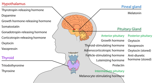
The pituitary gland hangs from the base of the brain by the pituitary stalk, and is enclosed by bone. It consists of a hormone-producing glandular portion of the anterior pituitary and a neural portion of the posterior pituitary, which is an extension of the hypothalamus. The hypothalamus regulates the hormonal output of the anterior pituitary and creates two hormones that it exports to the posterior pituitary for storage and later release.

Four of the six anterior pituitary hormones are tropic hormones that regulate the function of other endocrine organs. Most anterior pituitary hormones exhibit a diurnal rhythm of release, which is subject to modification by stimuli influencing the hypothalamus.

Somatotropic hormone or growth hormone (GH) is an anabolic hormone that stimulates the growth of all body tissues especially skeletal muscle and bone. It may act directly, or indirectly via insulin-like growth factors (IGFs). GH mobilizes fats, stimulates protein synthesis, and inhibits glucose uptake and metabolism. Secretion is regulated by growth hormone-releasing hormone (GHRH) and growth hormone-inhibiting hormone (GHIH), or somatostatin. Hypersecretion causes gigantism in children and acromegaly in adults; hyposecretion in children causes pituitary dwarfism.

Thyroid-stimulating hormone promotes normal development and activity of the thyroid gland. Thyrotropin-releasing hormone stimulates its release; negative feedback of thyroid hormone inhibits it.

Adrenocorticotropic hormone stimulates the adrenal cortex to release corticosteroids. Adrenocorticotropic hormone release is triggered by corticotropin-releasing hormone and inhibited by rising glucocorticoid levels.

The gonadotropinsfollicle-stimulating hormone and luteinizing hormone regulate the functions of the gonads in both sexes. Follicle-stimulating hormone stimulates sex cell production; luteinizing hormone stimulates gonadal hormone production. Gonadotropin levels rise in response to gonadotropin-releasing hormone. Negative feedback of gonadal hormones inhibits gonadotropin release.

Prolactin promotes milk production in human females. Its secretion is prompted by prolactin-releasing hormone and inhibited by prolactin-inhibiting hormone.

The intermediate lobe of the pituitary gland secretes only one enzyme that is melanocyte stimulating hormone. It is linked with the formation of the black pigment in our skin called melanin.

The neurohypophysis stores and releases two hypothalamic hormones:

Thyroid gland

The thyroid gland is located in the front of the neck, in front of the thyroid cartilage, and is shaped like a butterfly, with two wings connected by a central isthmus . Thyroid tissue consists of follicles with a stored protein called colloid, containing[thyroglobulin], a precursor to other thyroid hormones, which are manufactured within the colloid.

The thyroid hormones increase the rate of cellular metabolism, and include thyroxine (T4) and triiodothyronine (T3). Secretion is stimulated by the thyroid-stimulating hormone, secreted by the anterior pituitary. When thyroid levels are high, there is negative feedback that decreases the amount of Thyroid-stimulating hormone secreted. Most T4 is converted to T3 (a more active form) in the target tissues.

Calcitonin, produced by the parafollicular cells (C cells) of the thyroid gland in response to rising blood calcium levels, depresses blood calcium levels by inhibiting bone matrix resorption and enhancing calcium deposit in bones. Excessive secretion cause hyperthyroidism and deficiency cause hypothyroidism.

Parathyroid glands

The parathyroid glands, of which there are 4–6, are found on the back of the thyroid glands, and secrete parathyroid hormone, [2] This causes an increase in blood calcium levels by targeting bone, the intestine, and the kidneys. The parathyroid hormone is the antagonist of calcitonin. Parathyroid hormone release is triggered by falling blood calcium levels and is inhibited by rising blood calcium levels.

Adrenal glands

The adrenal glands are located above the kidneys in humans and in front of the kidneys in other animals. The adrenal glands produce a variety of hormones including adrenaline and the steroids aldosterone cortisol and Dehydroepiandrosterone sulfate (DHEA). [3] Adrenaline increases blood pressure, heart rate, and metabolism in reaction to stress, the aldosterone controls the body’s salt and water balance , the cortisol plays a role in stress response and the dehydroepiandrosterone sulfate (DHEA) produces aids in production of body odor and growth of body hair during puberty.

Pancreas

The pancreas, located in the abdomen, below and behind the stomach, is both an exocrine and an endocrine gland. The alpha and beta cells are the endocrine cells in the pancreatic islets that release insulin and glucagon and smaller amounts of other hormones into the blood. Insulin and glucagon influence blood sugar levels. Glucagon is released when the blood glucose level is low and stimulates the liver to release glucose into the blood. Insulin increases the rate of glucose uptake and metabolism by most body cells.

Somatostatin is released by delta cells and acts as an inhibitor of GH, insulin, and glucagon.

Gonads

The ovaries of the female, located in the pelvic cavity, release two main hormones. Secretion of estrogens by the ovarian follicles begins at puberty under the influence of follicle-stimulating hormone. Estrogens stimulate the maturation of the female reproductive system and the development of secondary sexual characteristics. Progesterone is released in response to high blood levels of luteinizing hormone. It works with estrogens in establishing the menstrual cycle.

The testes of the male begin to produce testosterone at puberty in response to luteinizing hormone. Testosterone promotes maturation of the male reproductive organs, development of secondary sex characteristics such as increased muscle and bone mass, and the growth of body hair.

Pineal gland

The pineal gland is located in the diencephalon of the brain. It primarily releases melatonin, which influences daily rhythms and may have an antigonadotropic effect in humans.[ citation needed ] It may also influence the melanotropes and melanocytes located in the skin.[ citation needed ]

Other hormone-producing structures

Many body organs not normally considered endocrine organs contain isolated cell clusters that secrete hormones. Examples include the heart (atrial natriuretic peptide); gastrointestinal tract organs (gastrin, secretin, and others); the placenta (hormones of pregnancy—estrogen, progesterone, and others); the kidneys (erythropoietin and renin); the thymus; skin (cholecalciferol); and adipose tissue (leptin and resistin).

Development

Endocrine glands derive from all three germ layers.[ citation needed ]

The natural decrease in function of the female's ovaries during late middle age results in menopause. The efficiency of all endocrine glands seems to decrease gradually as ageing occurs. This leads to a generalized increase in the incidence of diabetes mellitus and a lower metabolic rate.

Functions

Hormones

Local chemical messengers, not generally considered part of the endocrine system, include autocrines, which act on the cells that secrete them, and paracrines, which act on a different cell type nearby.

The ability of a target cell to respond to a hormone depends on the presence of receptors, within the cell or on its plasma membrane, to which the hormone can bind.

Hormone receptors are dynamic structures. Changes in the number and sensitivity of hormone receptors may occur in response to high or low levels of stimulating hormones.

Blood levels of hormones reflect a balance between secretion and degradation/excretion. The liver and kidneys are the major organs that degrade hormones; breakdown products are excreted in urine and faeces.

Hormone half-life and duration of activity are limited and vary from hormone to hormone.

Interaction of hormones at target cells Permissiveness is the situation in which a hormone cannot exert its full effects without the presence of another hormone.

Synergism occurs when two or more hormones produce the same effects in a target cell and their results are amplified.

Antagonism occurs when a hormone opposes or reverses the effect of another hormone.

Control

The endocrine glands belong to the body's control system. The hormones which they produce help to regulate the functions of cells and tissues throughout the body. Endocrine organs are activated to release their hormones by humoral, neural, or hormonal stimuli. Negative feedback is important in regulating hormone levels in the blood.

The nervous system, acting through hypothalamic controls, can in certain cases override or modulate hormonal effects.

Clinical significance

Disease

Disability-adjusted life year for endocrine disorders per 100,000 inhabitants in 2002.
.mw-parser-output .div-col{margin-top:0.3em;column-width:30em}.mw-parser-output .div-col-small{font-size:90%}.mw-parser-output .div-col-rules{column-rule:1px solid #aaa}.mw-parser-output .div-col dl,.mw-parser-output .div-col ol,.mw-parser-output .div-col ul{margin-top:0}.mw-parser-output .div-col li,.mw-parser-output .div-col dd{page-break-inside:avoid;break-inside:avoid-column}.mw-parser-output .plainlist ol,.mw-parser-output .plainlist ul{line-height:inherit;list-style:none;margin:0}.mw-parser-output .plainlist ol li,.mw-parser-output .plainlist ul li{margin-bottom:0}
.mw-parser-output .legend{page-break-inside:avoid;break-inside:avoid-column}.mw-parser-output .legend-color{display:inline-block;min-width:1.25em;height:1.25em;line-height:1.25;margin:1px 0;text-align:center;border:1px solid black;background-color:transparent;color:black}.mw-parser-output .legend-text{}
no data
less than 80
80-160
160-240
240-320
320-400
400-480
480-560
560-640
640-720
720-800
800-1000
more than 1000 Endocrine disorders world map - DALY - WHO2002.svg
Disability-adjusted life year for endocrine disorders per 100,000 inhabitants in 2002.
  no data
  less than 80
  80–160
  160–240
  240–320
  320–400
  400–480
  480–560
  560–640
  640–720
  720–800
  800–1000
  more than 1000

Diseases of the endocrine glands are common, [5] including conditions such as diabetes mellitus, thyroid disease, and obesity.

Endocrine disease is characterized by irregulated hormone release (a productive pituitary adenoma), inappropriate response to signalling (hypothyroidism), lack of a gland (diabetes mellitus type 1, diminished erythropoiesis in chronic kidney failure), or structural enlargement in a critical site such as the thyroid (toxic multinodular goitre). Hypofunction of endocrine glands can occur as a result of the loss of reserve, hyposecretion, agenesis, atrophy, or active destruction. Hyperfunction can occur as a result of hypersecretion, loss of suppression, hyperplastic, or neoplastic change, or hyperstimulation.

Endocrinopathies are classified as primary, secondary, or tertiary. Primary endocrine disease inhibits the action of downstream glands. Secondary endocrine disease is indicative of a problem with the pituitary gland. Tertiary endocrine disease is associated with dysfunction of the hypothalamus and its releasing hormones.[ citation needed ]

As the thyroid, and hormones have been implicated in signaling distant tissues to proliferate, for example, the estrogen receptor has been shown to be involved in certain breast cancers. Endocrine, paracrine, and autocrine signaling have all been implicated in proliferation, one of the required steps of oncogenesis. [6]

Other common diseases that result from endocrine dysfunction include Addison's disease, Cushing's disease and Grave's disease. Cushing's disease and Addison's disease are pathologies involving the dysfunction of the adrenal gland. Dysfunction in the adrenal gland could be due to primary or secondary factors and can result in hypercortisolism or hypocortisolism. Cushing's disease is characterized by the hypersecretion of the adrenocorticotropic hormone due to a pituitary adenoma that ultimately causes endogenous hypercortisolism by stimulating the adrenal glands. [7] Some clinical signs of Cushing's disease include obesity, moon face, and hirsutism. [8] Addison's disease is an endocrine disease that results from hypocortisolism caused by adrenal gland insufficiency. Adrenal insufficiency is significant because it is correlated with decreased ability to maintain blood pressure and blood sugar, a defect that can prove to be fatal. [9]

Graves' disease involves the hyperactivity of the thyroid gland which produces the T3 and T4 hormones. [8] Graves' disease effects range from excess sweating, fatigue, heat intolerance and high blood pressure to swelling of the eyes that causes redness, puffiness and in rare cases reduced or double vision.[ citation needed ]

Graves' disease is the most common cause of hyperthyroidism; hyposecretion causes cretinism in infants and myxoedema in adults.

Hyperparathyroidism results in hypercalcemia and its effects and in extreme bone wasting. Hypoparathyroidism leads to hypocalcemia, evidenced by tetany seizure and respiratory paralysis. Hyposecretion of insulin results in diabetes mellitus; cardinal signs are polyuria, polydipsia, and polyphagia.

Related Research Articles

<span class="mw-page-title-main">Adrenal gland</span> Endocrine gland

The adrenal glands are endocrine glands that produce a variety of hormones including adrenaline and the steroids aldosterone and cortisol. They are found above the kidneys. Each gland has an outer cortex which produces steroid hormones and an inner medulla. The adrenal cortex itself is divided into three main zones: the zona glomerulosa, the zona fasciculata and the zona reticularis.

<span class="mw-page-title-main">Endocrinology</span> Branch of medicine dealing the endocrine system

Endocrinology is a branch of biology and medicine dealing with the endocrine system, its diseases, and its specific secretions known as hormones. It is also concerned with the integration of developmental events proliferation, growth, and differentiation, and the psychological or behavioral activities of metabolism, growth and development, tissue function, sleep, digestion, respiration, excretion, mood, stress, lactation, movement, reproduction, and sensory perception caused by hormones. Specializations include behavioral endocrinology and comparative endocrinology.

<span class="mw-page-title-main">Endocrine system</span> Hormone-producing glands of a body

The endocrine system is a messenger system in an organism comprising feedback loops of hormones that are released by internal glands directly into the circulatory system and that target and regulate distant organs. In vertebrates, the hypothalamus is the neural control center for all endocrine systems.

<span class="mw-page-title-main">Pituitary gland</span> Endocrine gland at the base of the brain

The pituitary gland is an endocrine gland in vertebrates. In humans, the pituitary gland is located at the base of the brain, protruding off the bottom of the hypothalamus. The human pituitary gland is oval shaped, about the size of a chickpea, and weighs 0.5 grams (0.018 oz) on average.

<span class="mw-page-title-main">Hypothalamus</span> Area of the brain below the thalamus

The hypothalamus is a part of the brain that contains a number of small nuclei with a variety of functions. One of the most important functions is to link the nervous system to the endocrine system via the pituitary gland. The hypothalamus is located below the thalamus and is part of the limbic system. In the terminology of neuroanatomy, it forms the ventral part of the diencephalon. All vertebrate brains contain a hypothalamus. In humans, it is the size of an almond.

<span class="mw-page-title-main">Anterior pituitary</span> Anterior lobe of the pituitary gland

A major organ of the endocrine system, the anterior pituitary is the glandular, anterior lobe that together with the posterior lobe makes up the pituitary gland (hypophysis). The anterior pituitary regulates several physiological processes, including stress, growth, reproduction, and lactation. Proper functioning of the anterior pituitary and of the organs it regulates can often be ascertained via blood tests that measure hormone levels.

<span class="mw-page-title-main">Posterior pituitary</span> Posterior lobe of the pituitary gland

The posterior pituitary is the posterior lobe of the pituitary gland which is part of the endocrine system. The posterior pituitary is not glandular as is the anterior pituitary. Instead, it is largely a collection of axonal projections from the hypothalamus that terminate behind the anterior pituitary, and serve as a site for the secretion of neurohypophysial hormones directly into the blood. The hypothalamic–neurohypophyseal system is composed of the hypothalamus, posterior pituitary, and these axonal projections.

<span class="mw-page-title-main">Adrenal insufficiency</span> Medical condition

Adrenal insufficiency is a condition in which the adrenal glands do not produce adequate amounts of steroid hormones. The adrenal glands—also referred to as the adrenal cortex—normally secrete glucocorticoids, mineralocorticoids, and androgens. These hormones are important in regulating blood pressure, electrolytes, and metabolism as a whole. Deficiency of these hormones leads to symptoms ranging from abdominal pain, vomiting, muscle weakness and fatigue, low blood pressure, depression, mood and personality changes to organ failure and shock. Adrenal crisis may occur if a person having adrenal insufficiency experiences stresses, such as an accident, injury, surgery, or severe infection; this is a life-threatening medical condition resulting from severe deficiency of cortisol in the body. Death may quickly follow.

<span class="mw-page-title-main">Parathyroid chief cell</span>

Parathyroid chief cells are one of the two cell types of the parathyroid glands, along with oxyphil cells. The chief cells are much more prevalent in the parathyroid gland than the oxyphil cells. It is perceived that oxyphil cells may be derived from chief cells at puberty, as they are not present at birth like chief cells.

<span class="mw-page-title-main">Hypopituitarism</span> Medical condition

Hypopituitarism is the decreased (hypo) secretion of one or more of the eight hormones normally produced by the pituitary gland at the base of the brain. If there is decreased secretion of one specific pituitary hormone, the condition is known as selective hypopituitarism. If there is decreased secretion of most or all pituitary hormones, the term panhypopituitarism is used.

Neuroendocrine cells are cells that receive neuronal input and, as a consequence of this input, release messenger molecules (hormones) into the blood. In this way they bring about an integration between the nervous system and the endocrine system, a process known as neuroendocrine integration. An example of a neuroendocrine cell is a cell of the adrenal medulla, which releases adrenaline to the blood. The adrenal medullary cells are controlled by the sympathetic division of the autonomic nervous system. These cells are modified postganglionic neurons. Autonomic nerve fibers lead directly to them from the central nervous system. The adrenal medullary hormones are kept in vesicles much in the same way neurotransmitters are kept in neuronal vesicles. Hormonal effects can last up to ten times longer than those of neurotransmitters. Sympathetic nerve fiber impulses stimulate the release of adrenal medullary hormones. In this way the sympathetic division of the autonomic nervous system and the medullary secretions function together.

Releasing hormones and inhibiting hormones are hormones whose main purpose is to control the release of other hormones, either by stimulating or inhibiting their release. They are also called liberins and statins (respectively), or releasing factors and inhibiting factors. The principal examples are hypothalamic-pituitary hormones that can be classified from several viewpoints: they are hypothalamic hormones, they are hypophysiotropic hormones, and they are tropic hormones.

Neuroendocrinology is the branch of biology which studies the interaction between the nervous system and the endocrine system; i.e. how the brain regulates the hormonal activity in the body. The nervous and endocrine systems often act together in a process called neuroendocrine integration, to regulate the physiological processes of the human body. Neuroendocrinology arose from the recognition that the brain, especially the hypothalamus, controls secretion of pituitary gland hormones, and has subsequently expanded to investigate numerous interconnections of the endocrine and nervous systems.

<span class="mw-page-title-main">Hypothalamic–pituitary–gonadal axis</span> Concept of regarding the hypothalamus, pituitary gland and gonadal glands as a single entity

The hypothalamic–pituitary–gonadal axis refers to the hypothalamus, pituitary gland, and gonadal glands as if these individual endocrine glands were a single entity. Because these glands often act in concert, physiologists and endocrinologists find it convenient and descriptive to speak of them as a single system.

<span class="mw-page-title-main">Multiple endocrine neoplasia type 1</span> Medical condition

Multiple endocrine neoplasia type 1 (MEN-1) is one of a group of disorders, the multiple endocrine neoplasias, that affect the endocrine system through development of neoplastic lesions in pituitary, parathyroid gland and pancreas. Individuals suffering from this disorder are prone to developing multiple endocrine and nonendocrine tumors. It was first described by Paul Wermer in 1954.

Somatostatinomas are a tumor of the delta cells of the endocrine pancreas that produces somatostatin. Increased levels of somatostatin inhibit pancreatic hormones and gastrointestinal hormones. Thus, somatostatinomas are associated with mild diabetes mellitus, steatorrhoea and gallstones, and achlorhydria. Somatostatinomas are commonly found in the head of pancreas. Only ten percent of somatostatinomas are functional tumours [9], and 60–70% of tumours are malignant. Nearly two-thirds of patients with malignant somatostatinomas will present with metastatic disease.

An endocrine bone disease is a bone disease associated with a disorder of the endocrine system. An example is osteitis fibrosa cystica.

Hypothalamic disease is a disorder presenting primarily in the hypothalamus, which may be caused by damage resulting from malnutrition, including anorexia and bulimia eating disorders, genetic disorders, radiation, surgery, head trauma, lesion, tumour or other physical injury to the hypothalamus. The hypothalamus is the control center for several endocrine functions. Endocrine systems controlled by the hypothalamus are regulated by antidiuretic hormone (ADH), corticotropin-releasing hormone, gonadotropin-releasing hormone, growth hormone-releasing hormone, oxytocin, all of which are secreted by the hypothalamus. Damage to the hypothalamus may impact any of these hormones and the related endocrine systems. Many of these hypothalamic hormones act on the pituitary gland. Hypothalamic disease therefore affects the functioning of the pituitary and the target organs controlled by the pituitary, including the adrenal glands, ovaries and testes, and the thyroid gland.

The fetal endocrine system is one of the first systems to develop during prenatal development of a human individual. The endocrine system arises from all three embryonic germ layers. The endocrine glands that produce the steroid hormones, such as the gonads and adrenal cortex, arise from the mesoderm. In contrast, endocrine glands that arise from the endoderm and ectoderm produce the amine, peptide, and protein hormones.

Pulsatile secretion is a biochemical phenomenon observed in a wide variety of cell and tissue types, in which chemical products are secreted in a regular temporal pattern. The most common cellular products observed to be released in this manner are intercellular signaling molecules such as hormones or neurotransmitters. Examples of hormones that are secreted pulsatilely include insulin, thyrotropin, TRH, gonadotropin-releasing hormone (GnRH) and growth hormone (GH). In the nervous system, pulsatility is observed in oscillatory activity from central pattern generators. In the heart, pacemakers are able to work and secrete in a pulsatile manner. A pulsatile secretion pattern is critical to the function of many hormones in order to maintain the delicate homeostatic balance necessary for essential life processes, such as development and reproduction. Variations of the concentration in a certain frequency can be critical to hormone function, as evidenced by the case of GnRH agonists, which cause functional inhibition of the receptor for GnRH due to profound downregulation in response to constant (tonic) stimulation. Pulsatility may function to sensitize target tissues to the hormone of interest and upregulate receptors, leading to improved responses. This heightened response may have served to improve the animal's fitness in its environment and promote its evolutionary retention.

References

  1. Clarke, I. J. (January 2015). "Hypothalamus as an endocrine organ". Comprehensive Physiology. 5 (1): 217–253. doi:10.1002/cphy.c140019. ISBN   9780470650714. ISSN   2040-4603. PMID   25589270.
  2. Endocrinology: Tissue Histology. Archived 2010-02-04 at the Wayback Machine University of Nebraska at Omaha.
  3. "Adrenal gland". Medline Plus/Merriam-Webster Dictionary. Retrieved 11 February 2015.
  4. "Mortality and Burden of Disease Estimates for WHO Member States in 2002" (xls). World Health Organization. 2002.
  5. Kasper (2005). Harrison's Principles of Internal Medicine . McGraw Hill. pp.  2074. ISBN   978-0-07-139140-5.
  6. Bhowmick NA, Chytil A, Plieth D, Gorska AE, Dumont N, Shappell S, Washington MK, Neilson EG, Moses HL (2004). "TGF-beta signaling in fibroblasts modulates the oncogenic potential of adjacent epithelia". Science. 303 (5659): 848–51. Bibcode:2004Sci...303..848B. doi:10.1126/science.1090922. PMID   14764882. S2CID   1703215.
  7. Buliman A, Tataranu LG, Paun DL, Mirica A, Dumitrache C (2016). "Cushing's disease: a multidisciplinary overview of the clinical features, diagnosis, and treatment". Journal of Medicine & Life. 9 (1): 12–18.
  8. 1 2 Vander, Arthur (2008). Vander's Human Physiology: the mechanisms of body function. Boston: McGraw-Hill Higher Education. pp. 345-347
  9. Inder, Warrick J.; Meyer, Caroline; Hunt, Penny J. (2015-06-01). "Management of hypertension and heart failure in patients with Addison's disease". Clinical Endocrinology. 82 (6): 789–792. doi:10.1111/cen.12592. ISSN   1365-2265. PMID   25138826. S2CID   13552007.