
The pituitary gland or hypophysis is an endocrine gland in vertebrates. In humans, the pituitary gland is located at the base of the brain, protruding off the bottom of the hypothalamus. The human pituitary gland is oval shaped, about 1 cm in diameter, 0.5–1 gram (0.018–0.035 oz) in weight on average, and about the size of a kidney bean.

The anterior pituitary is a major organ of the endocrine system. The anterior pituitary is the glandular, anterior lobe that together with the makes up the pituitary gland (hypophysis) which, in humans, is located at the base of the brain, protruding off the bottom of the hypothalamus.
A chromophil biological cell is a cell which is easily stainable by absorbing chromium salts used in histology to increase the visual contrast of samples for microscopy.
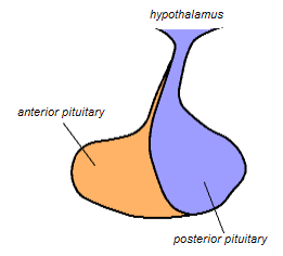
The posterior pituitary is the posterior lobe of the pituitary gland which is part of the endocrine system. The posterior pituitary is not glandular as is the anterior pituitary. Instead, it is largely a collection of axonal projections from the hypothalamus that terminate behind the anterior pituitary, and serve as a site for the secretion of neurohypophysial hormones directly into the blood. The hypothalamic–neurohypophyseal system is composed of the hypothalamus, posterior pituitary, and these axonal projections.
Thyrotropic cells are endocrine cells in the anterior pituitary which produce thyroid-stimulating hormone (TSH) in response to thyrotropin-releasing hormone (TRH) from the hypothalamus. Thyrotropes comprise around 5% of the anterior pituitary lobe cells.

Gonadotropic cells are endocrine cells in the anterior pituitary that produce the gonadotropins, such as the follicle-stimulating hormone (FSH) and luteinizing hormone (LH). Release of FSH and LH by gonadotropes is regulated by gonadotropin-releasing hormone (GnRH) from the hypothalamus.
Corticotropic cells, are basophilic cells in the anterior pituitary that produce pro-opiomelanocortin (POMC) which undergoes cleavage to adrenocorticotropin (ACTH), β-lipotropin (β-LPH), and melanocyte-stimulating hormone (MSH). These cells are stimulated by corticotropin releasing hormone (CRH) and make up 15–20% of the cells in the anterior pituitary. The release of ACTH from the corticotropic cells is controlled by CRH, which is formed in the cell bodies of parvocellular neurosecretory cells within the paraventricular nucleus of the hypothalamus and passes to the corticotropes in the anterior pituitary via the hypophyseal portal system. Adrenocorticotropin hormone stimulates the adrenal cortex to release glucocorticoids and plays an important role in the stress response.

Parathyroid oxyphil cells are one out of the two types of cells found in the parathyroid gland, the other being parathyroid chief cell. Oxyphil cells are only found in a select few number of species and humans are one of them.

Pituitary adenomas are tumors that occur in the pituitary gland. Most pituitary tumors are benign, approximately 35% are invasive and just 0.1% to 0.2% are carcinomas. Pituitary adenomas represent from 10% to 25% of all intracranial neoplasms, with an estimated prevalence rate in the general population of approximately 17%.

Basophilic is a technical term used by pathologists. It describes the appearance of cells, tissues and cellular structures as seen through the microscope after a histological section has been stained with a basic dye. The most common such dye is haematoxylin.

The endocrine system is a network of glands and organs located throughout the body. It is similar to the nervous system in that it plays a vital role in controlling and regulating many of the body's functions. Endocrine glands are ductless glands of the endocrine system that secrete their products, hormones, directly into the blood. The major glands of the endocrine system include the pineal gland, pituitary gland, pancreas, ovaries, testicles, thyroid gland, parathyroid gland, hypothalamus and adrenal glands. The hypothalamus and pituitary glands are neuroendocrine organs.
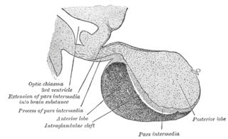
The pars intermedia is one of the three parts of the anterior pituitary. It is a section of tissue sometimes called a middle or intermediate lobe, between the pars distalis, and the posterior pituitary. It is a small region that is largely without blood supply. The cells in the pars intermedia are large and pale. They surround follicles that contain a colloidal matrix.
Neuroendocrine cells are cells that receive neuronal input and, as a consequence of this input, release messenger molecules (hormones) into the blood. In this way they bring about an integration between the nervous system and the endocrine system, a process known as neuroendocrine integration. An example of a neuroendocrine cell is a cell of the adrenal medulla, which releases adrenaline to the blood. The adrenal medullary cells are controlled by the sympathetic division of the autonomic nervous system. These cells are modified postganglionic neurons. Autonomic nerve fibers lead directly to them from the central nervous system. The adrenal medullary hormones are kept in vesicles much in the same way neurotransmitters are kept in neuronal vesicles. Hormonal effects can last up to ten times longer than those of neurotransmitters. Sympathetic nerve fiber impulses stimulate the release of adrenal medullary hormones. In this way the sympathetic division of the autonomic nervous system and the medullary secretions function together.

In cellular neuroscience, Nissl bodies are discrete granular structures in neurons that consist of rough endoplasmic reticulum, a collection of parallel, membrane-bound cisternae studded with ribosomes on the cytosolic surface of the membranes. Nissl bodies were named after Franz Nissl, a German neuropathologist who invented the staining method bearing his name. The term "Nissl bodies" generally refers to discrete clumps of rough endoplasmic reticulum and free ribosomes in nerve cells. Masses of rough endoplasmic reticulum also occur in some non-neuronal cells, where they are referred to as ergastoplasm, basophilic bodies, or chromophilic substance. While these organelles differ in some ways from Nissl bodies in neurons, large amounts of rough endoplasmic reticulum are generally linked to the copious production of proteins.
Pituicytes are glial cells of the posterior pituitary. Their main role is to assist in the storage and release of neurohypophysial hormones.
Acidophile is a term used by histologists to describe a particular staining pattern of cells and tissues when using haematoxylin and eosin stains. Specifically, the name refers to structures which "love" acid, and take it up readily. More specifically, acidophilia can be described by cationic groups of most often proteins in the cell readily reacting with acidic stains.
A chromophobe is a histological structure that does not stain readily, and thus appears relatively pale under the microscope.

In the anterior pituitary, the term "acidophil" is used to describe two different types of cells which stain well with acidic dyes.
A melanotroph is a cell in the pituitary gland that generates melanocyte-stimulating hormone (α‐MSH) from its precursor pro-opiomelanocortin. Chronic stress can induce the secretion of α‐MSH in melanotrophs and lead to their subsequent degeneration.
In hematology, myelopoiesis in the broadest sense of the term is the production of bone marrow and of all cells that arise from it, namely, all blood cells. In a narrower sense, myelopoiesis also refers specifically to the regulated formation of myeloid leukocytes (myelocytes), including eosinophilic granulocytes, basophilic granulocytes, neutrophilic granulocytes, and monocytes.
