
The endocrine system is a messenger system in an organism comprising feedback loops of hormones that are released by internal glands directly into the circulatory system and that target and regulate distant organs. In vertebrates, the hypothalamus is the neural control center for all endocrine systems.

The pituitary gland or hypophysis is an endocrine gland in vertebrates. In humans, the pituitary gland is located at the base of the brain, protruding off the bottom of the hypothalamus. The human pituitary gland is oval shaped, about 1 cm in diameter, 0.5–1 gram (0.018–0.035 oz) in weight on average, and about the size of a kidney bean.

The anterior pituitary is a major organ of the endocrine system. The anterior pituitary is the glandular, anterior lobe that together with the makes up the pituitary gland (hypophysis) which, in humans, is located at the base of the brain, protruding off the bottom of the hypothalamus.
A chromophil biological cell is a cell which is easily stainable by absorbing chromium salts used in histology to increase the visual contrast of samples for microscopy.
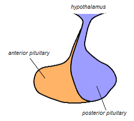
The posterior pituitary is the posterior lobe of the pituitary gland which is part of the endocrine system. The posterior pituitary is not glandular as is the anterior pituitary. Instead, it is largely a collection of axonal projections from the hypothalamus that terminate behind the anterior pituitary, and serve as a site for the secretion of neurohypophysial hormones directly into the blood. The hypothalamic–neurohypophyseal system is composed of the hypothalamus, posterior pituitary, and these axonal projections.

Chromaffin cells, also called pheochromocytes, are neuroendocrine cells found mostly in the medulla of the adrenal glands in mammals. These cells serve a variety of functions such as serving as a response to stress, monitoring carbon dioxide and oxygen concentrations in the body, maintenance of respiration and the regulation of blood pressure. They are in close proximity to pre-synaptic sympathetic ganglia of the sympathetic nervous system, with which they communicate, and structurally they are similar to post-synaptic sympathetic neurons. In order to activate chromaffin cells, the splanchnic nerve of the sympathetic nervous system releases acetylcholine, which then binds to nicotinic acetylcholine receptors on the adrenal medulla. This causes the release of catecholamines. The chromaffin cells release catecholamines: ~80% of adrenaline (epinephrine) and ~20% of noradrenaline (norepinephrine) into systemic circulation for systemic effects on multiple organs, and can also send paracrine signals. Hence they are called neuroendocrine cells.

The supraoptic nucleus (SON) is a nucleus of magnocellular neurosecretory cells in the hypothalamus of the mammalian brain. The nucleus is situated at the base of the brain, adjacent to the optic chiasm. In humans, the SON contains about 3,000 neurons.
A prolactin cell is a cell in the anterior pituitary which produces prolactin in response to hormonal signals including dopamine, thyrotropin-releasing hormone and estrogen, which are stimulatory. Prolactin is responsible for actions needed for body homeostasis, the development of breasts, and for lactation. The inhibitory effects of dopamine override the stimulatory effects of TRH in non-pregnant, non-lactating sexually mature females. Depending on the sex of the individual, prolactin cells account for 20% - 50% of all cells in the anterior pituitary gland. The inhibitory effects of dopamine override the stimulatory effects of TRH in non-pregnant, non-lactating sexually mature females. Other regulators include oxytocin and progesterone.

Parafollicular cells, also called C cells, are neuroendocrine cells in the thyroid. They are called C cells because the primary function of these cells is to secrete calcitonin. They are located adjacent to the thyroid follicles and reside in the connective tissue. These cells are large and have a pale stain compared with the follicular cells. In teleost and avian species these cells occupy a structure outside the thyroid gland named the ultimopharyngeal body.

Parathyroid oxyphil cells are one out of the two types of cells found in the parathyroid gland, the other being parathyroid chief cell. Oxyphil cells are only found in a select few number of species and humans are one of them.

Basophilic is a technical term used by pathologists. It describes the appearance of cells, tissues and cellular structures as seen through the microscope after a histological section has been stained with a basic dye. The most common such dye is haematoxylin.
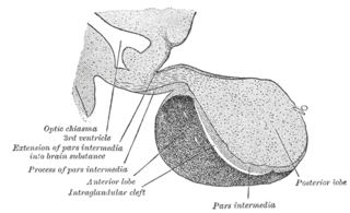
The pars intermedia is one of the three parts of the anterior pituitary. It is a section of tissue sometimes called a middle or intermediate lobe, between the pars distalis, and the posterior pituitary. It is a small region that is largely without blood supply. The cells in the pars intermedia are large and pale. They surround follicles that contain a colloidal matrix.
Neuroendocrine cells are cells that receive neuronal input and, as a consequence of this input, release messenger molecules (hormones) into the blood. In this way they bring about an integration between the nervous system and the endocrine system, a process known as neuroendocrine integration. An example of a neuroendocrine cell is a cell of the adrenal medulla, which releases adrenaline to the blood. The adrenal medullary cells are controlled by the sympathetic division of the autonomic nervous system. These cells are modified postganglionic neurons. Autonomic nerve fibers lead directly to them from the central nervous system. The adrenal medullary hormones are kept in vesicles much in the same way neurotransmitters are kept in neuronal vesicles. Hormonal effects can last up to ten times longer than those of neurotransmitters. Sympathetic nerve fiber impulses stimulate the release of adrenal medullary hormones. In this way the sympathetic division of the autonomic nervous system and the medullary secretions function together.
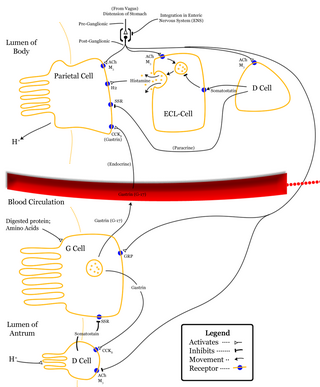
Enterochromaffin-like cells or ECL cells are a type of neuroendocrine cell found in the gastric glands of the gastric mucosa beneath the epithelium, in particular in the vicinity of parietal cells, that aid in the production of gastric acid via the release of histamine. They are also considered a type of enteroendocrine cell.
Neuroendocrinology is the branch of biology which studies the interaction between the nervous system and the endocrine system; i.e. how the brain regulates the hormonal activity in the body. The nervous and endocrine systems often act together in a process called neuroendocrine integration, to regulate the physiological processes of the human body. Neuroendocrinology arose from the recognition that the brain, especially the hypothalamus, controls secretion of pituitary gland hormones, and has subsequently expanded to investigate numerous interconnections of the endocrine and nervous systems.
Acidophile is a term used by histologists to describe a particular staining pattern of cells and tissues when using haematoxylin and eosin stains. Specifically, the name refers to structures which "love" acid, and take it up readily. More specifically, acidophilia can be described by cationic groups of most often proteins in the cell readily reacting with acidic stains.
A chromophobe is a histological structure that does not stain readily, and thus appears relatively pale under the microscope.

An anterior pituitary basophil is a type of cell in the anterior pituitary which manufactures hormones.
A melanotroph is a cell in the pituitary gland that generates melanocyte-stimulating hormone (α‐MSH) from its precursor pro-opiomelanocortin. Chronic stress can induce the secretion of α‐MSH in melanotrophs and lead to their subsequent degeneration.
A somatomammotroph or somatomammotrophic cell, also known as a somatolactotroph or somatolactotrophic cell, is a type of cell of the anterior pituitary gland that produces both somatotropin and prolactin. Cells that secrete only somatotropin or only prolactin are known as somatotrophs and mammotrophs, respectively.
