Herring bodies

Last updated
Neurosecretory body
Details
Location Posterior pituitary
Identifiers
Latin corpusculum neurosecretorium
TH H3.08.02.2.00039
Anatomical terms of microanatomy

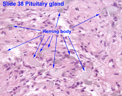
Herring bodies or neurosecretory bodies are structures found in the posterior pituitary. They represent the terminal end of the axons from the hypothalamus, and hormones are temporarily stored in these locations. They are neurosecretory terminals. [1]

Antidiuretic hormone (ADH) and oxytocin are both stored in Herring bodies, but are not stored simultaneously in the same Herring body. [2]

In addition, each Herring body also contains ATP and a type of neurophysin. Neurophysins are binding proteins, of which there are two types: neurophysin I and neurophysin II, which bind to oxytocin and ADH, respectively. Neurophysin and its hormone become a complex considered a single protein and stored in the neurohypophysis. Upon stimulation by the hypothalamus, secretory granules release stored hormones into the bloodstream. Fibers from supraoptic nuclei are concerned with ADH secretion; paraventricular nuclei with oxytocin. [3]

This anatomical structure was first described by Percy Theodore Herring in 1908.

Related Research Articles

<span class="mw-page-title-main">Pituitary gland</span> Endocrine gland at the base of the brain

In vertebrate anatomy, the pituitary gland, or hypophysis, is an endocrine gland, about the size of a chickpea and weighing, on average, 0.5 grams (0.018 oz) in humans. It is a protrusion off the bottom of the hypothalamus at the base of the brain. The hypophysis rests upon the hypophyseal fossa of the sphenoid bone in the center of the middle cranial fossa and is surrounded by a small bony cavity covered by a dural fold.

<span class="mw-page-title-main">Hypothalamus</span> Area of the brain below the thalamus

The hypothalamus is a part of the brain that contains a number of small nuclei with a variety of functions. One of the most important functions is to link the nervous system to the endocrine system via the pituitary gland. The hypothalamus is located below the thalamus and is part of the limbic system. In the terminology of neuroanatomy, it forms the ventral part of the diencephalon. All vertebrate brains contain a hypothalamus. In humans, it is the size of an almond.

<span class="mw-page-title-main">Vasopressin</span> Mammalian hormone released from the pituitary gland

Human vasopressin, also called antidiuretic hormone (ADH), arginine vasopressin (AVP) or argipressin, is a hormone synthesized from the AVP gene as a peptide prohormone in neurons in the hypothalamus, and is converted to AVP. It then travels down the axon terminating in the posterior pituitary, and is released from vesicles into the circulation in response to extracellular fluid hypertonicity (hyperosmolality). AVP has two primary functions. First, it increases the amount of solute-free water reabsorbed back into the circulation from the filtrate in the kidney tubules of the nephrons. Second, AVP constricts arterioles, which increases peripheral vascular resistance and raises arterial blood pressure.

<span class="mw-page-title-main">Posterior pituitary</span> Posterior lobe of the pituitary gland

The posterior pituitary is the posterior lobe of the pituitary gland which is part of the endocrine system. The posterior pituitary is not glandular as is the anterior pituitary. Instead, it is largely a collection of axonal projections from the hypothalamus that terminate behind the anterior pituitary, and serve as a site for the secretion of neurohypophysial hormones directly into the blood. The hypothalamic–neurohypophyseal system is composed of the hypothalamus, posterior pituitary, and these axonal projections.

<span class="mw-page-title-main">Supraoptic nucleus</span> ADH secreting nucleus of the hypothalamus.

The supraoptic nucleus (SON) is a nucleus of magnocellular neurosecretory cells in the hypothalamus of the mammalian brain. The nucleus is situated at the base of the brain, adjacent to the optic chiasm. In humans, the SON contains about 3,000 neurons.

<span class="mw-page-title-main">Paraventricular nucleus of hypothalamus</span>

The paraventricular nucleus is a nucleus in the hypothalamus. Anatomically, it is adjacent to the third ventricle and many of its neurons project to the posterior pituitary. These projecting neurons secrete oxytocin and a smaller amount of vasopressin, otherwise the nucleus also secretes corticotropin-releasing hormone (CRH) and thyrotropin-releasing hormone (TRH). CRH and TRH are secreted into the hypophyseal portal system and act on different targets neurons in the anterior pituitary. PVN is thought to mediate many diverse functions through these different hormones, including osmoregulation, appetite, and the response of the body to stress.

Magnocellular neurosecretory cells are large neuroendocrine cells within the supraoptic nucleus and paraventricular nucleus of the hypothalamus. They are also found in smaller numbers in accessory cell groups between these two nuclei, the largest one being the circular nucleus. There are two types of magnocellular neurosecretory cells, oxytocin-producing cells and vasopressin-producing cells, but a small number can produce both hormones. These cells are neuroendocrine neurons, are electrically excitable, and generate action potentials in response to afferent stimulation. Vasopressin is produced from the vasopressin-producing cells via the AVP gene, a molecular output of circadian pathways.

A neurohormone is any hormone produced and released by neuroendocrine cells into the blood. By definition of being hormones, they are secreted into the circulation for systemic effect, but they can also have a role of neurotransmitter or other roles such as autocrine (self) or paracrine (local) messenger.

Neuroendocrine cells are cells that receive neuronal input and, as a consequence of this input, release messenger molecules (hormones) into the blood. In this way they bring about an integration between the nervous system and the endocrine system, a process known as neuroendocrine integration. An example of a neuroendocrine cell is a cell of the adrenal medulla, which releases adrenaline to the blood. The adrenal medullary cells are controlled by the sympathetic division of the autonomic nervous system. These cells are modified postganglionic neurons. Autonomic nerve fibers lead directly to them from the central nervous system. The adrenal medullary hormones are kept in vesicles much in the same way neurotransmitters are kept in neuronal vesicles. Hormonal effects can last up to ten times longer than those of neurotransmitters. Sympathetic nerve fiber impulses stimulate the release of adrenal medullary hormones. In this way the sympathetic division of the autonomic nervous system and the medullary secretions function together.

Neurophysin I is a carrier protein with a size of 10 KDa and contains 90 to 97 amino acids. It is a cleavage product of preprooxyphysin. It is a neurohypophysial hormone that is transported in vesicles with oxytocin, the other cleavage product, along axons, from magnocellular neurons of the hypothalamus to the posterior lobe of the pituitary. Although it is stored in neurosecretory granules with oxytocin and released with oxytocin, its biological action is unclear.

Neuroendocrinology is the branch of biology which studies the interaction between the nervous system and the endocrine system; i.e. how the brain regulates the hormonal activity in the body. The nervous and endocrine systems often act together in a process called neuroendocrine integration, to regulate the physiological processes of the human body. Neuroendocrinology arose from the recognition that the brain, especially the hypothalamus, controls secretion of pituitary gland hormones, and has subsequently expanded to investigate numerous interconnections of the endocrine and nervous systems.

Neurophysins are carrier proteins which transport the hormones oxytocin and vasopressin to the posterior pituitary from the paraventricular and supraoptic nucleus of the hypothalamus, respectively. Inside the neurosecretory granules, the analogous neurophysin I and II form stabilizing complexes via covalent interactions. Stabilizing neurophysin-hormone complexes that are formed within neurosecretory granules located in the posterior pituitary gland aid in intra-axonal transport. During intra-axonal transport, the neurophysin's are believed to prevent the bound hormone from leaking into the cytoplasmic space and proteolytic digestion via enzymes. However, due to the low concentration of neurophysin in the blood, it is likely the protein-hormone complex dissociates, indicating the neurophysin does not aid in transporting the hormone through the circulatory system.

<span class="mw-page-title-main">Vasotocin</span> Chemical compound

Vasotocin is an oligopeptide homologous to oxytocin and vasopressin found in all non-mammalian vertebrates and possibly in mammals during the fetal stage of development. Arginine vasotocin (AVT), a hormone produced by neurosecretory cells within the posterior pituitary gland (neurohypophysis) of the brain, is a major endocrine regulator of water balance and osmotic homoeostasis and is involved in social and sexual behavior in non-mammalian vertebrates. In mammals, it appears to have biological properties similar to those of oxytocin and vasopressin. It has been found to have effects on the regulation of REM sleep. Evidence for the existence of endogenous vasotocin in mammals is limited and no mammalian gene encoding vasotocin has been confirmed.

<span class="mw-page-title-main">Neurophysin II</span>

Neurophysin II is a carrier protein with a size of 19,687.3 Da and is made up of a dimer of two virtually identical chains of amino acids. Neurophysin II is a cleavage product of the AVP gene. It is a neurohypophysial hormone that is transported in vesicles with vasopressin, the other cleavage product, along axons, from magnocellular neurons of the hypothalamus to the posterior lobe of the pituitary. Although it is stored in neurosecretory granules with vasopressin and released with vasopressin into the bloodstream, its biological action is unclear. Neurophysin II is also known as a stimulator of prolactin secretion.

Hypothalamic–pituitary hormones are hormones that are produced by the hypothalamus and pituitary gland. Although the organs in which they are produced are relatively small, the effects of these hormones cascade throughout the body. They can be classified as a hypothalamic–pituitary axis of which the adrenal (HPA), gonadal (HPG), thyroid (HPT), somatotropic (HPS), and prolactin (HPP) axes are branches.

Parvocellular neurosecretory cells are small neurons that produce hypothalamic releasing and inhibiting hormones. The cell bodies of these neurons are located in various nuclei of the hypothalamus or in closely related areas of the basal brain, mainly in the medial zone of the hypothalamus. All or most of the axons of the parvocellular neurosecretory cells project to the median eminence, at the base of the brain, where their nerve terminals release the hypothalamic hormones. These hormones are then immediately absorbed into the blood vessels of the hypothalamo-pituitary portal system, which carry them to the anterior pituitary gland, where they regulate the secretion of hormones into the systemic circulation.

<span class="mw-page-title-main">Neurohypophysial hormone</span>

The neurohypophysial hormones form a family of structurally and functionally related peptide hormones. Their representatives in humans are oxytocin and vasopressin. They are named after the location of their release into the blood, the neurohypophysis.

<span class="mw-page-title-main">Howard Sachs (scientist)</span>

Howard Sachs, was a biochemist who helped pioneer the study of neuroendocrinology. His discoveries concerning the production of the hormone vasopressin laid the foundation for the field of hormone biosynthesis.

<span class="mw-page-title-main">Oxytocin/neurophysin i prepropeptide</span> Protein-coding gene in the species Homo sapiens

Oxytocin/neurophysin I prepropeptide is a protein that in humans is encoded by the OXT gene.

<span class="mw-page-title-main">AVP gene</span> Gene

Arginine Vasopressin (AVP) Gene is a gene whose product is proteolytically cleaved to produce vasopressin, neurophysin II, and a glycoprotein called copeptin. AVP and other AVP-like peptides are found in mammals, as well as mollusks, arthropods, nematodes, and other invertebrate species. In humans, AVP is present on chromosome 20 and plays a role in homeostatic regulation. The products of AVP have many functions that include vasoconstriction, regulating the balance of water in the body, and regulating responses to stress. Expression of AVP is regulated by the Transcription Translation Feedback Loop (TTFL), which is an important part of the circadian system that controls the expression of clock genes. AVP has important implications in the medical field as its products have significant roles throughout body.

References

  1. Kwang W. Jeon (18 August 2005). International Review of Cytology: A Survey of Cell Biology. Gulf Professional Publishing. pp. 143–. ISBN   978-0-12-364649-1 . Retrieved 26 May 2011.
  2. Histology at KUMC endo-endo08
  3. Mescher, Anthony L. (2013). Junqueira's Basic Histology: Text and Atlas (13th ed.). McGraw-Hill Medical. ISBN   978-0071780339.