Clinical data | |
---|---|
Pronunciation | /ˌɒksɪˈtoʊsɪn/ |
ATC code | |
Physiological data | |
Source tissues | Pituitary gland |
Target tissues | Wide spread |
Receptors | Oxytocin receptor |
Antagonists | Atosiban |
Precursor | Oxytocin/neurophysin I prepropeptide |
Metabolism | Liver and other oxytocinases |
Legal status | |
Legal status | |
Pharmacokinetic data | |
Protein binding | 30% |
Metabolism | Liver and other oxytocinases |
Elimination half-life | 1–6 min (IV) ~2 h (intranasal) [2] [3] |
Excretion | Biliary and kidney |
Identifiers | |
| |
CAS Number | |
PubChem CID | |
IUPHAR/BPS | |
DrugBank | |
ChemSpider | |
UNII | |
KEGG | |
ChEBI | |
ChEMBL | |
CompTox Dashboard (EPA) | |
ECHA InfoCard | 100.000.045 |
Chemical and physical data | |
Formula | C43H66N12O12S2 |
Molar mass | 1007.19 g·mol−1 |
3D model (JSmol) | |
| |
| |
(verify) |
Oxytocin is a peptide hormone and neuropeptide normally produced in the hypothalamus and released by the posterior pituitary. [4] Present in animals since early stages of evolution, in humans it plays roles in behavior that include social bonding, love, reproduction, childbirth, and the period after childbirth. [5] [6] [7] [8] Oxytocin is released into the bloodstream as a hormone in response to sexual activity and during childbirth. [9] [10] It is also available in pharmaceutical form. In either form, oxytocin stimulates uterine contractions to speed up the process of childbirth.
In its natural form, it also plays a role in maternal bonding and milk production. [10] [11] Production and secretion of oxytocin is controlled by a positive feedback mechanism, where its initial release stimulates production and release of further oxytocin. For example, when oxytocin is released during a contraction of the uterus at the start of childbirth, this stimulates production and release of more oxytocin and an increase in the intensity and frequency of contractions. This process compounds in intensity and frequency and continues until the triggering activity ceases. A similar process takes place during lactation and during sexual activity.
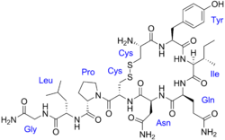
Oxytocin is derived by enzymatic splitting from the peptide precursor encoded by the human OXT gene. The deduced structure of the active nonapeptide is:
The term "oxytocin" derives from the Greek "ὠκυτόκος" (ōkutókos), based on ὀξύς (oxús), meaning "sharp" or "swift", and τόκος (tókos), meaning "childbirth". [12] [13] The adjective form is "oxytocic", which refers to medicines which stimulate uterine contractions, to speed up the process of childbirth. Colloquially, it has been referred to as the "cuddle hormone" or the "moral molecule" which have been considered misnomers. [14]
The uterine-contracting properties of the principle that would later be named oxytocin were discovered by British pharmacologist Henry Hallett Dale in 1906, [15] [16] and its milk ejection property was described by Ott and Scott in 1910 [17] and by Schafer and Mackenzie in 1911. [18] In 1909 the first clinical use of oxytocin was performed by William Blair-Bell to induce childbirth in patients with complications. [19] [20]
By the 1920s, oxytocin and vasopressin had been isolated from pituitary tissue and given their current names. Oxytocin's molecular structure was determined in 1952. [21] In the early 1950s, American biochemist Vincent du Vigneaud found that oxytocin is made up of nine amino acids, and he identified its amino acid sequence, the first polypeptide hormone to be sequenced. [22] In 1953, du Vigneaud carried out the synthesis of oxytocin, the first polypeptide hormone to be synthesized. [23] [24] [25] Du Vigneaud was awarded the Nobel Prize in Chemistry in 1955 for his work. [26] Further work on different synthetic routes for oxytocin, as well as the preparation of analogues of the hormone (e.g. 4-deamido-oxytocin) was performed in the following decade by Iphigenia Photaki. [27]
Estrogen has been found to increase the secretion of oxytocin and to increase the expression of its receptor, the oxytocin receptor, in the brain. [32] In women, a single dose of estradiol has been found to be sufficient to increase circulating oxytocin concentrations. [33]
Oxytocin and vasopressin are the only known hormones released by the human posterior pituitary gland to act at a distance. However, oxytocin neurons make other peptides, including corticotropin-releasing hormone and dynorphin, for example, that act locally. The magnocellular neurons that make oxytocin are adjacent to magnocellular neurons that make vasopressin and are similar in many respects.
The oxytocin peptide is synthesized as an inactive precursor protein from the OXT gene. [34] [35] [36] This precursor protein also includes the oxytocin carrier protein neurophysin I. [37] The inactive precursor protein is progressively hydrolyzed into smaller fragments (one of which is neurophysin I) via a series of enzymes. The last hydrolysis that releases the active oxytocin nonapeptide is catalyzed by peptidylglycine alpha-amidating monooxygenase (PAM). [38]
The activity of the PAM enzyme system is dependent upon vitamin C (ascorbate), which is a necessary vitamin cofactor. By chance, sodium ascorbate by itself was found to stimulate the production of oxytocin from ovarian tissue over a range of concentrations in a dose-dependent manner. [39] Many of the same tissues (e.g. ovaries, testes, eyes, adrenals, placenta, thymus, pancreas) where PAM (and oxytocin by default) is found are also known to store higher concentrations of vitamin C. [40]
Oxytocin is known to be metabolized by the oxytocinase, leucyl/cystinyl aminopeptidase. [41] [42] Other oxytocinases are also known to exist. [41] [43] Amastatin, bestatin (ubenimex), leupeptin, and puromycin have been found to inhibit the enzymatic degradation of oxytocin, though they also inhibit the degradation of various other peptides, such as vasopressin, met-enkephalin, and dynorphin A. [43] [44] [45] [46]
In the hypothalamus, oxytocin is made in magnocellular neurosecretory cells of the supraoptic and paraventricular nuclei, [47] and is stored in Herring bodies at the axon terminals in the posterior pituitary. It is then released into the blood from the posterior lobe (neurohypophysis) of the pituitary gland. These axons (likely, but dendrites have not been ruled out) have collaterals that innervate neurons in the nucleus accumbens, a brain structure where oxytocin receptors are expressed. [48] The endocrine effects of hormonal oxytocin, and the cognitive or behavioral effects of oxytocin neuropeptides are thought to be coordinated through its common release through these collaterals. [48] Oxytocin is also produced by some neurons in the paraventricular nucleus that project to other parts of the brain and to the spinal cord. [49] Depending on the species, oxytocin receptor-expressing cells are located in other areas, including the amygdala and bed nucleus of the stria terminalis.
In the pituitary gland, oxytocin is packaged in large, dense-core vesicles, where it is bound to neurophysin I as shown in the inset of the figure; neurophysin is a large peptide fragment of the larger precursor protein molecule from which oxytocin is derived by enzymatic cleavage.
Secretion of oxytocin from the neurosecretory nerve endings is regulated by the electrical activity of the oxytocin cells in the hypothalamus. These cells generate action potentials that propagate down axons to the nerve endings in the pituitary; the endings contain large numbers of oxytocin-containing vesicles, which are released by exocytosis when the nerve terminals are depolarised.
Endogenous oxytocin concentrations in the brain have been found to be as much as 1000-fold higher than peripheral levels. [50] Outside the brain, oxytocin-containing cells have been identified in several diverse tissues, including in females in the corpus luteum [51] [52] and the placenta; [53] in males in the testicles' interstitial cells of Leydig; [54] and in both sexes in the retina, [55] the adrenal medulla, [56] the thymus [57] and the pancreas. [58] The finding of significant amounts of this classically "neurohypophysial" hormone outside the central nervous system raises many questions regarding its possible importance in these diverse tissues.
The Leydig cells in some species have been shown to possess the biosynthetic machinery to manufacture testicular oxytocin de novo, to be specific, in rats (which can synthesize vitamin C endogenously), and in guinea pigs, which, like humans, require an exogenous source of vitamin C in their diets. [59] Oxytocin is synthesized by corpora lutea of several species, including ruminants and primates. Along with estrogen, it is involved in inducing the endometrial synthesis of prostaglandin F2α to cause regression of the corpus luteum. [60]
Virtually all vertebrates have an oxytocin-like nonapeptide hormone that supports reproductive functions and a vasopressin-like nonapeptide hormone involved in water regulation. The two genes are usually located close to each other (less than 15,000 bases apart) on the same chromosome, and are transcribed in opposite directions (however, in fugu, [61] the homologs are further apart and transcribed in the same direction). The two genes are believed to result from a gene duplication event; the ancestral gene is estimated to be about 500 million years old and is found in cyclostomata (modern members of the Agnatha). [62]
A 2023 study found that zebrafish utilize oxytocin in reaction to the fear of other fish. It found that zebrafish that have had oxytocin production removed by gene editing cannot respond to the fear of other fish. When oxytocin is injected back into the fish, they react again in a way that suggests they may have empathy in regards to this emotion. Furthermore, because the same regions of the brain are involved as in mammals, the study suggests oxytocin-based empathy may have evolved from a common ancestor many millions of years ago. [63]
Oxytocin has peripheral (hormonal) actions and also has actions in the brain. Its actions are mediated by specific oxytocin receptors. The oxytocin receptor is a G-protein-coupled receptor, OT-R, which requires magnesium and cholesterol and is expressed in myometrial cells. [64] It belongs to the rhodopsin-type (class I) group of G-protein-coupled receptors. [62]
Studies have looked at oxytocin's role in various behaviors, including orgasm, social recognition, pair bonding, anxiety, in-group bias, situational lack of honesty, autism, and maternal behaviors. [16] Oxytocin is believed to have a significant role in social learning. There are indicators that oxytocin may help to decrease noise in the brain's auditory system, increase perception of social cues and support more targeted social behavior. It may also enhance reward responses. However, its effects may be influenced by context, such as the presence of familiar or unfamiliar individuals. [65] [66] In addition to its oxytocin receptor agonism, oxytocin has been found to act as a PAM of the μ- and κ-opioid receptors and this may be involved in its analgesic effects. [67] [68] [69] [70] [71] [72]
The peripheral actions of oxytocin mainly reflect secretion from the pituitary gland. The behavioral effects of oxytocin are thought to reflect release from centrally projecting oxytocin neurons, different from those that project to the pituitary gland, or that are collaterals from them. [48] Oxytocin receptors are expressed by neurons in many parts of the brain and spinal cord, including the amygdala, ventromedial hypothalamus, septum, nucleus accumbens, and brainstem, although the distribution differs markedly between species. [62] Furthermore, the distribution of these receptors changes during development and has been observed to change after parturition in the montane vole. [62]
In the prairie vole, oxytocin released into the brain of the female during sexual activity is important for forming a pair bond with her sexual partner. Vasopressin appears to have a similar effect in males. [99] Oxytocin has a role in social behaviors in many species, so it likely also does in humans. In a 2003 study, both humans and dog oxytocin levels in the blood rose after a five to 24 minute petting session. This possibly plays a role in the emotional bonding between humans and dogs. [100]
Oxytocin is not only correlated with the preferences of individuals to associate with members of their own group, but it is also evident during conflicts between members of different groups. During conflict, individuals receiving nasally administered oxytocin demonstrate more frequent defense-motivated responses toward in-group members than out-group members. Further, oxytocin was correlated with participant desire to protect vulnerable in-group members, despite that individual's attachment to the conflict. [106] Similarly, it has been demonstrated that when oxytocin is administered, individuals alter their subjective preferences in order to align with in-group ideals over out-group ideals. [107] These studies demonstrate that oxytocin is associated with intergroup dynamics. Further, oxytocin influences the responses of individuals in a particular group to those of another group. The in-group bias is evident in smaller groups; however, it can also be extended to groups as large as one's entire country leading toward a tendency of strong national zeal. A study done in the Netherlands showed that oxytocin increased the in-group favoritism of their nation while decreasing acceptance of members of other ethnicities and foreigners. [108] People also show more affection for their country's flag while remaining indifferent to other cultural objects when exposed to oxytocin. [109] It has thus been hypothesized that this hormone may be a factor in xenophobic tendencies secondary to this effect. Thus, oxytocin appears to affect individuals at an international level where the in-group becomes a specific "home" country and the out-group grows to include all other countries.
Oxytocin is typically remembered for the effect it has on prosocial behaviors, such as its role in facilitating trust and attachment between individuals. [115] [ qualify evidence ] However, oxytocin has a more complex role than solely enhancing prosocial behaviors. There is consensus that oxytocin modulates fear and anxiety; that is, it does not directly elicit fear or anxiety. [116] Two dominant theories explain the role of oxytocin in fear and anxiety. One theory states that oxytocin increases approach/avoidance to certain social stimuli and the second theory states that oxytocin increases the salience of certain social stimuli, causing animals (including humans) to pay closer attention to socially relevant stimuli. [117] [118] [119]
Nasally administered oxytocin has been reported to reduce fear, possibly by inhibiting the amygdala (which is thought to be responsible for fear responses). [120] Indeed, studies in rodents have shown oxytocin can efficiently inhibit fear responses by activating an inhibitory circuit within the amygdala. [121] [122] Some researchers have argued oxytocin has a general enhancing effect on all social emotions, since intranasal administration of oxytocin also increases envy and Schadenfreude . [123] Individuals who receive an intranasal dose of oxytocin identify facial expressions of disgust more quickly than individuals who do not receive oxytocin. [117] [ qualify evidence ] Facial expressions of disgust are evolutionarily linked to the idea of contagion. Thus, oxytocin increases the salience of cues that imply contamination, which leads to a faster response because these cues are especially relevant for survival. In another study, after administration of oxytocin, individuals displayed an enhanced ability to recognize expressions of fear compared to the individuals who received the placebo. [124] Oxytocin modulates fear responses by enhancing the maintenance of social memories. Rats who are genetically modified to have a surplus of oxytocin receptors display a greater fear response to a previously conditioned stressor. Oxytocin enhances the aversive social memory, leading the rat to display a greater fear response when the aversive stimulus is encountered again. [116]
Oxytocin produces antidepressant-like effects in animal models of depression, [125] and a deficit of it may be involved in the pathophysiology of depression in humans. [126] The antidepressant-like effects of oxytocin are not blocked by a selective antagonist of the oxytocin receptor, suggesting that these effects are not mediated by the oxytocin receptor. [33] In accordance, unlike oxytocin, the selective non-peptide oxytocin receptor agonist WAY-267,464 does not produce antidepressant-like effects, at least in the tail suspension test. [127] In contrast to WAY-267,464, carbetocin, a close analogue of oxytocin and peptide oxytocin receptor agonist, notably does produce antidepressant-like effects in animals. [127] As such, the antidepressant-like effects of oxytocin may be mediated by modulation of a different target, perhaps the vasopressin V1A receptor where oxytocin is known to weakly bind as an agonist. [128] [129]
Oxytocin mediates the antidepressant-like effects of sexual activity. [130] [131] A drug for sexual dysfunction, sildenafil enhances electrically evoked oxytocin release from the pituitary gland. [132] In accordance, it may have promise as an antidepressant. [125] [133]
It has been shown that oxytocin differentially affects males and females. Females who are administered oxytocin are overall faster in responding to socially relevant stimuli than males who received oxytocin. [117] [134] Additionally, after the administration of oxytocin, females show increased amygdala activity in response to threatening scenes; however, males do not show increased amygdala activation. This phenomenon can be explained by looking at the role of gonadal hormones, specifically estrogen, which modulate the enhanced threat processing seen in females. Estrogen has been shown to stimulate the release of oxytocin from the hypothalamus and promote receptor binding in the amygdala. [134]
It has also been shown that testosterone directly suppresses oxytocin in mice. [135] This has been hypothesized to have evolutionary significance. With oxytocin suppressed, activities such as hunting and attacking invaders would be less mentally difficult as oxytocin is strongly associated with empathy. [136]
Because oxytocin plays a role in social bonding, maternal behaviors and emotional connections between people, it is also informally referred to as the "love hormone". [137] This term is not a medical or scientific name but is often used to describe oxytocin's effects on human behavior and emotions.
Oxytocin is a peptide of nine amino acids (a nonapeptide) in the sequence cysteine-tyrosine-isoleucine-glutamine-asparagine-cysteine-proline-leucine-glycine-amide (Cys – Tyr – Ile – Gln – Asn – Cys – Pro – Leu – Gly – NH2, or CYIQNCPLG-NH2); its C-terminus has been converted to a primary amide and a disulfide bridge joins the cysteine moieties. [165] Oxytocin has a molecular mass of 1007 Da, and one international unit (IU) of oxytocin is the equivalent of 1.68 μg of pure peptide. [166]
While the structure of oxytocin is highly conserved in placental mammals, a novel structure of oxytocin was reported in 2011 in marmosets, tamarins, and other new world primates. Genomic sequencing of the gene for oxytocin revealed a single in-frame mutation (thymine for cytosine) which results in a single amino acid substitution at the 8-position (proline for leucine). [167] Since this original Lee et al. paper, two other laboratories have confirmed Pro8-OT and documented additional oxytocin structural variants in this primate taxon. Vargas-Pinilla et al. sequenced the coding regions of the OXT gene in other genera in new world primates and identified the following variants in addition to Leu8- and Pro8-OT: Ala8-OT, Thr8-OT, and Val3/Pro8-OT. [168] Ren et al. identified a variant further, Phe2-OT in howler monkeys. [169]
Recent advances in analytical instrumental techniques highlighted the importance of liquid chromatography (LC) coupled with mass spectrometry (MS) for measuring oxytocin levels in various samples derived from biological sources. Most of these studies optimized the oxytocin quantification in electrospray ionization (ESI) positive mode, using [M+H]+ as the parent ion at mass-to-charge ratio (m/z) 1007.4 and the fragment ions as diagnostic peaks at m/z 991.0, [170] m/z 723.2 [171] and m/z 504.2. [172] These important ion selections paved the way for the development of current methods of oxytocin quantification using MS instrumentation.
The structure of oxytocin is very similar to that of vasopressin. Both are nonapeptides with a single disulfide bridge, differing only by two substitutions in the amino acid sequence (differences from oxytocin bolded for clarity): Cys – Tyr – Phe – Gln – Asn – Cys – Pro – Arg – Gly – NH2. [165] Oxytocin and vasopressin were isolated and their total synthesis reported in 1954, [173] work for which Vincent du Vigneaud was awarded the 1955 Nobel Prize in Chemistry with the citation: "for his work on biochemically important sulphur compounds, especially for the first synthesis of a polypeptide hormone." [174]
Oxytocin and vasopressin are the only known hormones released by the human posterior pituitary gland to act at a distance. However, oxytocin neurons make other peptides, including corticotropin-releasing hormone and dynorphin, for example, that act locally. The magnocellular neurosecretory cells that make oxytocin are adjacent to magnocellular neurosecretory cells that make vasopressin. These are large neuroendocrine neurons which are excitable and can generate action potentials. [175]
Small-molecule oxytocin receptor agonists like LIT-001 may prove to be useful in the treatment of social deficits, for instance in autism. [176] [177] [178]
The hypothalamus is a small part of the vertebrate brain that contains a number of nuclei with a variety of functions. One of the most important functions is to link the nervous system to the endocrine system via the pituitary gland. The hypothalamus is located below the thalamus and is part of the limbic system. It forms the basal part of the diencephalon. All vertebrate brains contain a hypothalamus. In humans, it is about the size of an almond.
The hypothalamic–pituitary–adrenal axis is a complex set of direct influences and feedback interactions among three components: the hypothalamus, the pituitary gland, and the adrenal glands. These organs and their interactions constitute the HPS axis.
Human vasopressin, also called antidiuretic hormone (ADH), arginine vasopressin (AVP) or argipressin, is a hormone synthesized from the AVP gene as a peptide prohormone in neurons in the hypothalamus, and is converted to AVP. It then travels down the axon terminating in the posterior pituitary, and is released from vesicles into the circulation in response to extracellular fluid hypertonicity (hyperosmolality). AVP has two primary functions. First, it increases the amount of solute-free water reabsorbed back into the circulation from the filtrate in the kidney tubules of the nephrons. Second, AVP constricts arterioles, which increases peripheral vascular resistance and raises arterial blood pressure.
The supraoptic nucleus (SON) is a nucleus of magnocellular neurosecretory cells in the hypothalamus of the mammalian brain. The nucleus is situated at the base of the brain, adjacent to the optic chiasm. In humans, the SON contains about 3,000 neurons.
The paraventricular nucleus of hypothalamus is a nucleus in the hypothalamus, that lies next to the third ventricle. Many of its neurons project to the posterior pituitary where they secrete oxytocin, and a smaller amount of vasopressin. Other secretions are corticotropin-releasing hormone (CRH) and thyrotropin-releasing hormone (TRH). CRH and TRH are secreted into the hypophyseal portal system, and target different neurons in the anterior pituitary. Dysfunctions of the PVN can cause hypersomnia in mice. In humans, the dysfunction of the PVN and the other nuclei around it can lead to drowsiness for up to 20 hours per day. The PVN is thought to mediate many diverse functions through different hormones, including osmoregulation, appetite, wakefulness, and the response of the body to stress.
Magnocellular neurosecretory cells are large neuroendocrine cells within the supraoptic nucleus and paraventricular nucleus of the hypothalamus. They are also found in smaller numbers in accessory cell groups between these two nuclei, the largest one being the circular nucleus. There are two types of magnocellular neurosecretory cells, oxytocin-producing cells and vasopressin-producing cells, but a small number can produce both hormones. These cells are neuroendocrine neurons, are electrically excitable, and generate action potentials in response to afferent stimulation. Vasopressin is produced from the vasopressin-producing cells via the AVP gene, a molecular output of circadian pathways.
Gonadotropin-releasing hormone (GnRH) is a releasing hormone responsible for the release of follicle-stimulating hormone (FSH) and luteinizing hormone (LH) from the anterior pituitary. GnRH is a tropic peptide hormone synthesized and released from GnRH neurons within the hypothalamus. GnRH is inhibited by testosterone. The peptide belongs to gonadotropin-releasing hormone family. It constitutes the initial step in the hypothalamic–pituitary–gonadal axis.
The arcuate nucleus of the hypothalamus (ARH), or ARC, is also known as the infundibular nucleus to distinguish it from the arcuate nucleus of the medulla oblongata in the brainstem. The arcuate nucleus is an aggregation of neurons in the mediobasal hypothalamus, adjacent to the third ventricle and the median eminence. The arcuate nucleus includes several important and diverse populations of neurons that help mediate different neuroendocrine and physiological functions, including neuroendocrine neurons, centrally projecting neurons, and astrocytes. The populations of neurons found in the arcuate nucleus are based on the hormones they secrete or interact with and are responsible for hypothalamic function, such as regulating hormones released from the pituitary gland or secreting their own hormones. Neurons in this region are also responsible for integrating information and providing inputs to other nuclei in the hypothalamus or inputs to areas outside this region of the brain. These neurons, generated from the ventral part of the periventricular epithelium during embryonic development, locate dorsally in the hypothalamus, becoming part of the ventromedial hypothalamic region. The function of the arcuate nucleus relies on its diversity of neurons, but its central role is involved in homeostasis. The arcuate nucleus provides many physiological roles involved in feeding, metabolism, fertility, and cardiovascular regulation.
Neuropeptides are chemical messengers made up of small chains of amino acids that are synthesized and released by neurons. Neuropeptides typically bind to G protein-coupled receptors (GPCRs) to modulate neural activity and other tissues like the gut, muscles, and heart.
Vasoactive intestinal peptide, also known as vasoactive intestinal polypeptide or VIP, is a peptide hormone that is vasoactive in the intestine. VIP is a peptide of 28 amino acid residues that belongs to a glucagon/secretin superfamily, the ligand of class II G protein–coupled receptors. VIP is produced in many tissues of vertebrates including the gut, pancreas, cortex, and suprachiasmatic nuclei of the hypothalamus in the brain. VIP stimulates contractility in the heart, causes vasodilation, increases glycogenolysis, lowers arterial blood pressure and relaxes the smooth muscle of trachea, stomach and gallbladder. In humans, the vasoactive intestinal peptide is encoded by the VIP gene.
Neuroendocrinology is the branch of biology which studies the interaction between the nervous system and the endocrine system; i.e. how the brain regulates the hormonal activity in the body. The nervous and endocrine systems often act together in a process called neuroendocrine integration, to regulate the physiological processes of the human body. Neuroendocrinology arose from the recognition that the brain, especially the hypothalamus, controls secretion of pituitary gland hormones, and has subsequently expanded to investigate numerous interconnections of the endocrine and nervous systems.
Vasopressin receptor 1A (V1AR), or arginine vasopressin receptor 1A is one of the three major receptor types for vasopressin, and is present throughout the brain, as well as in the periphery in the liver, kidney, and vasculature.
Vasopressin V1b receptor (V1BR) also known as vasopressin 3 receptor (VPR3) or antidiuretic hormone receptor 1B is a protein that in humans is encoded by the AVPR1B gene.
Vasotocin is an oligopeptide homologous to oxytocin and vasopressin found in all non-mammalian vertebrates and possibly in mammals during the fetal stage of development. Arginine vasotocin (AVT), a hormone produced by neurosecretory cells within the posterior pituitary gland (neurohypophysis) of the brain, is a major endocrine regulator of water balance and osmotic homoeostasis and is involved in social and sexual behavior in non-mammalian vertebrates. In mammals, it appears to have biological properties similar to those of oxytocin and vasopressin. It has been found to have effects on the regulation of REM sleep. Evidence for the existence of endogenous vasotocin in mammals is limited and no mammalian gene encoding vasotocin has been confirmed.
The oxytocin receptor, also known as OXTR, is a protein which functions as receptor for the hormone and neurotransmitter oxytocin. In humans, the oxytocin receptor is encoded by the OXTR gene which has been localized to human chromosome 3p25.
A serenic, or anti-aggressive drug, is a type of drug which reduces the capacity for aggression.
Parental experience, as well as changing hormone levels during pregnancy and postpartum, cause changes in the parental brain. Displaying maternal sensitivity towards infant cues, processing those cues and being motivated to engage socially with her infant and attend to the infant's needs in any context could be described as mothering behavior and is regulated by many systems in the maternal brain. Research has shown that hormones such as oxytocin, prolactin, estradiol and progesterone are essential for the onset and the maintenance of maternal behavior in rats, and other mammals as well. Mothering behavior has also been classified within the basic drives.
Endocrinology of parenting has been the subject of considerable study with focus both on human females and males and on females and males of other mammalian species. Parenting as an adaptive problem in mammals involves specific endocrine signals that were naturally selected to respond to infant cues and environmental inputs. Infants across species produce a number of cues to inform caregivers of their needs. These include visual cues, like facial characteristics, or in some species smiling, auditory cues, such as vocalizations, olfactory cues, and tactile stimulation. A commonly mentioned hormone in parenting is oxytocin, however many other hormones relay key information that results in variations in behavior. These include estrogen, progesterone, prolactin, cortisol, and testosterone. While hormones are not necessary for the expression of maternal behavior, they may influence it.
Synthetic oxytocin, sold under the brand name Pitocin among others, is a medication made from the peptide oxytocin. As a medication, it is used to cause contraction of the uterus to start labor, increase the speed of labor, and to stop bleeding following delivery. For this purpose, it is given by injection either into a muscle or into a vein.
The arginine vasopressin (AVP) gene is a gene whose product is proteolytically cleaved to produce vasopressin, neurophysin II, and a glycoprotein called copeptin. AVP and other AVP-like peptides are found in mammals, as well as mollusks, arthropods, nematodes, and other invertebrate species. In humans, AVP is present on chromosome 20 and plays a role in homeostatic regulation. The products of AVP have many functions that include vasoconstriction, regulating the balance of water in the body, and regulating responses to stress. Expression of AVP is regulated by the transcription translation feedback loop (TTFL), which is an important part of the circadian system that controls the expression of clock genes. AVP has important implications in the medical field as its products have significant roles throughout body.