Vasopressin

Last updated
Vasopressin
Vasopressin labeled.png
Arginine vasopressin3d.png
Clinical data
Pronunciation /ˌvzˈprɛsɪn/
Other namesAntidiuretic hormone (ADH); arginine vasopressin (AVP); argipressin
ATC code
Physiological data
Source tissues Supraoptic nucleus; paraventricular nucleus of hypothalamus
Target tissuesSystem-wide
Receptors V1A, V1B, V2, OXTR
Agonists Felypressin, desmopressin
Antagonists Diuretics
Metabolism Predominantly in the liver and kidneys
Pharmacokinetic data
Protein binding 1%
Metabolism Predominantly in the liver and kidneys
Elimination half-life 10–20 minutes
Excretion Urine
Identifiers
  • 1-{[(4R,7S,10S,13S,16S,19R)-19-Amino-7-(2-amino-2-oxoethyl)-10-(3-amino-3-oxopropyl)-13-benzyl-16-(4-hydroxybenzyl)-6,9,12,15,18-pentaoxo-1,2-dithia-5,8,11,14,17-pentaazacycloicosan-4-yl]carbonyl}-L-p rolyl-L-arginylglycinamide
CAS Number
PubChem CID
IUPHAR/BPS
DrugBank
ChemSpider
UNII
KEGG
ChEBI
ChEMBL
Chemical and physical data
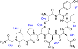
Formula C46H65N15O12S2
Molar mass 1084.24 g·mol−1
3D model (JSmol)
Density 1.6±0.1 g/cm3
  • c1ccc(cc1)C[C@H]2C(=O)N[C@H](C(=O)N[C@H](C(=O)N[C@@H](CSSC[C@@H](C(=O)N[C@H](C(=O)N2)Cc3ccc(cc3)O)N)C(=O)N4CCC[C@H]4C(=O)N[C@@H](CCCN=C(N)N)C(=O)NCC(=O)N)CC(=O)N)CCC(=O)N
  • InChI=1S/C46H65N15O12S2 /c47-27-22-74-75-23-33(45(73)61-17-5-9-34(61)44(72)56-28(8-4-16-53-46(51)52)39(67)54-21-37(50)65)60-43(71)32(20-36(49)64)59-40(68)29(14-15-35(48)63)55-41(69)31(18-24-6-2-1-3-7-24)58-42(70)30(57-38(27)66)19-25-10-12-26(62)13-11-25/h1-3,6-7,10-13,27-34,62H,4-5,8-9,14-23,47H2,(H2,48,63)(H2,49,64)(H2,50,65)(H,54,67)(H,55,69)(H,56,72)(H,57,66)(H,58,70)(H,59,68)(H,60,71)(H4,51,52,53)/t27-,28-,29-,30-,31-,32-,33-,34-/m0/s1 Yes check.svgY
  • Key:KBZOIRJILGZLEJ-LGYYRGKSSA-N Yes check.svgY
AVP
Arginine vasopressin3d.png
Identifiers
Aliases AVP , ADH, ARVP, AVP-NPII, AVRP, VP, arginine vasopressin, Vasopressin
External IDs OMIM: 192340 MGI: 88121 HomoloGene: 417 GeneCards: AVP
Orthologs
SpeciesHumanMouse
Entrez
Ensembl
UniProt
RefSeq (mRNA)

NM_000490

NM_009732

RefSeq (protein)

NP_000481

NP_033862

Location (UCSC) Chr 20: 3.08 – 3.08 Mb Chr 2: 130.42 – 130.42 Mb
PubMed search [3] [4]
Wikidata
View/Edit Human View/Edit Mouse

Human vasopressin, also called antidiuretic hormone (ADH), arginine vasopressin (AVP) or argipressin, [5] is a hormone synthesized from the AVP gene as a peptide prohormone in neurons in the hypothalamus, [6] and is converted to AVP. It then travels down the axon terminating in the posterior pituitary, and is released from vesicles into the circulation in response to extracellular fluid hypertonicity (hyperosmolality). AVP has two primary functions. First, it increases the amount of solute-free water reabsorbed back into the circulation from the filtrate in the kidney tubules of the nephrons. Second, AVP constricts arterioles, which increases peripheral vascular resistance and raises arterial blood pressure. [7] [8] [9]

Contents

A third function is possible. Some AVP may be released directly into the brain from the hypothalamus, and may play an important role in social behavior, sexual motivation and pair bonding, and maternal responses to stress. [10]

Vasopressin induces differentiation of stem cells into cardiomyocytes and promotes heart muscle homeostasis. [11]

It has a very short half-life, between 16 and 24 minutes. [9]

Physiology

Function

Vasopressin regulates the tonicity of body fluids. It is released from the posterior pituitary in response to hypertonicity and causes the kidneys to reabsorb solute-free water and return it to the circulation from the tubules of the nephron, thus returning the tonicity of the body fluids toward normal. An incidental consequence of this renal reabsorption of water is concentrated urine and reduced urine volume. AVP released in high concentrations may also raise blood pressure by inducing moderate vasoconstriction. [12]

AVP also may have a variety of neurological effects on the brain. It may influence pair-bonding in voles. The high-density distributions of vasopressin receptor AVPr1a in prairie vole ventral forebrain regions have been shown to facilitate and coordinate reward circuits during partner preference formation, critical for pair bond formation. [13]

A very similar substance, lysine vasopressin (LVP) or lypressin, has the same function in pigs and its synthetic version was used in human AVP deficiency, although it has been largely replaced by desmopressin. [14]

Kidney

Vasopressin has three main effects which are:

  1. Increasing the water permeability of distal convoluted tubule (DCT) and cortical collecting tubules (CCT), as well as outer and inner medullary collecting duct (OMCD & IMCD) in the kidney, thus allowing water reabsorption and excretion of more concentrated urine, i.e., antidiuresis. This occurs through increased transcription and insertion of water channels (Aquaporin-2) into the apical membrane of collecting tubule and collecting duct epithelial cells. [15] Aquaporins allow water to move down their osmotic gradient and out of the nephron, increasing the amount of water re-absorbed from the filtrate (forming urine) back into the bloodstream. This effect is mediated by V2 receptors. Vasopressin also increases the concentration of calcium in the collecting duct cells, by episodic release from intracellular stores. Vasopressin, acting through cAMP, also increases transcription of the aquaporin-2 gene, thus increasing the total number of aquaporin-2 molecules in collecting duct cells. [16]
  2. Increasing permeability of the inner medullary portion of the collecting duct to urea by regulating the cell surface expression of urea transporters, [17] which facilitates its reabsorption into the medullary interstitium as it travels down the concentration gradient created by removing water from the connecting tubule, cortical collecting duct, and outer medullary collecting duct.
  3. Acute increase of sodium absorption across the ascending loop of Henle. This adds to the countercurrent multiplication which aids in proper water reabsorption later in the distal tubule and collecting duct. [18]

Central nervous system

Vasopressin released within the brain may have several actions:

  • Vasopressin is released into the brain in a circadian rhythm by neurons of the suprachiasmatic nucleus. [19]
  • Vasopressin released from posterior pituitary is associated with nausea. [20]
  • Recent evidence suggests that vasopressin may have analgesic effects. The analgesia effects of vasopressin were found to be dependent on both stress and sex. [21]

Regulation

Gene regulation

Vasopressin is regulated by AVP gene expression which is managed by major clock controlled genes. In this circadian circuit known as the transcription-translation feedback loop (TTFL), Per2 protein accumulates and is phosphorylated by CK1E. Per2 subsequently inhibits the transcription factors Clock and BMAL1 in order to reduce Per2 protein levels in the cell. [22] At the same time, Per2 also inhibits the transcription factors for the AVP gene in order to regulate its expression, the expression of vasopressin, and other AVP gene products. [23]

Many factors influence the secretion of vasopressin:

  • Ethanol (alcohol) reduces the calcium-dependent secretion of AVP by blocking voltage-gated calcium channels in neurohypophyseal nerve terminals in rats. [24]
  • Angiotensin II stimulates AVP secretion, in keeping with its general pressor and pro-volumic effects on the body. [25]
  • Atrial natriuretic peptide inhibits AVP secretion, in part by inhibiting Angiotensin II-induced stimulation of AVP secretion. [25]
  • Cortisol inhibits secretion of antidiuretic hormone. [26]

Production and secretion

The physiological stimulus for secretion of vasopressin is increased osmolality of the plasma, monitored by the hypothalamus. A decreased arterial blood volume, (such as can occur in cirrhosis, nephrosis, and heart failure), stimulates secretion, even in the face of decreased osmolality of the plasma: it supersedes osmolality, but with a milder effect. In other words, vasopressin secretion is also stimulated in the presence of hypoosmolality (hyponatremia) when the arterial blood volume is low by the unloading of baroreceptors. [27]

The AVP that is measured in peripheral blood is almost all derived from secretion from the posterior pituitary gland (except in cases of AVP-secreting tumours). Vasopressin is produced by magnocellular neurosecretory neurons in the paraventricular nucleus of hypothalamus (PVN) and supraoptic nucleus (SON). It then travels down the axon through the infundibulum within neurosecretory granules that are found within Herring bodies, localized swellings of the axons and nerve terminals. These carry the peptide directly to the posterior pituitary gland, where it is stored until released into the blood.

There are other sources of AVP, beyond the hypothalamic magnocellular neurons. For example, AVP is also synthesized by parvocellular neurosecretory neurons of the PVN, transported and released at the median eminence, from which it travels through the hypophyseal portal system to the anterior pituitary, where it stimulates corticotropic cells synergistically with CRH to produce ACTH (by itself it is a weak secretagogue). [28]

Vasopressin during surgery and anaesthesia

Vasopressin concentration is used to measure surgical stress for evaluation of surgical techniques. Plasma vasopressin concentration is elevated by noxious stimuli, [29] [30] predominantly during abdominal surgery, [31] [32] [33] especially at gut manipulation, traction of viscera, [34] [35] [36] as well as abdominal insufflation with carbon dioxide during laparoscopic surgery. [37] [38]

Receptors

Types of AVP receptors and their actions:

Type Second messenger system LocationsActionsAgonistsAntagonists
AVPR1A Phosphatidylinositol/calcium Liver, kidney, peripheral vasculature, brain Vasoconstriction, glycogen breakdown, [39] platelet aggregation, and release of factor VIII and von Willebrand factor; social recognition, [40] circadian tau [41] Felypressin
AVPR1B or AVPR3 Phosphatidylinositol/calcium Pituitary gland, brain Adrenocorticotropic hormone secretion in response to stress; [42] social interpretation of olfactory cues [43]
AVPR2 Adenylate cyclase/cAMP Basolateral membrane of the cells lining the collecting ducts of the kidneys (especially the cortical and outer medullary collecting ducts)Insertion of aquaporin-2 (AQP2) channels (water channels). This allows water to be reabsorbed down an osmotic gradient, and so the urine is more concentrated. Release of von Willebrand factor and surface expression of P-selectin through exocytosis of Weibel-Palade bodies from endothelial cells [44] [45] AVP, desmopressin "-vaptan" diuretics, i.e. tolvaptan

Structure and relation to oxytocin

Chemical structure of the arginine vasopressin (argipressin) with an arginine at the 8th amino acid position. Lysine vasopressin differs only in having a lysine in this position. Vasopressin labeled.png
Chemical structure of the arginine vasopressin (argipressin) with an arginine at the 8th amino acid position. Lysine vasopressin differs only in having a lysine in this position.
Chemical structure of oxytocin. Differs from AVP at only the 3rd and 8th position. Oxytocin with labels.png
Chemical structure of oxytocin. Differs from AVP at only the 3rd and 8th position.

The vasopressins are peptides consisting of nine amino acids (nonapeptides). The amino acid sequence of arginine vasopressin (argipressin) is Cys-Tyr-Phe-Gln-Asn-Cys-Pro-Arg-Gly-NH2, with the cysteine residues forming a disulfide bond and the C-terminus of the sequence converted to a primary amide. [46] Lysine vasopressin (lypressin) has a lysine in place of the arginine as the eighth amino acid, and is found in pigs and some related animals, whereas arginine vasopressin is found in humans. [47]

The structure of oxytocin is very similar to that of the vasopressins: It is also a nonapeptide with a disulfide bridge and its amino acid sequence differs at only two positions. The two genes are located on the same chromosome separated by a relatively small distance of less than 15,000 bases in most species. The magnocellular neurons that secrete vasopressin are adjacent to magnocellular neurons that secrete oxytocin, and are similar in many respects. The similarity of the two peptides can cause some cross-reactions: oxytocin has a slight antidiuretic function, and high levels of AVP can cause uterine contractions. [48] [49]

Comparison of vasopressin and oxytocin neuropeptide families:

Vertebrate Vasopressin Family
Cys-Tyr-Phe-Gln-Asn-Cys-Pro-Arg-Gly-NH2 Argipressin (AVP, ADH)Most mammals
Cys-Tyr-Phe-Gln-Asn-Cys-Pro-Lys-Gly-NH2 Lypressin (LVP) Pigs, hippos, warthogs, some marsupials
Cys-Phe-Phe-Gln-Asn-Cys-Pro-Arg-Gly-NH2 Phenypressin Some marsupials
Cys-Tyr-Ile-Gln-Asn-Cys-Pro-Arg-Gly-NH2 VasotocinNon-mammals
Vertebrate Oxytocin Family
Cys-Tyr-Ile-Gln-Asn-Cys-Pro-Leu-Gly-NH2 Oxytocin (OXT)Most mammals, ratfish
Cys-Tyr-Ile-Gln-Asn-Cys-Pro-Pro-Gly-NH2Prol-Oxytocin Some New World monkeys, northern tree shrews
Cys-Tyr-Ile-Gln-Asn-Cys-Pro-Ile-Gly-NH2 Mesotocin Most marsupials, all birds, reptiles, amphibians, lungfishes, coelacanths
Cys-Tyr-Ile-Gln-Ser-Cys-Pro-Ile-Gly-NH2 Seritocin Frogs
Cys-Tyr-Ile-Ser-Asn-Cys-Pro-Ile-Gly-NH2 Isotocin Bony fishes
Cys-Tyr-Ile-Ser-Asn-Cys-Pro-Gln-Gly-NH2 Glumitocin skates
Cys-Tyr-Ile-Asn/Gln-Asn-Cys-Pro-Leu/Val-Gly-NH2Various tocins Sharks
Invertebrate VP/OT Superfamily
Cys-Leu-Ile-Thr-Asn-Cys-Pro-Arg-Gly-NH2 Inotocin Locust
Cys-Phe-Val-Arg-Asn-Cys-Pro-Thr-Gly-NH2 Annetocin Earthworm
Cys-Phe-Ile-Arg-Asn-Cys-Pro-Lys-Gly-NH2 Lys-Connopressin Geography & imperial cone snail, pond snail, sea hare, leech
Cys-Ile-Ile-Arg-Asn-Cys-Pro-Arg-Gly-NH2 Arg-Connopressin Striped cone snail
Cys-Tyr-Phe-Arg-Asn-Cys-Pro-Ile-Gly-NH2 Cephalotocin Octopus
Cys-Phe-Trp-Thr-Ser-Cys-Pro-Ile-Gly-NH2 Octopressin Octopus
†Vasotocin is the evolutionary progenitor of all the vertebrate neurohypophysial hormones. [50]

Medical use

Vasopressin is used to manage anti-diuretic hormone deficiency. Vasopressin is used to treat diabetes insipidus related to low levels of antidiuretic hormone. It is available as Pressyn. [51]

Vasopressin has off-label uses and is used in the treatment of vasodilatory shock, gastrointestinal bleeding, ventricular tachycardia and ventricular fibrillation.

Vasopressin agonists are used therapeutically in various conditions, and its long-acting synthetic analogue desmopressin is used in conditions featuring low vasopressin secretion, as well as for control of bleeding (in some forms of von Willebrand disease and in mild haemophilia A) and in extreme cases of bedwetting by children. Terlipressin and related analogues are used as vasoconstrictors in certain conditions. Use of vasopressin analogues for esophageal varices commenced in 1970. [52]

Vasopressin infusions are also used as second line therapy for septic shock patients not responding to fluid resuscitation or infusions of catecholamines (e.g., dopamine or norepinephrine) to increase the blood pressure while sparing the use of catecholamines. These argipressins have much shorter elimination half-life (around 20 minutes) comparing to synthetic non-arginine vasopresines with much longer elimination half-life of many hours. Further, argipressins act on V1a, V1b, and V2 receptors which consequently lead to higher eGFR and lower vascular resistance in the lungs. A number of injectable arginine vasopressins are currently in clinical use in the United States and in Europe.

Pharmacokinetics

Vasopressin is administered through an intravenous device, intramuscular injection or a subcutaneous injection. The duration of action depends on the mode of administration and ranges from thirty minutes to two hours. It has a half life of ten to twenty minutes. It is widely distributed throughout the body and remains in the extracellular fluid. It is degraded by the liver and excreted through the kidneys. [51] Arginin vasopressins for use in septic shock are intended for intravenous use only.

Side effects

The most common side effects during treatment with vasopressin are dizziness, angina, chest pain, abdominal cramps, heartburn, nausea, vomiting, trembling, fever, water intoxication, pounding sensation in the head, diarrhoea, sweating, paleness, and flatulence. The most severe adverse reactions are myocardial infarction and hypersensitivity. [51]

Contraindications

The use of lysine vasopressin is contraindicated in the presence of hypersensitivity to beef or pork proteins, increased BUN and chronic kidney failure. It is recommended that it be cautiously used in instances of perioperative polyuria, sensitivity to the drug, asthma, seizures, heart failure, a comatose state, migraine headaches, and cardiovascular disease. [51]

Interactions

Deficiency

Decreased AVP release (neurogenic — i.e. due to alcohol intoxication or tumour) or decreased renal sensitivity to AVP (nephrogenic, i.e. by mutation of V2 receptor or AQP) leads to diabetes insipidus, a condition featuring hypernatremia (increased blood sodium concentration), polyuria (excess urine production), and polydipsia (thirst).

Excess

Syndrome of Inappropriate Antidiuretic Hormone secretion (SIADH) in turn can be caused by a number of problems. Some forms of cancer can cause SIADH, particularly small cell lung carcinoma but also a number of other tumors. A variety of diseases affecting the brain or the lung (infections, bleeding) can be the driver behind SIADH. A number of drugs have been associated with SIADH, such as certain antidepressants (serotonin reuptake inhibitors and tricyclic antidepressants), the anticonvulsant carbamazepine, oxytocin (used to induce and stimulate labor), and the chemotherapy drug vincristine. It has also been associated with fluoroquinolones (including ciprofloxacin and moxifloxacin). [9] Finally, it can occur without a clear explanation. [53] Hyponatremia can be treated pharmaceutically through the use of vasopressin receptor antagonists. [53]

History

Vasopressin was elucidated and synthesized for the first time by Vincent du Vigneaud.

Animal studies

Evidence for an effect of AVP on monogamy vs polygamy comes from experimental studies in several species, which indicate that the precise distribution of vasopressin and vasopressin receptors in the brain is associated with species-typical patterns of social behavior. In particular, there are consistent differences between monogamous species and polygamous species in the distribution of AVP receptors, and sometimes in the distribution of vasopressin-containing axons, even when closely related species are compared. [54]

Human studies

Vasopressin has shown nootropic effects on pain perception and cognitive function. [55] Vasopressin also plays a role in autism, major depressive disorder, bipolar disorder, and schizophrenia. [56]

See also

Related Research Articles

<span class="mw-page-title-main">Diabetes insipidus</span> Condition characterized by large amounts of dilute urine and increased thirst

Diabetes insipidus (DI), alternately called arginine vasopressin deficiency (AVP-D) or arginine vasopressin resistance (AVP-R), is a condition characterized by large amounts of dilute urine and increased thirst. The amount of urine produced can be nearly 20 liters per day. Reduction of fluid has little effect on the concentration of the urine. Complications may include dehydration or seizures.

<span class="mw-page-title-main">Pituitary gland</span> Endocrine gland at the base of the brain

The pituitary gland is an endocrine gland in vertebrates. In humans, the pituitary gland is located at the base of the brain, protruding off the bottom of the hypothalamus. The human pituitary gland is oval shaped, about the size of a chickpea, and weighs 0.5 grams (0.018 oz) on average.

<span class="mw-page-title-main">Hypothalamus</span> Area of the brain below the thalamus

The hypothalamus is a small part of the brain that contains a number of nuclei with a variety of functions. One of the most important functions is to link the nervous system to the endocrine system via the pituitary gland. The hypothalamus is located below the thalamus and is part of the limbic system. It forms the ventral part of the diencephalon. All vertebrate brains contain a hypothalamus. In humans, it is the size of an almond.

<span class="mw-page-title-main">Secretin</span> Hormone involved in stomach, pancreas and liver secretions

Secretin is a hormone that regulates water homeostasis throughout the body and influences the environment of the duodenum by regulating secretions in the stomach, pancreas, and liver. It is a peptide hormone produced in the S cells of the duodenum, which are located in the intestinal glands. In humans, the secretin peptide is encoded by the SCT gene.

<span class="mw-page-title-main">Oxytocin</span> Peptide hormone and neuropeptide

Oxytocin is a peptide hormone and neuropeptide normally produced in the hypothalamus and released by the posterior pituitary. Present in animals since early stages of evolution, in humans it plays roles in behavior that include social bonding, reproduction, childbirth, and the period after childbirth. Oxytocin is released into the bloodstream as a hormone in response to sexual activity and during labour. It is also available in pharmaceutical form. In either form, oxytocin stimulates uterine contractions to speed up the process of childbirth. In its natural form, it also plays a role in maternal bonding and milk production. Production and secretion of oxytocin is controlled by a positive feedback mechanism, where its initial release stimulates production and release of further oxytocin. For example, when oxytocin is released during a contraction of the uterus at the start of childbirth, this stimulates production and release of more oxytocin and an increase in the intensity and frequency of contractions. This process compounds in intensity and frequency and continues until the triggering activity ceases. A similar process takes place during lactation and during sexual activity.

<span class="mw-page-title-main">Posterior pituitary</span> Posterior lobe of the pituitary gland

The posterior pituitary is the posterior lobe of the pituitary gland which is part of the endocrine system. The posterior pituitary is not glandular as is the anterior pituitary. Instead, it is largely a collection of axonal projections from the hypothalamus that terminate behind the anterior pituitary, and serve as a site for the secretion of neurohypophysial hormones directly into the blood. The hypothalamic–neurohypophyseal system is composed of the hypothalamus, posterior pituitary, and these axonal projections.

<span class="mw-page-title-main">Supraoptic nucleus</span> ADH secreting nucleus of the hypothalamus.

The supraoptic nucleus (SON) is a nucleus of magnocellular neurosecretory cells in the hypothalamus of the mammalian brain. The nucleus is situated at the base of the brain, adjacent to the optic chiasm. In humans, the SON contains about 3,000 neurons.

<span class="mw-page-title-main">Paraventricular nucleus of hypothalamus</span>

The paraventricular nucleus is a nucleus in the hypothalamus. Anatomically, it is adjacent to the third ventricle and many of its neurons project to the posterior pituitary. These projecting neurons secrete oxytocin and a smaller amount of vasopressin, otherwise the nucleus also secretes corticotropin-releasing hormone (CRH) and thyrotropin-releasing hormone (TRH). CRH and TRH are secreted into the hypophyseal portal system and act on different targets neurons in the anterior pituitary. PVN is thought to mediate many diverse functions through these different hormones, including osmoregulation, appetite, and the response of the body to stress.

Magnocellular neurosecretory cells are large neuroendocrine cells within the supraoptic nucleus and paraventricular nucleus of the hypothalamus. They are also found in smaller numbers in accessory cell groups between these two nuclei, the largest one being the circular nucleus. There are two types of magnocellular neurosecretory cells, oxytocin-producing cells and vasopressin-producing cells, but a small number can produce both hormones. These cells are neuroendocrine neurons, are electrically excitable, and generate action potentials in response to afferent stimulation. Vasopressin is produced from the vasopressin-producing cells via the AVP gene, a molecular output of circadian pathways.

<span class="mw-page-title-main">Desmopressin</span> Medication

Desmopressin, sold under the trade name DDAVP among others, is a medication used to treat diabetes insipidus, bedwetting, hemophilia A, von Willebrand disease, and high blood urea levels. In hemophilia A and von Willebrand disease, it should only be used for mild to moderate cases. It may be given in the nose, by injection into a vein, by mouth, or under the tongue.

The syndrome of inappropriate antidiuretic hormone secretion (SIADH), also known as the syndrome of inappropriate antidiuresis (SIAD), is characterized by a physiologically inappropriate release of antidiuretic hormone (ADH) either from the posterior pituitary gland, or an abnormal non-pituitary source. Unsuppressed ADH causes a physiologically inappropriate increase in solute-free water being reabsorbed by the tubules of the kidney to the venous circulation leading to hypotonic hyponatremia.

Neuroendocrinology is the branch of biology which studies the interaction between the nervous system and the endocrine system; i.e. how the brain regulates the hormonal activity in the body. The nervous and endocrine systems often act together in a process called neuroendocrine integration, to regulate the physiological processes of the human body. Neuroendocrinology arose from the recognition that the brain, especially the hypothalamus, controls secretion of pituitary gland hormones, and has subsequently expanded to investigate numerous interconnections of the endocrine and nervous systems.

<span class="mw-page-title-main">Vasopressin receptor 1B</span> Protein-coding gene in the species Homo sapiens

Vasopressin V1b receptor (V1BR) also known as vasopressin 3 receptor (VPR3) or antidiuretic hormone receptor 1B is a protein that in humans is encoded by the AVPR1B gene.

<span class="mw-page-title-main">Vasopressin receptor 2</span> Protein-coding gene in the species Homo sapiens

Vasopressin receptor 2 (V2R), or arginine vasopressin receptor 2, is a protein that acts as receptor for vasopressin. AVPR2 belongs to the subfamily of G-protein-coupled receptors. Its activity is mediated by the Gs type of G proteins, which stimulate adenylate cyclase.

The actions of vasopressin are mediated by stimulation of tissue-specific G protein-coupled receptors (GPCRs) called vasopressin receptors that are classified into the V1 (V1A), V2, and V3 (V1B) receptor subtypes. These three subtypes differ in localization, function and signal transduction mechanisms.

<span class="mw-page-title-main">Vasotocin</span> Chemical compound

Vasotocin is an oligopeptide homologous to oxytocin and vasopressin found in all non-mammalian vertebrates and possibly in mammals during the fetal stage of development. Arginine vasotocin (AVT), a hormone produced by neurosecretory cells within the posterior pituitary gland (neurohypophysis) of the brain, is a major endocrine regulator of water balance and osmotic homoeostasis and is involved in social and sexual behavior in non-mammalian vertebrates. In mammals, it appears to have biological properties similar to those of oxytocin and vasopressin. It has been found to have effects on the regulation of REM sleep. Evidence for the existence of endogenous vasotocin in mammals is limited and no mammalian gene encoding vasotocin has been confirmed.

A vasopressin receptor antagonist (VRA) is an agent that interferes with action at the vasopressin receptors. Most commonly VRAs are used in the treatment of hyponatremia, especially in patients with congestive heart failure, liver cirrhosis or SIADH.

<span class="mw-page-title-main">Neurohypophysial hormone</span>

The neurohypophysial hormones form a family of structurally and functionally related peptide hormones. Their representatives in humans are oxytocin and vasopressin. They are named after the location of their release into the blood, the neurohypophysis.

Copeptin is a 39-amino acid-long peptide derived from the C-terminus of pre-pro-hormone of arginine vasopressin, neurophysin II and copeptin. Arginine vasopressin (AVP), also known as the antidiuretic hormone (ADH), is encoded by the AVP gene and is involved in multiple cardiovascular and renal pathways and abnormal level of AVP are associated with various diseases. Hence measurement of AVP would be useful, but is not commonly carried out in clinical practice because of its very short half-life making it difficult to quantify. In contrast, copeptin can be immunologically tested with ease and therefore can be used as a vasopressin surrogate marker.

<span class="mw-page-title-main">AVP gene</span> Gene

The arginine vasopressin gene (AVP) is a gene whose product is proteolytically cleaved to produce vasopressin, neurophysin II, and a glycoprotein called copeptin. AVP and other AVP-like peptides are found in mammals, as well as mollusks, arthropods, nematodes, and other invertebrate species. In humans, AVP is present on chromosome 20 and plays a role in homeostatic regulation. The products of AVP have many functions that include vasoconstriction, regulating the balance of water in the body, and regulating responses to stress. Expression of AVP is regulated by the transcription translation feedback loop (TTFL), which is an important part of the circadian system that controls the expression of clock genes. AVP has important implications in the medical field as its products have significant roles throughout body.

References

  1. 1 2 3 GRCh38: Ensembl release 89: ENSG00000101200 - Ensembl, May 2017
  2. 1 2 3 GRCm38: Ensembl release 89: ENSMUSG00000037727 - Ensembl, May 2017
  3. "Human PubMed Reference:". National Center for Biotechnology Information, U.S. National Library of Medicine.
  4. "Mouse PubMed Reference:". National Center for Biotechnology Information, U.S. National Library of Medicine.
  5. Anderson DA (2012). Dorland's Illustrated Medical Dictionary (32nd ed.). Elsevier. ISBN   978-1-4160-6257-8.
  6. Sukhov RR, Walker LC, Rance NE, Price DL, Young WS 3rd (1993). "Vasopressin and oxytocin gene expression in the human hypothalamus". Journal of Comparative Neurology. 337 (2): 295–306. doi:10.1002/cne.903370210. PMC   9883978 . PMID   8277003. S2CID   35174328.
  7. Marieb E (2014). Anatomy & physiology. Glenview, IL: Pearson Education, Inc. ISBN   978-0-321-86158-0.
  8. Caldwell HK, Young WS III (2006). "Oxytocin and Vasopressin: Genetics and Behavioral Implications" (PDF). In Lajtha A, Lim R (eds.). Handbook of Neurochemistry and Molecular Neurobiology: Neuroactive Proteins and Peptides (3rd ed.). Berlin: Springer. pp. 573–607. ISBN   978-0-387-30348-2.
  9. 1 2 3 Babar SM (October 2013). "SIADH associated with ciprofloxacin". The Annals of Pharmacotherapy. 47 (10): 1359–63. doi:10.1177/1060028013502457. PMID   24259701. S2CID   36759747.
  10. Insel TR (March 2010). "The challenge of translation in social neuroscience: a review of oxytocin, vasopressin, and affiliative behavior". Neuron. 65 (6): 768–79. doi:10.1016/j.neuron.2010.03.005. PMC   2847497 . PMID   20346754.
  11. Costa A, Rossi E, Scicchitano BM, Coletti D, Moresi V, Adamo S (September 2014). "Neurohypophyseal Hormones: Novel Actors of Striated Muscle Development and Homeostasis". review. European Journal of Translational Myology. 24 (3): 3790. doi:10.4081/bam.2014.3.217. PMC   4756744 . PMID   26913138.
  12. Cuzzo B, Padala SA, Lappin SL (2024). "Physiology, Vasopressin". StatPearls. StatPearls Publishing. PMID   30252325.
  13. Lim MM, Young LJ (2004). "Vasopressin-dependent neural circuits underlying pair bond formation in the monogamous prairie vole". Neuroscience. 125 (1): 35–45. doi:10.1016/j.neuroscience.2003.12.008. PMID   15051143. S2CID   16210017.
  14. Chapman IM, Professor of Medicine, Discipline of Medicine, University of Adelaide, Royal Adelaide Hospital. "Central Diabetes Insipidus". MSD. Merck & Co. Inc.
  15. Boron WR, Boulpaep EL (2016-05-05). Medical Physiology (Third ed.). Philadelphia, PA: Elsevier. ISBN   978-1-4557-4377-3. OCLC   951680737.
  16. Wilson JL, Miranda CA, Knepper MA (2013). "Vasopressin and the Regulation of Aquaporin-2". Clinical and Experimental Nephrology. 17 (6): 10.1007/s10157-013–0789-5. doi:10.1007/s10157-013-0789-5. PMC   3775849 . PMID   23584881.
  17. Sands JM, Blount MA, Klein JD (2011). "Regulation of renal urea transport by vasopressin". Transactions of the American Clinical and Climatological Association. 122: 82–92. PMC   3116377 . PMID   21686211.
  18. Knepper MA, Kim GH, Fernández-Llama P, Ecelbarger CA (March 1999). "Regulation of thick ascending limb transport by vasopressin". Journal of the American Society of Nephrology. 10 (3): 628–34. doi: 10.1681/ASN.V103628 . PMID   10073614.
  19. Forsling ML, Montgomery H, Halpin D, Windle RJ, Treacher DF (May 1998). "Daily patterns of secretion of neurohypophysial hormones in man: effect of age". Experimental Physiology. 83 (3): 409–18. doi: 10.1113/expphysiol.1998.sp004124 . PMID   9639350. S2CID   2295415.
  20. Magtanong E (2017). "What Is Nausea? A Historical Analysis of Changing Views". Auton Neurosci. 202: 5–17. doi:10.1016/j.autneu.2016.07.003. PMC   5203950 . PMID   27450627.
  21. Wiltshire T, Maixner W, Diatchenko L (December 2011). "Relax, you won't feel the pain". Nature Neuroscience. 14 (12): 1496–7. doi:10.1038/nn.2987. PMID   22119947. S2CID   205434100.
  22. Dunlap JC (January 1999). "Molecular bases for circadian clocks". Cell. 96 (2): 271–90. doi: 10.1016/s0092-8674(00)80566-8 . PMID   9988221. S2CID   14991100.
  23. Jin X, Shearman LP, Weaver DR, Zylka MJ, de Vries GJ, Reppert SM (January 1999). "A molecular mechanism regulating rhythmic output from the suprachiasmatic circadian clock". Cell. 96 (1): 57–68. doi: 10.1016/s0092-8674(00)80959-9 . PMID   9989497. S2CID   6916996.
  24. Wang XM, Dayanithi G, Lemos JR, Nordmann JJ, Treistman SN (November 1991). "Calcium currents and peptide release from neurohypophysial terminals are inhibited by ethanol". The Journal of Pharmacology and Experimental Therapeutics. 259 (2): 705–11. PMID   1941619.
  25. 1 2 Matsukawa T, Miyamoto T (March 2011). "Angiotensin II-stimulated secretion of arginine vasopressin is inhibited by atrial natriuretic peptide in humans". American Journal of Physiology. Regulatory, Integrative and Comparative Physiology. 300 (3): R624–9. doi:10.1152/ajpregu.00324.2010. PMID   21123762.
  26. Collège des enseignants d'endocrinologie, diabète et maladie (2012-01-30). Endocrinologie, diabétologie et maladies métaboliques. Elsevier Masson. ISBN   978-2-294-72233-2.
  27. Garrahy A, Thompston CJ (2019). "General Principles, Diabetes, Metabolism, Obesity, Gastrointestinal Hormones, Aging, Endocrine Toxicology". Encyclopedia of Endocrine Diseases. 1 (2): 969–974.
  28. Salata RA, Jarrett DB, Verbalis JG, Robinson AG (March 1988). "Vasopressin stimulation of adrenocorticotropin hormone (ACTH) in humans. In vivo bioassay of corticotropin-releasing factor (CRF) which provides evidence for CRF mediation of the diurnal rhythm of ACTH". The Journal of Clinical Investigation. 81 (3): 766–74. doi:10.1172/JCI113382. PMC   442524 . PMID   2830315.
  29. Day TA, Sibbald JR (June 1990). "Noxious somatic stimuli excite neurosecretory vasopressin cells via A1 cell group". The American Journal of Physiology. 258 (6 Pt 2): R1516-20. doi:10.1152/ajpregu.1990.258.6.R1516. PMID   2360697.
  30. Höglund OV, Hagman R, Olsson K, Olsson U, Lagerstedt AS (October 2014). "Intraoperative changes in blood pressure, heart rate, plasma vasopressin, and urinary noradrenalin during elective ovariohysterectomy in dogs: repeatability at removal of the 1st and 2nd ovary". Veterinary Surgery. 43 (7): 852–9. doi:10.1111/j.1532-950X.2014.12264.x. PMID   25130060.
  31. Goldmann A, Hoehne C, Fritz GA, Unger J, Ahlers O, Nachtigall I, et al. (September 2008). "Combined vs. Isoflurane/Fentanyl anesthesia for major abdominal surgery: Effects on hormones and hemodynamics". Medical Science Monitor. 14 (9): CR445-52. PMID   18758414.
  32. Furuya K, Shimizu R, Hirabayashi Y, Ishii R, Fukuda H (May 1993). "Stress hormone responses to major intra-abdominal surgery during and immediately after sevoflurane-nitrous oxide anaesthesia in elderly patients". Canadian Journal of Anaesthesia. 40 (5 Pt 1): 435–9. doi: 10.1007/BF03009513 . PMID   8390330.
  33. Haas M, Glick SM (May 1978). "Radioimmunoassayable plasma vasopressin associated with surgery". Archives of Surgery. 113 (5): 597–600. doi:10.1001/archsurg.1978.01370170059011. PMID   646620.
  34. Nussey SS, Page SR, Ang VT, Jenkins JS (March 1988). "The response of plasma oxytocin to surgical stress". Clinical Endocrinology. 28 (3): 277–82. doi:10.1111/j.1365-2265.1988.tb01213.x. PMID   3168310. S2CID   37668345.
  35. Melville RJ, Forsling ML, Frizis HI, LeQuesne LP (December 1985). "Stimulus for vasopressin release during elective intra-abdominal operations". The British Journal of Surgery. 72 (12): 979–82. doi:10.1002/bjs.1800721215. PMID   4084755. S2CID   43764321.
  36. Moran WH, Miltenberger FW, Shuayb WA, Zimmermann B (July 1964). "The Relationship of Antidiuretic Hormone Secretion to Surgical Stress". Surgery. 56: 99–108. PMID   14175989.
  37. Gutt CN, Oniu T, Mehrabi A, Schemmer P, Kashfi A, Kraus T, et al. (2004). "Circulatory and respiratory complications of carbon dioxide insufflation". Digestive Surgery. 21 (2): 95–105. doi: 10.1159/000077038 . PMID   15010588. S2CID   3369276. ProQuest   223606053.
  38. Nguyen NT, Wolfe BM (February 2005). "The physiologic effects of pneumoperitoneum in the morbidly obese". Annals of Surgery. 241 (2): 219–226. doi:10.1097/01.sla.0000151791.93571.70. PMC   1356906 . PMID   15650630.
  39. Alberts B, Bray D, Hopkin K, Johnson AD, Lewis J, Roberts K (2014). Essential Cell Biology. Garland Science. ISBN   978-0-8153-4455-1.
  40. Bielsky IF, Hu SB, Szegda KL, Westphal H, Young LJ (March 2004). "Profound impairment in social recognition and reduction in anxiety-like behavior in vasopressin V1a receptor knockout mice". Neuropsychopharmacology. 29 (3): 483–93. doi: 10.1038/sj.npp.1300360 . PMID   14647484.
  41. Wersinger SR, Caldwell HK, Martinez L, Gold P, Hu SB, Young WS (August 2007). "Vasopressin 1a receptor knockout mice have a subtle olfactory deficit but normal aggression". Genes, Brain and Behavior. 6 (6): 540–51. doi: 10.1111/j.1601-183X.2006.00281.x . PMID   17083331. S2CID   29923520.
  42. Lolait SJ, Stewart LQ, Jessop DS, Young WS, O'Carroll AM (February 2007). "The hypothalamic-pituitary-adrenal axis response to stress in mice lacking functional vasopressin V1b receptors". Endocrinology. 148 (2): 849–56. doi:10.1210/en.2006-1309. PMC   2040022 . PMID   17122081.
  43. Wersinger SR, Kelliher KR, Zufall F, Lolait SJ, O'Carroll AM, Young WS (December 2004). "Social motivation is reduced in vasopressin 1b receptor null mice despite normal performance in an olfactory discrimination task". Hormones and Behavior. 46 (5): 638–45. doi:10.1016/j.yhbeh.2004.07.004. PMID   15555506. S2CID   38444963.
  44. Kanwar S, Woodman RC, Poon MC, Murohara T, Lefer AM, Davenpeck KL, et al. (October 1995). "Desmopressin induces endothelial P-selectin expression and leukocyte rolling in postcapillary venules". Blood. 86 (7): 2760–6. doi: 10.1182/blood.V86.7.2760.2760 . PMID   7545469.
  45. Kaufmann JE, Oksche A, Wollheim CB, Günther G, Rosenthal W, Vischer UM (July 2000). "Vasopressin-induced von Willebrand factor secretion from endothelial cells involves V2 receptors and cAMP". The Journal of Clinical Investigation. 106 (1): 107–16. doi:10.1172/JCI9516. PMC   314363 . PMID   10880054.
  46. Burtis CA, Ashwood ER, Bruns DE (2012). Tietz Textbook of Clinical Chemistry and Molecular Diagnostics (5th ed.). Elsevier Health Sciences. p. 1833. ISBN   978-1-4557-5942-2.
  47. Donaldson D (1994). "Polyuria and Disorders of Thirst". In Williams DL, Marks V (eds.). Scientific Foundations of Biochemistry in Clinical Practice (2nd ed.). Butterworth-Heinemann. pp. 76–102. doi:10.1016/B978-0-7506-0167-2.50010-8. ISBN   978-0-7506-0167-2.
  48. Li C, Wang W, Summer SN, Westfall TD, Brooks DP, Falk S, et al. (February 2008). "Molecular mechanisms of antidiuretic effect of oxytocin". Journal of the American Society of Nephrology. 19 (2): 225–32. doi:10.1681/ASN.2007010029. PMC   2396735 . PMID   18057218.
  49. Joo KW, Jeon US, Kim GH, Park J, Oh YK, Kim YS, et al. (October 2004). "Antidiuretic action of oxytocin is associated with increased urinary excretion of aquaporin-2". Nephrology, Dialysis, Transplantation. 19 (10): 2480–6. doi:10.1093/ndt/gfh413. PMID   15280526.
  50. Acher R, Chauvet J (July 1995). "The neurohypophysial endocrine regulatory cascade: precursors, mediators, receptors, and effectors". Frontiers in Neuroendocrinology. 16 (3): 237–89. doi:10.1006/frne.1995.1009. PMID   7556852. S2CID   12739464.
  51. 1 2 3 4 5 "Vasopressin" (PDF). F.A. Davis Company. 2017. Retrieved 2017-03-13.[ dead link ]
  52. Baum S, Nusbaum M (March 1971). "The control of gastrointestinal hemorrhage by selective mesenteric arterial infusion of vasopressin". Radiology. 98 (3): 497–505. doi:10.1148/98.3.497. PMID   5101576.
  53. 1 2 Verbalis JG, Goldsmith SR, Greenberg A, Schrier RW, Sterns RH (November 2007). "Hyponatremia treatment guidelines 2007: expert panel recommendations". The American Journal of Medicine. 120 (11 Suppl 1): S1–21. CiteSeerX   10.1.1.499.7585 . doi:10.1016/j.amjmed.2007.09.001. PMID   17981159.
  54. Young LJ (October 2009). "The neuroendocrinology of the social brain". Frontiers in Neuroendocrinology. 30 (4): 425–8. doi:10.1016/j.yfrne.2009.06.002. PMID   19596026. S2CID   31960688.
  55. Mavani GP, DeVita MV, Michelis MF (2015). "A review of the nonpressor and nonantidiuretic actions of the hormone vasopressin". Frontiers in Medicine. 2: 19. doi: 10.3389/fmed.2015.00019 . PMC   4371647 . PMID   25853137.
  56. Iovino M, Messana T, De Pergola G, Iovino E, Dicuonzo F, Guastamacchia E, et al. (2018). "The Role of Neurohypophyseal Hormones Vasopressin and Oxytocin in Neuropsychiatric Disorders". Endocrine, Metabolic & Immune Disorders Drug Targets. 18 (4): 341–347. doi:10.2174/1871530318666180220104900. PMID   29468985. S2CID   3465601.

Further reading