Corticotropin-like intermediate peptide

Last updated
pro-opiomelanocortin
Corticotropin-like intermediate peptide.svg
Identifiers
SymbolPOMC
NCBI gene 5443
HGNC 9201
OMIM 176830
RefSeq NM_000939
UniProt P01189
Other data
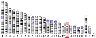
Locus Chr. 2 p23
Corticotropin-like intermediate peptide
Corticotropin-like intermediate peptide.svg
Names
IUPAC name
L-arginyl-L-prolyl-L-valyl-L-lysyl-L-valyl-L-tyrosyl-L-prolyl-L-asparaginyl-L-glycyl-L-alanyl-L-α-glutamyl-L-α-aspartyl-L-α-glutamyl-L-seryl-L-alanyl-L-α-glutamyl-L-alanyl-L-phenylalanyl-L-prolyl-L-leucyl-L-α-glutamyl-L-phenylalanine
Identifiers
3D model (JSmol)
ChemSpider
PubChem CID
  • InChI=1S/C112H165N27O36/c1-56(2)48-72(100(163)125-70(37-41-86(148)149)97(160)133-77(111(174)175)51-63-24-14-11-15-25-63)129-103(166)79-28-20-46-138(79)109(172)75(49-62-22-12-10-13-23-62)131-93(156)61(9)121-95(158)68(35-39-84(144)145)123-92(155)60(8)122-102(165)78(55-140)134-98(161)71(38-42-87(150)151)126-101(164)74(53-88(152)153)128-96(159)69(36-40-85(146)147)124-91(154)59(7)120-83(143)54-119-94(157)73(52-82(115)142)130-104(167)80-29-21-47-139(80)110(173)76(50-64-31-33-65(141)34-32-64)132-107(170)89(57(3)4)135-99(162)67(27-16-17-43-113)127-106(169)90(58(5)6)136-105(168)81-30-19-45-137(81)108(171)66(114)26-18-44-118-112(116)117/h10-15,22-25,31-34,56-61,66-81,89-90,140-141H,16-21,26-30,35-55,113-114H2,1-9H3,(H2,115,142)(H,119,157)(H,120,143)(H,121,158)(H,122,165)(H,123,155)(H,124,154)(H,125,163)(H,126,164)(H,127,169)(H,128,159)(H,129,166)(H,130,167)(H,131,156)(H,132,170)(H,133,160)(H,134,161)(H,135,162)(H,136,168)(H,144,145)(H,146,147)(H,148,149)(H,150,151)(H,152,153)(H,174,175)(H4,116,117,118)/t59-,60-,61-,66-,67-,68-,69-,70-,71-,72-,73-,74-,75-,76-,77-,78-,79-,80-,81-,89-,90-/m0/s1
    Key: ZYDMZKPAPSZILB-WKNDHWIVSA-N
  • Key: ZYDMZKPAPSZILB-UHFFFAOYAN
  • C[C@@H](C(=N[C@@H](CCC(=O)O)C(=N[C@@H](CC(=O)O)C(=N[C@@H](CCC(=O)O)C(=N[C@@H](CO)C(=N[C@@H](C)C(=N[C@@H](CCC(=O)O)C(=N[C@@H](C)C(=N[C@@H](CC1=CC=CC=C1)C(=O)N2CCC[C@H]2C(=N[C@@H](CC(C)C)C(=N[C@@H](CCC(=O)O)C(=N[C@@H](CC3=CC=CC=C3)C(=O)O)O)O)O)O)O)O)O)O)O)O)O)N=C(CN=C([C@H](CC(=N)O)N=C([C@@H]4CCCN4C(=O)[C@H](CC5=CC=C(C=C5)O)N=C([C@H](C(C)C)N=C([C@H](CCCCN)N=C([C@H](C(C)C)N=C([C@@H]6CCCN6C(=O)[C@H](CCCNC(=N)N)N)O)O)O)O)O)O)O
Properties
C112H165N27O36
Molar mass 2465.705 g·mol−1
Except where otherwise noted, data are given for materials in their standard state (at 25 °C [77 °F], 100 kPa).

Corticotropin-like intermediate [lobe] peptide (CLIP), also known as adrenocorticotropic hormone fragment 18-39 (ACTH(18-39)), is a naturally occurring, endogenous neuropeptide with a docosapeptide structure and the amino acid sequence Arg-Pro-Val-Lys-Val-Tyr-Pro-Asn-Gly-Ala-Glu-Asp-Glu-Ser-Ala-Glu-Ala-Phe-Pro-Leu-Glu-Phe. CLIP is generated as a proteolyic cleavage product of adrenocorticotropic hormone (ACTH), [1] [2] which in turn is a cleavage product of proopiomelanocortin (POMC). [3] Its physiological role has been investigated in various tissues, [4] [5] specifically in the central nervous system. [6] [7] [8] [9] [10]

It has been suggested to function as an insulin secretagogue in the pancreas. [11]

Related Research Articles

<span class="mw-page-title-main">Pituitary gland</span> Endocrine gland at the base of the brain

In vertebrate anatomy, the pituitary gland, or hypophysis, is an endocrine gland, about the size of a chickpea and weighing, on average, 0.5 grams (0.018 oz) in humans. It is a protrusion off the bottom of the hypothalamus at the base of the brain. The hypophysis rests upon the hypophyseal fossa of the sphenoid bone in the center of the middle cranial fossa and is surrounded by a small bony cavity covered by a dural fold. The anterior pituitary is a lobe of the gland that regulates several physiological processes including stress, growth, reproduction, and lactation. The intermediate lobe synthesizes and secretes melanocyte-stimulating hormone. The posterior pituitary is a lobe of the gland that is functionally connected to the hypothalamus by the median eminence via a small tube called the pituitary stalk.

<span class="mw-page-title-main">Adrenocorticotropic hormone</span> Pituitary hormone

Adrenocorticotropic hormone is a polypeptide tropic hormone produced by and secreted by the anterior pituitary gland. It is also used as a medication and diagnostic agent. ACTH is an important component of the hypothalamic-pituitary-adrenal axis and is often produced in response to biological stress. Its principal effects are increased production and release of cortisol by the cortex of the adrenal gland. ACTH is also related to the circadian rhythm in many organisms.

Endorphins are endogenous opioid neuropeptides and peptide hormones in humans and other animals. They are produced and stored in the pituitary gland. The classification of molecules as endorphins is based on their pharmacological activity, as opposed to a specific chemical formulation.

<span class="mw-page-title-main">Proopiomelanocortin</span> Mammalian protein found in Homo sapiens

Pro-opiomelanocortin (POMC) is a precursor polypeptide with 241 amino acid residues. POMC is synthesized in corticotrophs of the anterior pituitary from the 267-amino-acid-long polypeptide precursor pre-pro-opiomelanocortin (pre-POMC), by the removal of a 26-amino-acid-long signal peptide sequence during translation. POMC is part of the central melanocortin system.

<span class="mw-page-title-main">Corticotropin-releasing hormone</span> Mammalian protein found in Homo sapiens

Corticotropin-releasing hormone (CRH) is a peptide hormone involved in stress responses. It is a releasing hormone that belongs to corticotropin-releasing factor family. In humans, it is encoded by the CRH gene. Its main function is the stimulation of the pituitary synthesis of adrenocorticotropic hormone (ACTH), as part of the hypothalamic–pituitary–adrenal axis.

Corticotropin-releasing factor family, CRF family is a family of related neuropeptides in vertebrates. This family includes corticotropin-releasing hormone, urotensin-I, urocortin, and sauvagine. The family can be grouped into 2 separate paralogous lineages, with urotensin-I, urocortin and sauvagine in one group and CRH forming the other group. Urocortin and sauvagine appear to represent orthologues of fish urotensin-I in mammals and amphibians, respectively. The peptides have a variety of physiological effects on stress and anxiety, vasoregulation, thermoregulation, growth and metabolism, metamorphosis and reproduction in various species, and are all released as prohormones.

The melanocyte-stimulating hormones, known collectively as MSH, also known as melanotropins or intermedins, are a family of peptide hormones and neuropeptides consisting of α-melanocyte-stimulating hormone (α-MSH), β-melanocyte-stimulating hormone (β-MSH), and γ-melanocyte-stimulating hormone (γ-MSH) that are produced by cells in the pars intermedia of the anterior lobe of the pituitary gland.

<span class="mw-page-title-main">Paraventricular nucleus of hypothalamus</span>

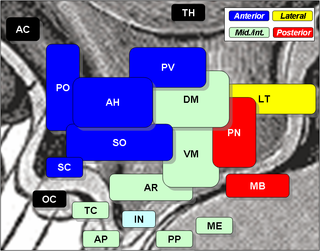
The paraventricular nucleus is a nucleus in the hypothalamus. Anatomically, it is adjacent to the third ventricle and many of its neurons project to the posterior pituitary. These projecting neurons secrete oxytocin and a smaller amount of vasopressin, otherwise the nucleus also secretes corticotropin-releasing hormone (CRH) and thyrotropin-releasing hormone (TRH). CRH and TRH are secreted into the hypophyseal portal system and act on different targets neurons in the anterior pituitary. PVN is thought to mediate many diverse functions through these different hormones, including osmoregulation, appetite, and the response of the body to stress.

Corticotropes are basophilic cells in the anterior pituitary that produce pro-opiomelanocortin (POMC) which undergoes cleavage to adrenocorticotropin (ACTH), β-lipotropin (β-LPH), and melanocyte-stimulating hormone (MSH). These cells are stimulated by corticotropin releasing hormone (CRH) and make up 15–20% of the cells in the anterior pituitary. The release of ACTH from the corticotropic cells is controlled by CRH, which is formed in the cell bodies of parvocellular neurosecretory cells within the paraventricular nucleus of the hypothalamus and passes to the corticotropes in the anterior pituitary via the hypophyseal portal system. Adrenocorticotropin hormone stimulates the adrenal cortex to release glucocorticoids and plays an important role in the stress response.

<span class="mw-page-title-main">Arcuate nucleus</span>

The arcuate nucleus of the hypothalamus is an aggregation of neurons in the mediobasal hypothalamus, adjacent to the third ventricle and the median eminence. The arcuate nucleus includes several important and diverse populations of neurons that help mediate different neuroendocrine and physiological functions, including neuroendocrine neurons, centrally projecting neurons, and astrocytes. The populations of neurons found in the arcuate nucleus are based on the hormones they secrete or interact with and are responsible for hypothalamic function, such as regulating hormones released from the pituitary gland or secreting their own hormones. Neurons in this region are also responsible for integrating information and providing inputs to other nuclei in the hypothalamus or inputs to areas outside this region of the brain. These neurons, generated from the ventral part of the periventricular epithelium during embryonic development, locate dorsally in the hypothalamus, becoming part of the ventromedial hypothalamic region. The function of the arcuate nucleus relies on its diversity of neurons, but its central role is involved in homeostasis. The arcuate nucleus provides many physiological roles involved in feeding, metabolism, fertility, and cardiovascular regulation.

<i>beta</i>-Endorphin Peptide hormone in Homo sapiens

Beta-Endorphin or β-Endorphin is an endogenous opioid neuropeptide and peptide hormone that is produced in certain neurons within the central nervous system and peripheral nervous system. It is one of three endorphins that are produced in humans, the others of which include α-endorphin and γ-endorphin.

<span class="mw-page-title-main">Adrenocorticotropic hormone deficiency</span> Medical condition

Adrenocorticotropic hormone deficiency is a result of a decreased or absent production of adrenocorticotropic hormone (ACTH) by the pituitary gland. It can be associated with TBX19.

The melanocortins are a family of neuropeptide hormones which are the ligands of the melanocortin receptors The melanocortin system consists of melanocortin receptors, ligands, and accessory proteins. The genes of the melanocortin system are found in chordates. Melanocortins were originally named so because their earliest known function was in melanogenesis. It is now known that the melanocortin system regulates diverse functions throughout the body, including inflammatory response, fibrosis, melanogenesis, steroidogenesis, energy homeostasis, sexual function, and exocrine gland function.

Delta-sleep-inducing peptide (DSIP) is a neuropeptide that when infused into the mesodiencephalic ventricle of recipient rabbits induces spindle and delta EEG activity and reduced motor activities.

Corticotropin-releasing hormone receptors (CRHRs), also known as corticotropin-releasing factor receptors (CRFRs) are a G protein-coupled receptor family that binds corticotropin-releasing hormone (CRH). There are two receptors in the family, designated as type 1 and 2, each encoded by a separate gene.

<span class="mw-page-title-main">ACTH receptor</span> Mammalian protein found in Homo sapiens

The adrenocorticotropic hormone receptor or ACTH receptor also known as the melanocortin receptor 2 or MC2 receptor is a type of melanocortin receptor (type 2) which is specific for ACTH. A G protein–coupled receptor located on the external cell plasma membrane, it is coupled to Gαs and upregulates levels of cAMP by activating adenylyl cyclase. The ACTH receptor plays a role in immune function and glucose metabolism.

<span class="mw-page-title-main">Antalarmin</span> Chemical compound

Antalarmin (CP-156,181) is a drug that acts as a CRH1 antagonist.

α-Melanocyte-stimulating hormone (α-MSH) is an endogenous peptide hormone and neuropeptide of the melanocortin family, with a tridecapeptide structure and the amino acid sequence Ac-Ser-Tyr-Ser-Met-Glu-His-Phe-Arg-Trp-Gly-Lys-Pro-Val-NH2. It is the most important of the melanocyte-stimulating hormones (MSHs) (also known as melanotropins) in stimulating melanogenesis, a process that in mammals (including humans) is responsible for pigmentation primarily of the hair and skin. It also plays a role in feeding behavior, energy homeostasis, sexual activity, and protection against ischemia and reperfusion injury.

<i>beta</i>-Melanocyte-stimulating hormone Chemical compound

β-Melanocyte-stimulating hormone (β-MSH) is an endogenous peptide hormone and neuropeptide. It is a melanocortin, specifically, one of the three types of melanocyte-stimulating hormone (MSH), and is produced from proopiomelanocortin (POMC). It is an agonist of the MC1, MC3, MC4, and MC5 receptors.

γ-Melanocyte-stimulating hormone (γ-MSH) is an endogenous peptide hormone and neuropeptide. It is a melanocortin, specifically, one of the three types of melanocyte-stimulating hormone (MSH), and is produced from proopiomelanocortin (POMC). It is an agonist of the MC1, MC3, MC4, and MC5 receptors. It exists in three forms, γ1-MSH, γ2-MSH, and γ3-MSH.

References

  1. Gianoulakis C, Seidah NG, Routhier R, Chrétien M (December 1979). "Biosynthesis and characterization of adrenocorticotropic hormone, alpha-melanocyte-stimulating hormone, and an NH2-terminal fragment of the adrenocorticotropic hormone/beta-lipotropin precursor from rat pars intermedia". The Journal of Biological Chemistry. 254 (23): 11903–6. doi: 10.1016/S0021-9258(19)86402-5 . PMID   227883.
  2. Lloyd D. Fricker (24 July 1991). Peptide biosynthesis and processing. CRC Press. p. 78. ISBN   978-0-8493-8852-1 . Retrieved 25 November 2011.
  3. Anthony W. Norman; Gerald Litwack (26 September 1997). Hormones . Academic Press. pp.  12. ISBN   978-0-12-521441-4 . Retrieved 25 November 2011.
  4. Marshall JB, Kapcala LP, Manning LD, McCullough AJ (November 1984). "Effect of corticotropin-like intermediate lobe peptide on pancreatic exocrine function in isolated rat pancreatic lobules". The Journal of Clinical Investigation. 74 (5): 1886–9. doi:10.1172/JCI111608. PMC   425369 . PMID   6209301.
  5. Zaphiropoulos A, Charnay Y, Vallet P, Constantinidis J, Bouras C (January 1991). "Immunohistochemical distribution of corticotropin-like intermediate lobe peptide (CLIP) immunoreactivity in the human brain". Brain Research Bulletin. 26 (1): 99–111. doi:10.1016/0361-9230(91)90194-O. PMID   1849784. S2CID   556547.
  6. Shojiro Inoué; Shojiro Inoué (1989). Biology of sleep substances. CRC Press. p. 136. ISBN   978-0-8493-4822-8 . Retrieved 25 November 2011.
  7. Chastrette N, Cespuglio R, Jouvet M (February 1990). "Proopiomelanocortin (POMC)-derived peptides and sleep in the rat. Part 1--Hypnogenic properties of ACTH derivatives". Neuropeptides. 15 (2): 61–74. doi:10.1016/0143-4179(90)90042-w. PMID   1981927. S2CID   41264103.
  8. Chastrette N, Cespuglio R, Lin YL, Jouvet M (February 1990). "Proopiomelanocortin (POMC)-derived peptides and sleep in the rat. Part 2--Aminergic regulatory processes". Neuropeptides. 15 (2): 75–88. doi:10.1016/0143-4179(90)90043-x. PMID   1964203. S2CID   13731355.
  9. Grigoriev VV, Petrova LN, Ivanova TA, Gabreliyan AV, Serkova TP (March 2009). "Effect of corticotropin-like intermediate lobe peptide on presynaptic and postsynaptic glutamate receptors and postsynaptic GABA receptors in rat brain". Bulletin of Experimental Biology and Medicine. 147 (3): 319–22. doi:10.1007/s10517-009-0499-x. PMID   19529852. S2CID   29237407.
  10. Seidenbecher T, Balschun D, Vogel D, Reymann KG (1993). "Neuronal transmission of hippocampal CA1 neurones is modulated by corticotropin-like intermediate lobe peptide [CLIP; ACTH(18-39)]". Peptides. 14 (6): 1221–4. doi:10.1016/0196-9781(93)90179-K. PMID   8134304. S2CID   7935999.
  11. Marshall, J. B.; Kapcala, L. P.; Manning, L. D.; McCullough, A. J. (November 1984). "Effect of corticotropin-like intermediate lobe peptide on pancreatic exocrine function in isolated rat pancreatic lobules". The Journal of Clinical Investigation. 74 (5): 1886–1889. doi:10.1172/JCI111608. ISSN   0021-9738. PMC   425369 . PMID   6209301.