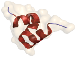
Osteocalcin

Last updated

BGLAP
Osteocalcin 1Q8H.png
Identifiers
Aliases BGLAP , BGP, OC, OCN, bone gamma-carboxyglutamate protein, Osteocalcin
External IDs OMIM: 112260; MGI: 88155; HomoloGene: 104130; GeneCards: BGLAP; OMA:BGLAP - orthologs
Orthologs
SpeciesHumanMouse
Entrez
Ensembl
UniProt
RefSeq (mRNA)

NM_199173

NM_031368
NM_001305448
NM_001305449
NM_001305450

RefSeq (protein)

NP_954642

NP_001292377
NP_001292378
NP_001292379
NP_112736

Location (UCSC) Chr 1: 156.24 – 156.24 Mb Chr 3: 88.28 – 88.28 Mb
PubMed search [3] [4]
Wikidata
View/Edit Human View/Edit Mouse

Osteocalcin, also known as bone gamma-carboxyglutamic acid-containing protein (BGLAP), is a small (49-amino-acid [5] ) noncollagenous protein hormone found in bone and dentin, first identified as a calcium-binding protein. [6]

Contents

Because osteocalcin has gla domains, its synthesis is vitamin K-dependent. In humans, osteocalcin is encoded by the BGLAP gene. [7] [8] Its receptors include GPRC6A, GPR158, and possibly a third, yet-to-be-identified receptor. [9] [10] There is evidence that GPR37 might be the third osteocalcin receptor. [11]

Function

Osteocalcin is secreted solely by osteoblasts and is thought to play a role in the body's metabolic regulation. [12] In its carboxylated form, calcium is bound directly to the bone and thus concentrates here.

In its uncarboxylated form, osteocalcin acts as a hormone in the body, signalling in the pancreas, fat, muscle, testes, and brain. [13]

An acute stress response (ASR), colloquially known as the fight-or-flight response, stimulates osteocalcin release from bone within minutes in mice, rats, and humans. Injections of high levels of osteocalcin alone can trigger an ASR in the presence of adrenal insufficiency. [18]

Use as a biochemical marker for bone formation

As osteoblasts produce osteocalcin, it is often used as a marker for the bone formation process. Higher serum osteocalcin levels have been observed to correlate relatively well with increases in bone mineral density during treatment with anabolic bone formation drugs for osteoporosis, such as teriparatide. In many studies, osteocalcin is used as a preliminary biomarker for the effectiveness of a given drug on bone formation. For instance, one study that aimed to study the efficacy of a glycoprotein called lactoferrin on bone formation used osteocalcin to measure osteoblast activity. [19]

Related Research Articles

<span class="mw-page-title-main">Parathyroid hormone</span> Mammalian protein found in humans

Parathyroid hormone (PTH), also called parathormone or parathyrin, is a peptide hormone secreted by the parathyroid glands that regulates the serum calcium concentration through its effects on bone, kidney, and intestine.

<span class="mw-page-title-main">Osteoblast</span> Cells secreting extracellular matrix

Osteoblasts are cells with a single nucleus that synthesize bone. However, in the process of bone formation, osteoblasts function in groups of connected cells. Individual cells cannot make bone. A group of organized osteoblasts together with the bone made by a unit of cells is usually called the osteon.

<span class="mw-page-title-main">Adipocyte</span> Cells that primarily compose adipose tissue, specialized in storing energy as fat

Adipocytes, also known as lipocytes and fat cells, are the cells that primarily compose adipose tissue, specialized in storing energy as fat. Adipocytes are derived from mesenchymal stem cells which give rise to adipocytes through adipogenesis. In cell culture, adipocyte progenitors can also form osteoblasts, myocytes and other cell types.

<span class="mw-page-title-main">Osteoprotegerin</span> Mammalian protein found in Homo sapiens

Osteoprotegerin (OPG), also known as osteoclastogenesis inhibitory factor (OCIF) or tumour necrosis factor receptor superfamily member 11B (TNFRSF11B), is a cytokine receptor of the tumour necrosis factor (TNF) receptor superfamily encoded by the TNFRSF11B gene.

<span class="mw-page-title-main">Osteocyte</span> Mature osteoblasts which helps in communication between cells and also in molecular synthesis

An osteocyte, an oblate shaped type of bone cell with dendritic processes, is the most commonly found cell in mature bone. It can live as long as the organism itself. The adult human body has about 42 billion of them. Osteocytes do not divide and have an average half life of 25 years. They are derived from osteoprogenitor cells, some of which differentiate into active osteoblasts. Osteoblasts/osteocytes develop in mesenchyme.

<span class="mw-page-title-main">Klotho (biology)</span> Human enzyme

Klotho is an enzyme that in humans is encoded by the KL gene. The three subfamilies of klotho are α-klotho, β-klotho, and γ-klotho. α-klotho activates FGF23, and β-klotho activates FGF19 and FGF21. When the subfamily is not specified, the word "klotho" typically refers to the α-klotho subfamily, because α-klotho was discovered before the other members.

<span class="mw-page-title-main">Osteopontin</span> Mammalian protein found in Homo sapiens

Osteopontin (OPN), also known as bone /sialoprotein I, early T-lymphocyte activation (ETA-1), secreted phosphoprotein 1 (SPP1), 2ar and Rickettsia resistance (Ric), is a protein that in humans is encoded by the SPP1 gene. The murine ortholog is Spp1. Osteopontin is a SIBLING (glycoprotein) that was first identified in 1986 in osteoblasts.

<span class="mw-page-title-main">Sclerostin</span> Protein-coding gene in the species Homo sapiens

Sclerostin is a protein that in humans is encoded by the SOST gene. It is a secreted glycoprotein with a C-terminal cysteine knot-like (CTCK) domain and sequence similarity to the DAN family of bone morphogenetic protein (BMP) antagonists. Sclerostin is produced primarily by the osteocyte but is also expressed in other tissues, and has anti-anabolic effects on bone formation.

<span class="mw-page-title-main">TRPV6</span> Protein-coding gene in the species Homo sapiens

TRPV6 is a membrane calcium (Ca2+) channel protein which is particularly involved in the first step in Ca2+absorption in the intestine.

<span class="mw-page-title-main">Calcitonin receptor</span> Protein found in humans

The calcitonin receptor (CT) is a G protein-coupled receptor that binds the peptide hormone calcitonin and is involved in maintenance of calcium homeostasis, particularly with respect to bone formation and metabolism.

<span class="mw-page-title-main">RUNX2</span> Protein-coding gene in humans

Runt-related transcription factor 2 (RUNX2) also known as core-binding factor subunit alpha-1 (CBF-alpha-1) is a protein that in humans is encoded by the RUNX2 gene. RUNX2 is a key transcription factor associated with osteoblast differentiation.

<span class="mw-page-title-main">Bone sialoprotein</span> Protein-coding gene in the species Homo sapiens

Bone sialoprotein (BSP) is a component of mineralized tissues such as bone, dentin, cementum and calcified cartilage. BSP is a significant component of the bone extracellular matrix and has been suggested to constitute approximately 8% of all non-collagenous proteins found in bone and cementum. BSP, a SIBLING protein, was originally isolated from bovine cortical bone as a 23-kDa glycopeptide with high sialic acid content.

<span class="mw-page-title-main">KiSS1-derived peptide receptor</span> Mammalian protein found in Homo sapiens

The KiSS1-derived peptide receptor is a G protein-coupled receptor which binds the peptide hormone kisspeptin (metastin). Kisspeptin is encoded by the metastasis suppressor gene KISS1, which is expressed in a variety of endocrine and gonadal tissues. Activation of the kisspeptin receptor is linked to the phospholipase C and inositol trisphosphate second messenger cascades inside the cell.

<span class="mw-page-title-main">Parathyroid hormone 1 receptor</span> Protein-coding gene in the species Homo sapiens

Parathyroid hormone/parathyroid hormone-related peptide receptor, also known as parathyroid hormone 1 receptor (PTH1R), is a protein that in humans is encoded by the PTH1R gene. PTH1R functions as a receptor for parathyroid hormone (PTH) and for parathyroid hormone-related protein (PTHrP), also called parathyroid hormone-like hormone (PTHLH).

<span class="mw-page-title-main">GPRC6A</span> Protein-coding gene in the species Homo sapiens

G protein-coupled receptor family C group 6 member A (GPRC6A) is a protein that in humans is encoded by the GPRC6A gene. This protein functions as a receptor of L-α-amino acids, cations, osteocalcin, and steroids. It is a membrane androgen receptor.

<span class="mw-page-title-main">DLK1</span> Protein-coding gene in the species Homo sapiens

Protein delta homolog 1, delta like non-canonical Notch ligand 1, fetal antigen 1 or preadipocyte factor 1 is a protein that in humans is encoded by the DLK1 gene.

<span class="mw-page-title-main">DMP1</span> Protein-coding gene in the species Homo sapiens

Dentin matrix acidic phosphoprotein 1 is a protein that in humans is encoded by the DMP1 gene.

<span class="mw-page-title-main">Sp7 transcription factor</span> Protein-coding gene in the species Homo sapiens

Transcription factor Sp7, also called osterix (Osx), is a protein that in humans is encoded by the SP7 gene. It is a member of the Sp family of zinc-finger transcription factors It is highly conserved among bone-forming vertebrate species It plays a major role, along with Runx2 and Dlx5 in driving the differentiation of mesenchymal precursor cells into osteoblasts and eventually osteocytes. Sp7 also plays a regulatory role by inhibiting chondrocyte differentiation maintaining the balance between differentiation of mesenchymal precursor cells into ossified bone or cartilage. Mutations of this gene have been associated with multiple dysfunctional bone phenotypes in vertebrates. During development, a mouse embryo model with Sp7 expression knocked out had no formation of bone tissue. Through the use of GWAS studies, the Sp7 locus in humans has been strongly associated with bone mass density. In addition there is significant genetic evidence for its role in diseases such as Osteogenesis imperfecta (OI).

<span class="mw-page-title-main">Adipogenesis</span>

Adipogenesis is the formation of adipocytes from stem cells. It involves 2 phases, determination, and terminal differentiation. Determination is mesenchymal stem cells committing to the adipocyte precursor cells, also known as lipoblasts or preadipocytes which lose the potential to differentiate to other types of cells such as chondrocytes, myocytes, and osteoblasts. Terminal differentiation is that preadipocytes differentiate into mature adipocytes. Adipocytes can arise either from preadipocytes resident in adipose tissue, or from bone-marrow derived progenitor cells that migrate to adipose tissue.

<span class="mw-page-title-main">Bone marrow adipose tissue</span>

Bone marrow adipose tissue (BMAT), sometimes referred to as marrow adipose tissue (MAT), is a type of fat deposit in bone marrow. It increases in states of low bone density, such as osteoporosis, anorexia nervosa/caloric restriction, skeletal unweighting such as that which occurs in space travel, and anti-diabetes therapies. BMAT decreases in anaemia, leukaemia, and hypertensive heart failure; in response to hormones such as oestrogen, leptin, and growth hormone; with exercise-induced weight loss or bariatric surgery; in response to chronic cold exposure; and in response to pharmacological agents such as bisphosphonates, teriparatide, and metformin.

References

  1. 1 2 3 GRCh38: Ensembl release 89: ENSG00000242252 Ensembl, May 2017
  2. 1 2 3 GRCm38: Ensembl release 89: ENSMUSG00000074489 Ensembl, May 2017
  3. "Human PubMed Reference:". National Center for Biotechnology Information, U.S. National Library of Medicine.
  4. "Mouse PubMed Reference:". National Center for Biotechnology Information, U.S. National Library of Medicine.
  5. Hauschka PV, Carr SA, Biemann K (February 1982). "Primary structure of monkey osteocalcin". Biochemistry. 21 (4): 638–642. doi:10.1021/bi00533a006. PMID   6978733.
  6. Hauschka PV, Reid ML (August 1978). "Timed appearance of a calcium-binding protein containing gamma-carboxyglutamic acid in developing chick bone". Developmental Biology. 65 (2): 426–434. doi:10.1016/0012-1606(78)90038-6. PMID   680371.
  7. Puchacz E, Lian JB, Stein GS, Wozney J, Huebner K, Croce C (May 1989). "Chromosomal localization of the human osteocalcin gene". Endocrinology. 124 (5): 2648–50. doi:10.1210/endo-124-5-2648. PMID   2785029.
  8. Cancela L, Hsieh CL, Francke U, Price PA (September 1990). "Molecular structure, chromosome assignment, and promoter organization of the human matrix Gla protein gene". The Journal of Biological Chemistry. 265 (25): 15040–8. doi: 10.1016/S0021-9258(18)77221-9 . PMID   2394711.
  9. Pi M, Wu Y, Quarles LD (July 2011). "GPRC6A mediates responses to osteocalcin in β-cells in vitro and pancreas in vivo". Journal of Bone and Mineral Research. 26 (7): 1680–3. doi:10.1002/jbmr.390. PMC   5079536 . PMID   21425331.
  10. Berger JM, Singh P, Khrimian L, Morgan DA, Chowdhury S, Arteaga-Solis E, et al. (September 2019). "Mediation of the Acute Stress Response by the Skeleton". Cell Metabolism. 30 (5): 890–902.e8. doi:10.1016/j.cmet.2019.08.012. PMC   6834912 . PMID   31523009.
  11. Qian Z, Li H, Yang H, Yang Q, Lu Z, Wang L, et al. (October 2021). "Osteocalcin attenuates oligodendrocyte differentiation and myelination via GPR37 signaling in the mouse brain". Science Advances. 7 (43): eabi5811. Bibcode:2021SciA....7.5811Q. doi:10.1126/sciadv.abi5811. PMC   8535816 . PMID   34678058.
  12. 1 2 3 Lee NK, Sowa H, Hinoi E, Ferron M, Ahn JD, Confavreux C, et al. (August 2007). "Endocrine regulation of energy metabolism by the skeleton". Cell. 130 (3): 456–69. doi:10.1016/j.cell.2007.05.047. PMC   2013746 . PMID   17693256.
  13. Karsenty G, Olson EN (March 2016). "Bone and Muscle Endocrine Functions: Unexpected Paradigms of Inter-organ Communication". Cell. 164 (6): 1248–1256. doi:10.1016/j.cell.2016.02.043. PMC   4797632 . PMID   26967290.
  14. Mera P, Laue K, Ferron M, Confavreux C, Wei J, Galán-Díez M, et al. (June 2016). "Osteocalcin Signaling in Myofibers Is Necessary and Sufficient for Optimum Adaptation to Exercise". Cell Metabolism. 23 (6): 1078–1092. doi:10.1016/j.cmet.2016.05.004. PMC   4910629 . PMID   27304508.
  15. Dance A (23 February 2022). "Fun facts about bones: More than just scaffolding". Knowable Magazine. doi: 10.1146/knowable-022222-1 . Retrieved 8 March 2022.
  16. Karsenty G, Oury F (January 2014). "Regulation of male fertility by the bone-derived hormone osteocalcin". Molecular and Cellular Endocrinology. 382 (1): 521–526. doi:10.1016/j.mce.2013.10.008. PMC   3850748 . PMID   24145129.
  17. Obri A, Khrimian L, Karsenty G, Oury F (March 2018). "Osteocalcin in the brain: from embryonic development to age-related decline in cognition". Nature Reviews. Endocrinology. 14 (3): 174–182. doi:10.1038/nrendo.2017.181. PMC   5958904 . PMID   29376523.
  18. Meyer-Berger J, Singh P, Khrimian L, Morgan D, Chowdhury S, Arteaga-Solis E, et al. (2019). "Mediation of the Acute Stress Response by the Skeleton". Cell Metabolism. 30 (5): 890–902.e8. doi:10.1016/j.cmet.2019.08.012. PMC   6834912 . PMID   31523009.
  19. Bharadwaj S, Naidu AG, Betageri GV, Prasadarao NV, Naidu AS (September 2009). "Milk ribonuclease-enriched lactoferrin induces positive effects on bone turnover markers in postmenopausal women". Osteoporosis International. 20 (9): 1603–11. doi:10.1007/s00198-009-0839-8. PMID   19172341. S2CID   10711802.

Further reading