Osteonectin

Last updated
SPARC
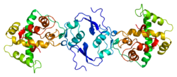
Protein SPARC PDB 1bmo.png
Available structures
PDB Ortholog search: PDBe RCSB
Identifiers
Aliases SPARC , BM-40, ON, OI17, secreted protein acidic and cysteine rich, ONT
External IDs OMIM: 182120 MGI: 98373 HomoloGene: 31132 GeneCards: SPARC
Orthologs
SpeciesHumanMouse
Entrez
Ensembl
UniProt
RefSeq (mRNA)

NM_003118
NM_001309443
NM_001309444

NM_009242
NM_001290817

RefSeq (protein)

NP_001296372
NP_001296373
NP_003109

NP_001277746
NP_033268

Location (UCSC) Chr 5: 151.66 – 151.69 Mb Chr 11: 55.29 – 55.31 Mb
PubMed search [3] [4]
Wikidata
View/Edit Human View/Edit Mouse

Osteonectin (ON) also known as secreted protein acidic and rich in cysteine (SPARC) or basement-membrane protein 40 (BM-40) is a protein that in humans is encoded by the SPARC gene.

Contents

Osteonectin is a glycoprotein in the bone that binds calcium. It is secreted by osteoblasts during bone formation, initiating mineralization and promoting mineral crystal formation. Osteonectin also shows affinity for collagen in addition to bone mineral calcium. A correlation between osteonectin over-expression and ampullary cancers and chronic pancreatitis has been found.

Gene

The human SPARC gene is 26.5 kb long, and contains 10 exons and 9 introns and is located on chromosome 5q31-q33.

Structure

Osteonectin is a 40 kDa acidic and cysteine-rich glycoprotein consisting of a single polypeptide chain that can be broken into 4 domains: 1) a Ca2+ binding domain near the glutamic acid-rich region at the amino terminus (domain I), 2) a cysteine-rich domain (II), 3) a hydrophilic region (domain III), and 4) an EF hand motif at the carboxy terminus region (domain IV). [5]

Function

Osteonectin is an acidic extracellular matrix glycoprotein that plays a vital role in bone mineralization, cell-matrix interactions, and collagen binding. Osteonectin also increases the production and activity of matrix metalloproteinases, a function important to invading cancer cells within bone. Additional functions of osteonectin beneficial to tumor cells include angiogenesis, proliferation and migration. Overexpression of osteonectin is reported in many human cancers such as breast, prostate, colon and pancreatic. [6]

This molecule has been implicated in several biological functions, including mineralization of bone and cartilage, inhibiting mineralization, modulation of cell proliferation, facilitation of acquisition of differentiated phenotype and promotion of cell attachment and spreading.

A number of phosphoproteins and glycoproteins are found in bone. The phosphate is bound to the protein backbone through phosphorylated serine or threonine amino acid residues. The best characterized of these bone proteins is osteonectin. It binds collagen and hydroxyapatite in separate domains, is found in relatively large amounts in immature bone, and promotes mineralization of collagen.

Tissue distribution

Fibroblasts, including periodontal fibroblasts, synthesize osteonectin. [7] This protein is synthesized by macrophages at sites of wound repair and platelet degranulation, so it may play an important role in wound healing. SPARC does not support cell attachment, and like tenascin, is anti-adhesive and an inhibitor of cell spreading. It disrupts focal adhesions in fibroblasts. It also regulates the proliferation of some cells, especially endothelial cells, mediated by its ability to bind to cytokines and growth factors. [8] Osteonectin has also been found to decrease DNA synthesis in cultured bone. [9]

High levels of immunodetectable osteonectin are found in active osteoblasts and marrow progenitor cells, odontoblasts, periodontal ligament and gingival cells, and some chondrocytes and hypertrophic chondrocytes. Osteonectin is also detectable in osteoid, bone matrix proper, and dentin. Osteonectin has been localized in a variety of tissues, but is found in greatest abundance in osseous tissue, tissues characterized by high turnover (such as intestinal epithelium), basement membranes, and certain neoplasms. Osteonectin is expressed by a wide variety of cells, including chondrocytes, fibroblasts, platelets, endothelial cells, epithelial cells, Leydig cells, Sertoli cells, luteal cells, adrenal cortical cells, and numerous neoplastic cell lines (such as SaOS-2 cells from human osteosarcoma).

Model organisms

Model organisms have been used in the study of SPARC function. A conditional knockout mouse line, called Sparctm1a(EUCOMM)Wtsi [18] [19] was generated as part of the International Knockout Mouse Consortium program — a high-throughput mutagenesis project to generate and distribute animal models of disease to interested scientists. [20] [21] [22]

Male and female animals underwent a standardized phenotypic screen to determine the effects of deletion. [16] [23] Twenty six tests were carried out on mutant mice and six significant abnormalities were observed. [16] Homozygous mutant animals had unusually white incisors, decreased bone mineral density, abnormal lens morphology, cataracts and a decreased length of long bones. [16]

Related Research Articles

<span class="mw-page-title-main">Fibronectin</span> Protein involved in cell adhesion, cell growth, cell migration and differentiation

Fibronectin is a high-molecular weight glycoprotein of the extracellular matrix that binds to membrane-spanning receptor proteins called integrins. It is approved for marketing as a topical solution in India by Central Drugs Standard Control organization in 2020 under the brand name FIBREGA for chronic wounds. Fibronectin also binds to other extracellular matrix proteins such as collagen, fibrin, and heparan sulfate proteoglycans.

<span class="mw-page-title-main">Extracellular matrix</span> Network of proteins and molecules outside cells that provides structural support for cells

In biology, the extracellular matrix (ECM), is a network consisting of extracellular macromolecules and minerals, such as collagen, enzymes, glycoproteins and hydroxyapatite that provide structural and biochemical support to surrounding cells. Because multicellularity evolved independently in different multicellular lineages, the composition of ECM varies between multicellular structures; however, cell adhesion, cell-to-cell communication and differentiation are common functions of the ECM.

<span class="mw-page-title-main">Osteoblast</span> Cells secreting extracellular matrix

Osteoblasts are cells with a single nucleus that synthesize bone. However, in the process of bone formation, osteoblasts function in groups of connected cells. Individual cells cannot make bone. A group of organized osteoblasts together with the bone made by a unit of cells is usually called the osteon.

<span class="mw-page-title-main">Wound healing</span> Series of events that restore integrity to damaged tissue after an injury

Wound healing refers to a living organism's replacement of destroyed or damaged tissue by newly produced tissue.

<span class="mw-page-title-main">Platelet-derived growth factor</span> Signaling glycoprotein regulating cell proliferation

Platelet-derived growth factor (PDGF) is one among numerous growth factors that regulate cell growth and division. In particular, PDGF plays a significant role in blood vessel formation, the growth of blood vessels from already-existing blood vessel tissue, mitogenesis, i.e. proliferation, of mesenchymal cells such as fibroblasts, osteoblasts, tenocytes, vascular smooth muscle cells and mesenchymal stem cells as well as chemotaxis, the directed migration, of mesenchymal cells. Platelet-derived growth factor is a dimeric glycoprotein that can be composed of two A subunits (PDGF-AA), two B subunits (PDGF-BB), or one of each (PDGF-AB).

<span class="mw-page-title-main">Human tooth development</span> Process by which teeth form

Tooth development or odontogenesis is the complex process by which teeth form from embryonic cells, grow, and erupt into the mouth. For human teeth to have a healthy oral environment, all parts of the tooth must develop during appropriate stages of fetal development. Primary (baby) teeth start to form between the sixth and eighth week of prenatal development, and permanent teeth begin to form in the twentieth week. If teeth do not start to develop at or near these times, they will not develop at all, resulting in hypodontia or anodontia.

<span class="mw-page-title-main">Osteopontin</span> Mammalian protein found in Homo sapiens

Osteopontin (OPN), also known as bone /sialoprotein I, early T-lymphocyte activation (ETA-1), secreted phosphoprotein 1 (SPP1), 2ar and Rickettsia resistance (Ric), is a protein that in humans is encoded by the SPP1 gene. The murine ortholog is Spp1. Osteopontin is a SIBLING (glycoprotein) that was first identified in 1986 in osteoblasts.

<span class="mw-page-title-main">Chondroblast</span> Mesenchymal progenitor cell that forms a chondrocyte

Chondroblasts, or perichondrial cells, is the name given to mesenchymal progenitor cells in situ which, from endochondral ossification, will form chondrocytes in the growing cartilage matrix. Another name for them is subchondral cortico-spongious progenitors. They have euchromatic nuclei and stain by basic dyes.

<span class="mw-page-title-main">Perlecan</span>

Perlecan (PLC) also known as basement membrane-specific heparan sulfate proteoglycan core protein (HSPG) or heparan sulfate proteoglycan 2 (HSPG2), is a protein that in humans is encoded by the HSPG2 gene. The HSPG2 gene codes for a 4,391 amino acid protein with a molecular weight of 468,829. It is one of the largest known proteins. The name perlecan comes from its appearance as a "string of pearls" in rotary shadowed images.

<span class="mw-page-title-main">Decorin</span> Protein-coding gene in humans

Decorin is a protein that in humans is encoded by the DCN gene.

<span class="mw-page-title-main">Biglycan</span> Protein-coding gene in humans

Biglycan is a small leucine-rich repeat proteoglycan (SLRP) which is found in a variety of extracellular matrix tissues, including bone, cartilage and tendon. In humans, biglycan is encoded by the BGN gene which is located on the X chromosome.

<span class="mw-page-title-main">Bone sialoprotein</span> Protein-coding gene in the species Homo sapiens

Bone sialoprotein (BSP) is a component of mineralized tissues such as bone, dentin, cementum and calcified cartilage. BSP is a significant component of the bone extracellular matrix and has been suggested to constitute approximately 8% of all non-collagenous proteins found in bone and cementum. BSP, a SIBLING protein, was originally isolated from bovine cortical bone as a 23-kDa glycopeptide with high sialic acid content.

<span class="mw-page-title-main">Integrin alpha 2</span> Mammalian protein found in Homo sapiens

Integrin alpha-2, or CD49b, is a protein which in humans is encoded by the CD49b gene.

<span class="mw-page-title-main">DMP1</span> Protein-coding gene in the species Homo sapiens

Dentin matrix acidic phosphoprotein 1 is a protein that in humans is encoded by the DMP1 gene.

<span class="mw-page-title-main">SPARCL1</span> Protein-coding gene in the species Homo sapiens

SPARC-like protein 1, also known as hevin, is a secreted protein with high structural similarity to SPARC. It interacts with the extracellular matrix to create intermediate states of cell adhesion. Due to its dynamic extracellular roles, being implicated in cancer metastasis and inflammation, it is considered a matricellular protein. In humans hevin is encoded by the SPARCL1 gene.

<span class="mw-page-title-main">Dermatopontin</span> Protein-coding gene in the species Homo sapiens

Dermatopontin also known as tyrosine-rich acidic matrix protein (TRAMP) is a protein that in humans is encoded by the DPT gene. Dermatopontin is a 22-kDa protein of the noncollagenous extracellular matrix (ECM) estimated to comprise 12 mg/kg of wet dermis weight. To date, homologues have been identified in five different mammals and 12 different invertebrates with multiple functions. In vertebrates, the primary function of dermatopontin is a structural component of the ECM, cell adhesion, modulation of TGF-β activity and cellular quiescence). It also has pathological involvement in heart attacks and decreased expression in leiomyoma and fibrosis. In invertebrate, dermatopontin homologue plays a role in hemagglutination, cell-cell aggregation, and expression during parasite infection.

Collagen receptors are membrane proteins that bind the extracellular matrix protein collagen, the most abundant protein in mammals. They control mainly cell proliferation, migration and adhesion, coagulation cascade activation and they affect ECM structure by regulation of MMP.

<span class="mw-page-title-main">FAM20C</span> Protein-coding gene in the species Homo sapiens

Family with sequence similarity 20, member C also known as FAM20C or DMP4 is a protein which in humans is encoded by the FAM20C gene. Fam20C, a Golgi localized protein kinase, is a serine kinase that phosphorylates both casein and other highly acidic proteins and members of the small integrin-binding ligand, the N-linked glycoproteins (SIBLING) family at the target motif SerXGlu.

Fibroblast-like synoviocytes (FLS) represent a specialised cell type located inside joints in the synovium. These cells play a crucial role in the pathogenesis of chronic inflammatory diseases, such as rheumatoid arthritis.

SNED1 is an extracellular matrix (ECM) protein expressed at low levels in a wide range of tissues. The gene encoding SNED1 is located in the human chromosome 2 at locus q37.3. The corresponding mRNA isolated from the spleen and is 6834bp in length, and the corresponding protein is 1413 amino-acid long. The mouse ortholog of SNED1 was cloned in 2004 from the embryonic kidney by Leimester et al. SNED1 present domains characteristic of ECM proteins, including an amino-terminal NIDO domain, several calcium binding EGF-like domains (EGF_CA), a Sushi domain also known as complement control protein (CCP) domain, and three type III fibronectin (FN3) domains in the carboxy-terminal region.

References

  1. 1 2 3 GRCh38: Ensembl release 89: ENSG00000113140 - Ensembl, May 2017
  2. 1 2 3 GRCm38: Ensembl release 89: ENSMUSG00000018593 - Ensembl, May 2017
  3. "Human PubMed Reference:". National Center for Biotechnology Information, U.S. National Library of Medicine.
  4. "Mouse PubMed Reference:". National Center for Biotechnology Information, U.S. National Library of Medicine.
  5. Villarreal XC, Mann KG, Long GL (Jul 1989). "Structure of human osteonectin based upon analysis of cDNA and genomic sequences". Biochemistry. 28 (15): 6483–91. doi:10.1021/bi00441a049. PMID   2790009.
  6. Guweidhi A, Kleeff J, Adwan H, Giese NA, Wente MN, Giese T, Büchler MW, Berger MR, Friess H (Aug 2005). "Osteonectin influences growth and invasion of pancreatic cancer cells". Annals of Surgery. 242 (2): 224–34. doi:10.1097/01.sla.0000171866.45848.68. PMC   1357728 . PMID   16041213.
  7. Wasi S, Otsuka K, Yao KL, Tung PS, Aubin JE, Sodek J, Termine JD (Jun 1984). "An osteonectinlike protein in porcine periodontal ligament and its synthesis by periodontal ligament fibroblasts". Canadian Journal of Biochemistry and Cell Biology. 62 (6): 470–8. doi:10.1139/o84-064. PMID   6380686.
  8. Young MF, Kerr JM, Ibaraki K, Heegaard AM, Robey PG (Aug 1992). "Structure, expression, and regulation of the major noncollagenous matrix proteins of bone". Clinical Orthopaedics and Related Research. 281 (281): 275–94. doi:10.1097/00003086-199208000-00042. PMID   1499220.
  9. Lane TF, Sage EH (Feb 1994). "The biology of SPARC, a protein that modulates cell-matrix interactions". FASEB Journal. 8 (2): 163–73. doi:10.1096/fasebj.8.2.8119487. PMID   8119487. S2CID   32958146.
  10. "Dysmorphology data for Sparc". Wellcome Trust Sanger Institute.
  11. "DEXA data for Sparc". Wellcome Trust Sanger Institute.
  12. "Radiography data for Sparc". Wellcome Trust Sanger Institute.
  13. "Eye morphology data for Sparc". Wellcome Trust Sanger Institute.
  14. "Salmonella infection data for Sparc". Wellcome Trust Sanger Institute.
  15. "Citrobacter infection data for Sparc". Wellcome Trust Sanger Institute.
  16. 1 2 3 4 Gerdin AK (2010). "The Sanger Mouse Genetics Programme: High throughput characterisation of knockout mice". Acta Ophthalmologica. 88: 925–7. doi:10.1111/j.1755-3768.2010.4142.x. S2CID   85911512.
  17. Mouse Resources Portal, Wellcome Trust Sanger Institute.
  18. "International Knockout Mouse Consortium".
  19. "Mouse Genome Informatics".
  20. Skarnes WC, Rosen B, West AP, Koutsourakis M, Bushell W, Iyer V, Mujica AO, Thomas M, Harrow J, Cox T, Jackson D, Severin J, Biggs P, Fu J, Nefedov M, de Jong PJ, Stewart AF, Bradley A (Jun 2011). "A conditional knockout resource for the genome-wide study of mouse gene function". Nature. 474 (7351): 337–42. doi:10.1038/nature10163. PMC   3572410 . PMID   21677750.
  21. Dolgin E (Jun 2011). "Mouse library set to be knockout". Nature. 474 (7351): 262–3. doi: 10.1038/474262a . PMID   21677718.
  22. Collins FS, Rossant J, Wurst W (Jan 2007). "A mouse for all reasons". Cell. 128 (1): 9–13. doi: 10.1016/j.cell.2006.12.018 . PMID   17218247. S2CID   18872015.
  23. van der Weyden L, White JK, Adams DJ, Logan DW (2011). "The mouse genetics toolkit: revealing function and mechanism". Genome Biology. 12 (6): 224. doi: 10.1186/gb-2011-12-6-224 . PMC   3218837 . PMID   21722353.

Further reading