Annexin A5

Last updated
ANXA5
Protein ANXA5 PDB 1a8a.png
Available structures
PDB Ortholog search: PDBe RCSB
Identifiers
Aliases ANXA5 , ANX5, ENX2, HEL-S-7, PP4, RPRGL3, annexin A5, CPB-I, VAC-alph
External IDs OMIM: 131230 MGI: 106008 HomoloGene: 20312 GeneCards: ANXA5
Orthologs
SpeciesHumanMouse
Entrez
Ensembl
UniProt
RefSeq (mRNA)

NM_001154

NM_009673

RefSeq (protein)

NP_001145

NP_033803

Location (UCSC) Chr 4: 121.67 – 121.7 Mb Chr 3: 36.5 – 36.53 Mb
PubMed search [3] [4]
Wikidata
View/Edit Human View/Edit Mouse

Annexin A5 (or annexin V) is a cellular protein in the annexin group. In flow cytometry, annexin V is commonly used to detect apoptotic cells by its ability to bind to phosphatidylserine, a marker of apoptosis when it is on the outer leaflet of the plasma membrane. The function of the protein is unknown; however, annexin A5 has been proposed to play a role in the inhibition of blood coagulation by competing for phosphatidylserine binding sites with prothrombin and also to inhibit the activity of phospholipase A1. These properties have been found by in vitro experiments.

Pathology

Antibodies directed against annexin A5 are found in patients with a disease called the antiphospholipid syndrome (APS), a thrombophilic disease associated with autoantibodies against phospholipid compounds.

Annexin A5 forms a shield around negatively charged phospholipid molecules. The formation of an annexin A5 shield blocks the entry of phospholipids into coagulation (clotting) reactions. In the antiphospholipid antibody syndrome, the formation of the shield is disrupted by antibodies. Without the shield, there is an increased quantity of phospholipid molecules on cell membranes, speeding up coagulation reactions and causing the blood-clotting characteristic of the antiphospholipid antibody syndrome.
Annexin A5 showed upregulation in papillary thyroid carcinoma. [5]

Laboratory use

Annexin A5 is used as a non-quantitative probe to detect cells that have expressed phosphatidylserine (PS) on the cell surface, an event found in apoptosis as well as other forms of cell death. [6] [7] [8] Platelets also expose PS and PE on their surface when activated, which serves as binding site for various coagulation factors.

The annexin A5 affinity assay typically uses a conjugate of annexin V and a fluorescent or enzymatic label, biotin or other tags, or a radioelement, in a suitable buffer (annexin V binding to aminophospholipids is Ca2+ dependent). The assay combines annexin V staining of PS and PE membrane events with the staining of DNA in the cell nucleus with propidium iodide (PI) or 7-Aminoactinomycin D (AAD-7), distinguishing viable cells from apoptotic cells and necrotic cells. [9] Detection occurs by flow cytometry or a fluorescence microscope.

Interactions

Annexin A5 has been shown to interact with Kinase insert domain receptor [10] and Integrin, beta 5. [11]

Related Research Articles

<span class="mw-page-title-main">Protein S</span>

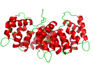
Protein S is a vitamin K-dependent plasma glycoprotein synthesized in the liver. In the circulation, Protein S exists in two forms: a free form and a complex form bound to complement protein C4b-binding protein (C4BP). In humans, protein S is encoded by the PROS1 gene. Protein S plays a role in coagulation.

<span class="mw-page-title-main">Factor V</span> Mammalian protein found in humans

Factor V is a protein of the coagulation system, rarely referred to as proaccelerin or labile factor. In contrast to most other coagulation factors, it is not enzymatically active but functions as a cofactor. Deficiency leads to predisposition for hemorrhage, while some mutations predispose for thrombosis.

<span class="mw-page-title-main">Annexin</span> Protein family

Annexin is a common name for a group of cellular proteins. They are mostly found in eukaryotic organisms.

<span class="mw-page-title-main">Dilute Russell's viper venom time</span>

Dilute Russell's viper venom time (dRVVT) is a laboratory test often used for detection of lupus anticoagulant (LA).

The prothrombinase enzyme complex consists of factor Xa (a serine protease) and factor Va (a protein cofactor). The complex assembles on negatively charged phospholipid membranes in the presence of calcium ions. The prothrombinase complex catalyzes the conversion of prothrombin (factor II), an inactive zymogen, to thrombin (factor IIa), an active serine protease. The activation of thrombin is a critical reaction in the coagulation cascade, which functions to regulate hemostasis in the body. To produce thrombin, the prothrombinase complex cleaves two peptide bonds in prothrombin, one after Arg271 and the other after Arg320. Although it has been shown that factor Xa can activate prothrombin when unassociated with the prothrombinase complex, the rate of thrombin formation is severely decreased under such circumstances. The prothrombinase complex can catalyze the activation of prothrombin at a rate 3 x 10⁵-fold faster than can factor Xa alone. Thus, the prothrombinase complex is required for the efficient production of activated thrombin and also for adequate hemostasis.

<span class="mw-page-title-main">Phospholipid scramblase</span> Protein

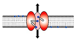
Scramblase is a protein responsible for the translocation of phospholipids between the two monolayers of a lipid bilayer of a cell membrane. In humans, phospholipid scramblases (PLSCRs) constitute a family of five homologous proteins that are named as hPLSCR1–hPLSCR5. Scramblases are members of the general family of transmembrane lipid transporters known as flippases. Scramblases are distinct from flippases and floppases. Scramblases, flippases, and floppases are three different types of enzymatic groups of phospholipid transportation enzymes. The inner-leaflet, facing the inside of the cell, contains negatively charged amino-phospholipids and phosphatidylethanolamine. The outer-leaflet, facing the outside environment, contains phosphatidylcholine and sphingomyelin. Scramblase is an enzyme, present in the cell membrane, that can transport (scramble) the negatively charged phospholipids from the inner-leaflet to the outer-leaflet, and vice versa.

In molecular biology, an annexin A5 affinity assay is a test to quantify the number of cells undergoing apoptosis. The assay uses the protein annexin A5 to tag apoptotic and dead cells, and the numbers are then counted using either flow cytometry or a fluorescence microscope.

<span class="mw-page-title-main">Phosphatidylethanolamine</span> Group of chemical compounds

Phosphatidylethanolamine (PE) is a class of phospholipids found in biological membranes. They are synthesized by the addition of cytidine diphosphate-ethanolamine to diglycerides, releasing cytidine monophosphate. S-Adenosyl methionine can subsequently methylate the amine of phosphatidylethanolamines to yield phosphatidylcholines.

<span class="mw-page-title-main">Annexin A1</span> Protein-coding gene in the species Homo sapiens

Annexin A1, also known as lipocortin I, is a protein that is encoded by the ANXA1 gene in humans.

<span class="mw-page-title-main">Annexin A2</span> Protein-coding gene in the species Homo sapiens

Annexin A2 also known as annexin II is a protein that in humans is encoded by the ANXA2 gene.

<span class="mw-page-title-main">Apolipoprotein H</span> Protein-coding gene in humans

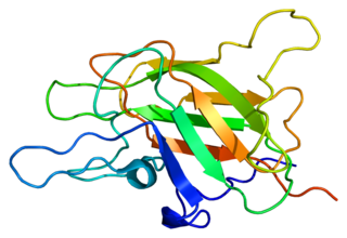
β2-glycoprotein 1, also known as beta-2 glycoprotein 1 and Apolipoprotein H (Apo-H), is a 38 kDa multifunctional plasma protein that in humans is encoded by the APOH gene. One of its functions is to bind cardiolipin. When bound, the structure of cardiolipin and β2-GP1 both undergo large changes in structure. Within the structure of Apo-H is a stretch of positively charged amino acids, Lys-Asn-Lys-Glu-Lys-Lys, are involved in phospholipid binding.

In autoimmune disease, anti-apolipoprotein H (AAHA) antibodies, also called anti-β2 glycoprotein I antibodies, comprise a subset of anti-cardiolipin antibodies and lupus anticoagulant. These antibodies are involved in sclerosis and are strongly associated with thrombotic forms of lupus. As a result, AAHA are strongly implicated in autoimmune deep vein thrombosis.

<span class="mw-page-title-main">Annexin A6</span> Protein-coding gene in the species Homo sapiens

Annexin A6 is a protein that in humans is encoded by the ANXA6 gene.

<span class="mw-page-title-main">Discoidin domain</span>

Discoidin domain is major protein domain of many blood coagulation factors.

<span class="mw-page-title-main">S100A10</span> Protein-coding gene in the species Homo sapiens

S100 calcium-binding protein A10 (S100A10), also known as p11, is a protein that is encoded by the S100A10 gene in humans and the S100a10 gene in other species. S100A10 is a member of the S100 family of proteins containing two EF-hand calcium-binding motifs. S100 proteins are localized in the cytoplasm and/or nucleus of a wide range of cells. They regulate a number of cellular processes such as cell cycle progression and differentiation. The S100 protein is implicated in exocytosis and endocytosis by reorganization of F-actin.

<span class="mw-page-title-main">Annexin A4</span> Protein-coding gene in the species Homo sapiens

Annexin A4 is a protein that in humans is encoded by the ANXA4 gene.

<span class="mw-page-title-main">Annexin A7</span> Protein-coding gene in the species Homo sapiens

Annexin A7 is a protein that in humans is encoded by the ANXA7 gene.

<span class="mw-page-title-main">Annexin A11</span> Protein-coding gene in the species Homo sapiens

Annexin A11 is a protein that in humans is encoded by the ANXA11 gene.

<span class="mw-page-title-main">Annexin A3</span> Protein-coding gene in the species Homo sapiens

Annexin A3 is a protein that in humans is encoded by the ANXA3 gene.

<span class="mw-page-title-main">ANXA8L2</span> Protein

Annexin A8-like protein 2 is a protein that in humans is encoded by the ANXA8L2 gene.

References

  1. 1 2 3 GRCh38: Ensembl release 89: ENSG00000164111 - Ensembl, May 2017
  2. 1 2 3 GRCm38: Ensembl release 89: ENSMUSG00000027712 - Ensembl, May 2017
  3. "Human PubMed Reference:". National Center for Biotechnology Information, U.S. National Library of Medicine.
  4. "Mouse PubMed Reference:". National Center for Biotechnology Information, U.S. National Library of Medicine.
  5. Sofiadis A, Becker S, Hellman U, Hultin-Rosenberg L, Dinets A, Hulchiy M, Zedenius J, Wallin G, Foukakis T, Höög A, Auer G, Lehtiö J, Larsson C (Apr 2012). "Proteomic profiling of follicular and papillary thyroid tumors". European Journal of Endocrinology. 166 (4): 657–67. doi:10.1530/EJE-11-0856. PMC   3315832 . PMID   22275472.
  6. Meers P and Mealy T (1994). "Phospholipid determinants for annexin V binding sites and the role of tryptophan". Biochemistry. 33 (19): 5829–37. doi:10.1021/bi00185a022. PMID   8180211.
  7. Koopman G, Reutelingsperger CP, Kuijten GA, Keehnen RM, Pals ST, van Oers MH (Sep 1994). "Annexin V for flow cytometric detection of phosphatidylserine expression on B cells undergoing apoptosis". Blood. 84 (5): 1415–20. doi:10.1182/blood.V84.5.1415.1415. PMID   8068938.
  8. Vermes I, Haanen C, Steffens-Nakken H, Reutelingsperger C (Jul 1995). "A novel assay for apoptosis. Flow cytometric detection of phosphatidylserine expression on early apoptotic cells using fluorescein labelled Annexin V". Journal of Immunological Methods. 184 (1): 39–51. doi:10.1016/0022-1759(95)00072-I. PMID   7622868.
  9. Annexin-FP488 fluorescent staining protocol at Interchim
  10. Wen Y, Edelman JL, Kang T, Sachs G (May 1999). "Lipocortin V may function as a signaling protein for vascular endothelial growth factor receptor-2/Flk-1". Biochemical and Biophysical Research Communications. 258 (3): 713–21. doi:10.1006/bbrc.1999.0678. PMID   10329451.
  11. Cardó-Vila M, Arap W, Pasqualini R (May 2003). "Alpha v beta 5 integrin-dependent programmed cell death triggered by a peptide mimic of annexin V". Molecular Cell. 11 (5): 1151–62. doi: 10.1016/S1097-2765(03)00138-2 . PMID   12769841.

Further reading