Calpain

Last updated • 6 min readFrom Wikipedia, The Free Encyclopedia
Calpain
PDB 1mdw EBI.jpg
Crystal structure of the peptidase core of Calpain II.
Identifiers
SymbolCalpain
Pfam PF00648
Pfam clan CL0125
InterPro IPR001300
SMART CysPc
PROSITE PDOC50203
MEROPS C2
SCOP2 1mdw / SCOPe / SUPFAM
Available protein structures:
Pfam   structures / ECOD  
PDB RCSB PDB; PDBe; PDBj
PDBsum structure summary
PDB 1tl9 A:55-354; 1kxr B:55-354; 1tlo A:55-354; 2ary B:55-354; 1zcm A:55-353; 1mdw B:45-344; 1u5i A:45-344; 1kfx L:45-344; 1kfu L:45-344; 1ziv A:42-337
calpain-1
Identifiers
EC no. 3.4.22.52
CAS no. 689772-75-6
Databases
IntEnz IntEnz view
BRENDA BRENDA entry
ExPASy NiceZyme view
KEGG KEGG entry
MetaCyc metabolic pathway
PRIAM profile
PDB structures RCSB PDB PDBe PDBsum
Search
PMC articles
PubMed articles
NCBI proteins
calpain-2
Identifiers
EC no. 3.4.22.53
CAS no. 702693-80-9
Databases
IntEnz IntEnz view
BRENDA BRENDA entry
ExPASy NiceZyme view
KEGG KEGG entry
MetaCyc metabolic pathway
PRIAM profile
PDB structures RCSB PDB PDBe PDBsum
Search
PMC articles
PubMed articles
NCBI proteins

A calpain ( /ˈkælpn/ ; [1] EC 3.4.22.52, EC 3.4.22.53) is a protein belonging to the family of calcium-dependent, non-lysosomal cysteine proteases (proteolytic enzymes) expressed ubiquitously in mammals and many other organisms. Calpains constitute the C2 family of protease clan CA in the MEROPS database. The calpain proteolytic system includes the calpain proteases, the small regulatory subunit CAPNS1, also known as CAPN4, and the endogenous calpain-specific inhibitor, calpastatin.

Contents

Discovery

The history of calpain's discovery originates in 1964, when calcium-dependent proteolytic activities caused by a "calcium-activated neutral protease" (CANP) were detected in brain, lens of the eye and other tissues. In the late 1960s the enzymes were isolated and characterised independently in both rat brain and skeletal muscle. These activities were caused by an intracellular cysteine protease not associated with the lysosome and having an optimum activity at neutral pH, which clearly distinguished it from the cathepsin family of proteases. The calcium-dependent activity, intracellular localization, and the limited, specific proteolysis on its substrates, highlighted calpain’s role as a regulatory, rather than a digestive, protease. When the sequence of this enzyme became known, [2] it was given the name "calpain", to recognize its common properties with two well-known proteins at the time, the calcium-regulated signalling protein, calmodulin, and the cysteine protease of papaya, papain. Shortly thereafter, the activity was found to be attributable to two main isoforms, dubbed μ ("mu")-calpain and m-calpain (or calpain I and II), that differed primarily in their calcium requirements in vitro. Their names reflect the fact that they are activated by micro- and nearly millimolar concentrations of Ca2+ within the cell, respectively. [3]

To date, these two isoforms remain the best characterised members of the calpain family. Structurally, these two heterodimeric isoforms share an identical small (28 kDa) subunit (CAPNS1 (formerly CAPN4)), but have distinct large (80 kDa) subunits, known as calpain 1 and calpain 2 (each encoded by the CAPN1 and CAPN2 genes, respectively).

Cleavage specificity

No specific amino acid sequence is uniquely recognized by calpains. Amongst protein substrates, tertiary structure elements rather than primary amino acid sequences are likely responsible for directing cleavage to a specific substrate. Amongst peptide and small-molecule substrates, the most consistently reported specificity is for small, hydrophobic amino acids (e.g. leucine, valine and isoleucine) at the P2 position, and large hydrophobic amino acids (e.g. phenylalanine and tyrosine) at the P1 position. [4] Arguably, the best currently available fluorogenic calpain substrate is (EDANS)-Glu-Pro-Leu-Phe=Ala-Glu-Arg-Lys-(DABCYL), with cleavage occurring at the Phe=Ala bond.

Extended family

The Human Genome Project has revealed that more than a dozen other calpain isoforms exist, some with multiple splice variants. [5] [6] [7] As the first calpain whose three-dimensional structure was determined, m-calpain is the type-protease for the C2 (calpain) family in the MEROPS database.

GeneProteinAliasesTissue expressionDisease linkage
CAPN1 Calpain 1Calpain-1 large subunit, Calpain mu-typeubiquitous
CAPN2 Calpain 2Calpain-2 large subunit, Calpain m-typeubiquitous
CAPN3 Calpain 3skeletal muscle retina and lens specificLimb Girdle muscular dystrophy 2A
CAPN5 Calpain 5ubiquitous (high in colon, small intestine and testis)might be linked to necrosis,
as it is an ortholog of the C. elegans necrosis gene tra-3
CAPN6 Calpain 6CAPNX, Calpamodulin
CAPN7 Calpain 7palBHubiquitous
CAPN8 Calpain 8exclusive to stomach mucosa and the GI tractmight be linked to colon polyp formation
CAPN9 Calpain 9exclusive to stomach mucosa and the GI tractmight be linked to colon polyp formation
CAPN10 Calpain 10susceptibility gene for type II diabetes
CAPN11 Calpain 11testis
CAPN12 Calpain 12ubiquitous but high in hair follicle
CAPN13 Calpain 13testis and lung
CAPN14 Calpain 14ubiquitous
CAPN17 Calpain 17Fish and amphibian-only
SOLH Calpain 15Sol H (homolog of the drosophila gene sol)
CAPNS1 Calpain small subunit 1Calpain 4
CAPNS2 Calpain small subunit 2

Function

Although the physiological role of calpains is still poorly understood, they have been shown to be active participants in processes such as cell mobility and cell cycle progression, as well as cell-type specific functions such as long-term potentiation in neurons and cell fusion in myoblasts. Under these physiological conditions, a transient and localized influx of calcium into the cell activates a small local population of calpains (for example, those close to Ca2+ channels), which then advance the signal transduction pathway by catalyzing the controlled proteolysis of its target proteins. [8] Additionally, phosphorylation by protein kinase A and dephosphorylation by alkaline phosphatase have been found to positively regulate the activity of μ-calpains by increasing random coils and decreasing β-sheets in its structure. Phosphorylation improves proteolytic activity and stimulates auto-activation of μ-calpains. However, increased calcium concentration overruns the effects of phosphorylation and dephosphorylation on calpain activity, and thus calpain activity ultimately depends on the presence of calcium. [9] Other reported roles of calpains are in cell function, helping to regulate clotting and the diameter of blood vessels, and playing a role in memory. Calpains have been implicated in apoptotic cell death, and appear to be an essential component of necrosis. Detergent fractionation revealed the cytosolic localization of calpain. [8]

Enhanced calpain activity, regulated by CAPNS1, significantly contributes to platelet hyperreactivity under hypoxic environment. [10]

In the brain, while μ-calpain is mainly located in the cell body and dendrites of neurons and to a lesser extent in axons and glial cells, m-calpain is found in glia and a small number in axons. [11] Calpain is also involved in skeletal muscle protein breakdown due to exercise and altered nutritional states. [12]

Clinical significance

Pathology

The structural and functional diversity of calpains in the cell is reflected in their involvement in the pathogenesis of a wide range of disorders. At least two well known genetic disorders and one form of cancer have been linked to tissue-specific calpains. When defective, the mammalian calpain 3 (also known as p94) is the gene product responsible for limb-girdle muscular dystrophy type 2A, [13] [14] calpain 10 has been identified as a susceptibility gene for type II diabetes mellitus, and calpain 9 has been identified as a tumour suppressor for gastric cancer. Moreover, the hyperactivation of calpains is implicated in a number of pathologies associated with altered calcium homeostasis such as Alzheimer's disease, [15] and cataract formation, as well as secondary degeneration resulting from acute cellular stress following myocardial ischemia, cerebral (neuronal) ischemia, traumatic brain injury and spinal cord injury. Excessive amounts of calpain can be activated due to Ca2+ influx after cerebrovascular accident (during the ischemic cascade) or some types of traumatic brain injury such as diffuse axonal injury. Increase in concentration of calcium in the cell results in calpain activation, which leads to unregulated proteolysis of both target and non-target proteins and consequent irreversible tissue damage. Excessively active calpain breaks down molecules in the cytoskeleton such as spectrin, microtubule subunits, microtubule-associated proteins, and neurofilaments. [16] [17] It may also damage ion channels, other enzymes, cell adhesion molecules, and cell surface receptors. [11] This can lead to degradation of the cytoskeleton and plasma membrane. Calpain may also break down sodium channels that have been damaged due to axonal stretch injury, [18] leading to an influx of sodium into the cell. This, in turn, leads to the neuron's depolarization and the influx of more Ca2+. A significant consequence of calpain activation is the development of cardiac contractile dysfunction that follows ischemic insult to the heart. Upon reperfusion of the ischemic myocardium, there is development of calcium overload or excess in the heart cell (cardiomyocytes). This increase in calcium leads to activation of calpain. [19] [ irrelevant citation ] Recently calpain has been implicated in promoting high altitude induced venous thrombosis by mediating platelet hyperactivation. [10]

Therapeutic inhibitors

The exogenous regulation of calpain activity is therefore of interest for the development of therapeutics in a wide array of pathological states. As a few of the many examples supporting the therapeutic potential of calpain inhibition in ischemia, calpain inhibitor AK275 protected against focal ischemic brain damage in rats when administered after ischemia, and MDL28170 significantly reduced the size of damaged infarct tissue in a rat focal ischemia model. Also, calpain inhibitors are known to have neuroprotective effects: PD150606, [20] SJA6017, [21] ABT-705253, [22] [23] and SNJ-1945. [24]

Calpain may be released in the brain for up to a month after a head injury, and may be responsible for a shrinkage of the brain sometimes found after such injuries. [25] However, calpain may also be involved in a "resculpting" process that helps repair damage after injury. [25]

See also

Related Research Articles

<span class="mw-page-title-main">Proteolysis</span> Breakdown of proteins into smaller polypeptides or amino acids

Proteolysis is the breakdown of proteins into smaller polypeptides or amino acids. Uncatalysed, the hydrolysis of peptide bonds is extremely slow, taking hundreds of years. Proteolysis is typically catalysed by cellular enzymes called proteases, but may also occur by intra-molecular digestion.

<span class="mw-page-title-main">Proteasome</span> Protein complexes which degrade unnecessary or damaged proteins by proteolysis

Proteasomes are protein complexes which degrade unneeded or damaged proteins by proteolysis, a chemical reaction that breaks peptide bonds. Enzymes that help such reactions are called proteases.

<span class="mw-page-title-main">Protease</span> Enzyme that cleaves other proteins into smaller peptides

A protease is an enzyme that catalyzes proteolysis, breaking down proteins into smaller polypeptides or single amino acids, and spurring the formation of new protein products. They do this by cleaving the peptide bonds within proteins by hydrolysis, a reaction where water breaks bonds. Proteases are involved in many biological functions, including digestion of ingested proteins, protein catabolism, and cell signaling.

<span class="mw-page-title-main">Excitotoxicity</span> Process that kills nerve cells

In excitotoxicity, nerve cells suffer damage or death when the levels of otherwise necessary and safe neurotransmitters such as glutamate become pathologically high, resulting in excessive stimulation of receptors. For example, when glutamate receptors such as the NMDA receptor or AMPA receptor encounter excessive levels of the excitatory neurotransmitter, glutamate, significant neuronal damage might ensue. Excess glutamate allows high levels of calcium ions (Ca2+) to enter the cell. Ca2+ influx into cells activates a number of enzymes, including phospholipases, endonucleases, and proteases such as calpain. These enzymes go on to damage cell structures such as components of the cytoskeleton, membrane, and DNA. In evolved, complex adaptive systems such as biological life it must be understood that mechanisms are rarely, if ever, simplistically direct. For example, NMDA in subtoxic amounts induces neuronal survival of otherwise toxic levels of glutamate.

<span class="mw-page-title-main">Diffuse axonal injury</span> Medical condition

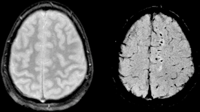
Diffuse axonal injury (DAI) is a brain injury in which scattered lesions occur over a widespread area in white matter tracts as well as grey matter. DAI is one of the most common and devastating types of traumatic brain injury and is a major cause of unconsciousness and persistent vegetative state after severe head trauma. It occurs in about half of all cases of severe head trauma and may be the primary damage that occurs in concussion. The outcome is frequently coma, with over 90% of patients with severe DAI never regaining consciousness. Those who awaken from the coma often remain significantly impaired.

<span class="mw-page-title-main">Cathepsin</span> Family of proteases

Cathepsins are proteases found in all animals as well as other organisms. There are approximately a dozen members of this family, which are distinguished by their structure, catalytic mechanism, and which proteins they cleave. Most of the members become activated at the low pH found in lysosomes. Thus, the activity of this family lies almost entirely within those organelles. There are, however, exceptions such as cathepsin K, which works extracellularly after secretion by osteoclasts in bone resorption. Cathepsins have a vital role in mammalian cellular turnover.

<span class="mw-page-title-main">Cysteine protease</span> Class of enzymes

Cysteine proteases, also known as thiol proteases, are hydrolase enzymes that degrade proteins. These proteases share a common catalytic mechanism that involves a nucleophilic cysteine thiol in a catalytic triad or dyad.

<span class="mw-page-title-main">Sterol regulatory element-binding protein</span> Protein family

Sterol regulatory element-binding proteins (SREBPs) are transcription factors that bind to the sterol regulatory element DNA sequence TCACNCCAC. Mammalian SREBPs are encoded by the genes SREBF1 and SREBF2. SREBPs belong to the basic-helix-loop-helix leucine zipper class of transcription factors. Unactivated SREBPs are attached to the nuclear envelope and endoplasmic reticulum membranes. In cells with low levels of sterols, SREBPs are cleaved to a water-soluble N-terminal domain that is translocated to the nucleus. These activated SREBPs then bind to specific sterol regulatory element DNA sequences, thus upregulating the synthesis of enzymes involved in sterol biosynthesis. Sterols in turn inhibit the cleavage of SREBPs and therefore synthesis of additional sterols is reduced through a negative feed back loop.

In molecular biology, the Signal Peptide Peptidase (SPP) is a type of protein that specifically cleaves parts of other proteins. It is an intramembrane aspartyl protease with the conserved active site motifs 'YD' and 'GxGD' in adjacent transmembrane domains (TMDs). Its sequences is highly conserved in different vertebrate species. SPP cleaves remnant signal peptides left behind in membrane by the action of signal peptidase and also plays key roles in immune surveillance and the maturation of certain viral proteins.

Taicatoxin (TCX) is a snake toxin that blocks voltage-dependent L-type calcium channels and small conductance Ca2+-activated K+ channels. The name taicatoxin (TAIpan + CAlcium + TOXIN) is derived from its natural source, the taipan snake, the site of its action, calcium channels, and from its function as a toxin. Taicatoxin was isolated from the venom of Australian taipan snake, Oxyuranus scutellatus scutellatus. TCX is a secreted protein, produced in the venom gland of the snake.

Phosphodiesterase 1, PDE1, EC 3.1.4.1, systematic name oligonucleotide 5-nucleotidohydrolase) is a phosphodiesterase enzyme also known as calcium- and calmodulin-dependent phosphodiesterase. It is one of the 11 families of phosphodiesterase (PDE1-PDE11). Phosphodiesterase 1 has three subtypes, PDE1A, PDE1B and PDE1C which divide further into various isoforms. The various isoforms exhibit different affinities for cAMP and cGMP.

Calpain-2 is an intracellular heterodimeric calcium-activated cysteine protease. This enzyme catalyses the following chemical reaction

<span class="mw-page-title-main">Calpain-2 catalytic subunit</span> Protein-coding gene in the species Homo sapiens

Calpain-2 catalytic subunit is a protein that in humans is encoded by the CAPN2 gene.

<span class="mw-page-title-main">Proteinase K</span> Broad-spectrum serine protease

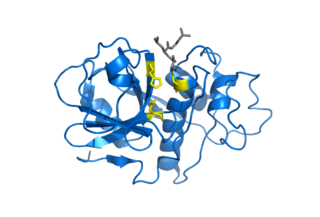
In molecular biology, Proteinase K is a broad-spectrum serine protease. The enzyme was discovered in 1974 in extracts of the fungus Parengyodontium album. Proteinase K is able to digest hair (keratin), hence, the name "Proteinase K". The predominant site of cleavage is the peptide bond adjacent to the carboxyl group of aliphatic and aromatic amino acids with blocked alpha amino groups. It is commonly used for its broad specificity. This enzyme belongs to Peptidase family S8 (subtilisin). The molecular weight of Proteinase K is 28,900 daltons.

<span class="mw-page-title-main">Calpain-1 catalytic subunit</span> Protein-coding gene in the species Homo sapiens

Calpain-1 catalytic subunit(CANP 1) is a protein that in humans is encoded by the CAPN1 gene.

<span class="mw-page-title-main">Calpastatin</span> Protein-coding gene in the species Homo sapiens

Calpastatin is a protein that in humans is encoded by the CAST gene.

<span class="mw-page-title-main">MG132</span> Chemical compound

MG132 is a potent, reversible, and cell-permeable proteasome inhibitor (Ki = 4 nM). It belongs to the class of synthetic peptide aldehydes. It reduces the degradation of ubiquitin-conjugated proteins in mammalian cells and permeable strains of yeast by the 26S complex without affecting its ATPase or isopeptidase activities. MG132 activates c-Jun N-terminal kinase (JNK1), which initiates apoptosis. MG132 also inhibits NF-κB activation with an IC50 of 3 μM and prevents β-secretase cleavage.

<span class="mw-page-title-main">Acid-sensing ion channel</span> Class of transport proteins

Acid-sensing ion channels (ASICs) are neuronal voltage-insensitive sodium channels activated by extracellular protons permeable to Na+. ASIC1 also shows low Ca2+ permeability. ASIC proteins are a subfamily of the ENaC/Deg superfamily of ion channels. These genes have splice variants that encode for several isoforms that are marked by a suffix. In mammals, acid-sensing ion channels (ASIC) are encoded by five genes that produce ASIC protein subunits: ASIC1, ASIC2, ASIC3, ASIC4, and ASIC5. Three of these protein subunits assemble to form the ASIC, which can combine into both homotrimeric and heterotrimeric channels typically found in both the central nervous system and peripheral nervous system. However, the most common ASICs are ASIC1a and ASIC1a/2a and ASIC3. ASIC2b is non-functional on its own but modulates channel activity when participating in heteromultimers and ASIC4 has no known function. On a broad scale, ASICs are potential drug targets due to their involvement in pathological states such as retinal damage, seizures, and ischemic brain injury.

<span class="mw-page-title-main">Oligopeptidase</span>

An Oligopeptidase is an enzyme that cleaves peptides but not proteins. This property is due to its structure: the active site of this enzyme is located at the end of a narrow cavity which can only be reached by peptides.

<span class="mw-page-title-main">Asparagine endopeptidase</span> Class of enzymes

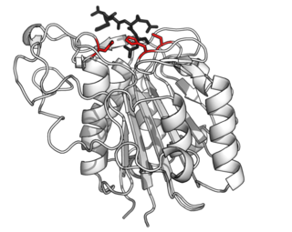
Asparagine endopeptidase is a proteolytic enzyme from C13 peptidase family which hydrolyses a peptide bond using the thiol group of a cysteine residue as a nucleophile. It is also known as asparaginyl endopeptidase, citvac, proteinase B, hemoglobinase, PRSC1 gene product or LGMN, vicilin peptidohydrolase and bean endopeptidase. In humans it is encoded by the LGMN gene.

References

  1. "the definition of calpain". Dictionary.com. Retrieved 23 April 2018.
  2. Ohno S, Emori Y, Imajoh S, Kawasaki H, Kisaragi M, Suzuki K (1984). "Evolutionary origin of a calcium-dependent protease by fusion of genes for a thiol protease and a calcium-binding protein?". Nature. 312 (5994): 566–70. Bibcode:1984Natur.312..566O. doi:10.1038/312566a0. PMID   6095110. S2CID   4359635.
  3. Glass JD, Culver DG, Levey AI, Nash NR (April 2002). "Very early activation of m-calpain in peripheral nerve during Wallerian degeneration". J. Neurol. Sci. 196 (1–2): 9–20. doi:10.1016/S0022-510X(02)00013-8. PMID   11959150. S2CID   22674283.
  4. Cuerrier D, Moldoveanu T, Davies PL (December 2005). "Determination of peptide substrate specificity for mu-calpain by a peptide library-based approach: the importance of primed side interactions". J. Biol. Chem. 280 (49): 40632–41. doi: 10.1074/jbc.M506870200 . PMID   16216885.
  5. Thompson V (2002-02-12). "Calpain Nomenclature". College of Agriculture and Life Sciences at the University of Arizona. Retrieved 2010-08-06.
  6. Huang Y, Wang KK (August 2001). "The calpain family and human disease". Trends Mol Med. 7 (8): 355–62. doi:10.1016/S1471-4914(01)02049-4. PMID   11516996.
  7. Suzuki K, Hata S, Kawabata Y, Sorimachi H (February 2004). "Structure, activation, and biology of calpain". Diabetes. 53. Suppl 1: S12–8. doi: 10.2337/diabetes.53.2007.s12 . PMID   14749260.
  8. 1 2 Jaguva Vasudevan, AA; Perkovic, M; Bulliard, Y; Cichutek, K; Trono, D; Häussinger, D; Münk, C (August 2013). "Prototype foamy virus Bet impairs the dimerization and cytosolic solubility of human APOBEC3G". Journal of Virology. 87 (16): 9030–40. doi:10.1128/JVI.03385-12. PMC   3754047 . PMID   23760237.
  9. Du, Manting; Li, Xin; Li, Zheng; Shen, Qingwu; Wang, Ying; Li, Guixia; Zhang, Dequan (2018-06-30). "Phosphorylation regulated by protein kinase A and alkaline phosphatase play positive roles in μ-calpain activity". Food Chemistry. 252: 33–39. doi:10.1016/j.foodchem.2018.01.103. ISSN   0308-8146. PMID   29478550. S2CID   3538480.
  10. 1 2 Tyagi, T.; Ahmad, S.; Gupta, N.; Sahu, A.; Ahmad, Y.; Nair, V.; Chatterjee, T.; Bajaj, N.; Sengupta, S.; Ganju, L.; Singh, S. B.; Ashraf, M. Z. (Feb 2014). "Altered expression of platelet proteins and calpain activity mediate hypoxia-induced prothrombotic phenotype". Blood. 123 (8): 1250–60. doi: 10.1182/blood-2013-05-501924 . PMID   24297866.
  11. 1 2 Lenzlinger PM, Saatman KE, Raghupathi R, Mcintosh TK (2000). "Chapter 1: Overview of basic mechanisms underlying neuropathological consequences of head trauma". In Newcomb JK, Miller LS, Hayes RL (eds.). Head trauma: basic, preclinical, and clinical directions. New York: Wiley-Liss. ISBN   978-0-471-36015-5.
  12. Belcastro AN, Albisser TA, Littlejohn B (October 1996). "Role of calcium-activated neutral protease (calpain) with diet and exercise". Can J Appl Physiol. 21 (5): 328–46. doi:10.1139/h96-029. PMID   8905185.
  13. Richard I, Broux O, Allamand V, et al. (April 1995). "Mutations in the proteolytic enzyme calpain 3 cause limb-girdle muscular dystrophy type 2A". Cell. 81 (1): 27–40. doi: 10.1016/0092-8674(95)90368-2 . PMID   7720071. S2CID   17565219.
  14. Ono Y, Shimada H, Sorimachi H, et al. (July 1998). "Functional defects of a muscle-specific calpain, p94, caused by mutations associated with limb-girdle muscular dystrophy type 2A". J. Biol. Chem. 273 (27): 17073–8. doi: 10.1074/jbc.273.27.17073 . PMID   9642272.
  15. Yamashima T (2013). "Reconsider Alzheimer's disease by the 'calpain-cathepsin hypothesis'--a perspective review". Progress in Neurology. 105: 1–23. doi:10.1016/j.pneurobio.2013.02.004. PMID   23499711. S2CID   39292302.
  16. Liu J, Liu MC, Wang KK (April 2008). "Calpain in the CNS: from synaptic function to neurotoxicity". Sci. Signal. 1 (14): re 1. doi:10.1126/stke.114re1. PMID   18398107. S2CID   21992464.
  17. Castillo MR, Babson JR (October 1998). "Ca2+-dependent mechanisms of cell injury in cultured cortical neurons". Neuroscience. 86 (4): 1133–44. doi:10.1016/S0306-4522(98)00070-0. PMID   9697120. S2CID   54228571.
  18. Iwata A, Stys PK, Wolf JA, et al. (May 2004). "Traumatic axonal injury induces proteolytic cleavage of the voltage-gated sodium channels modulated by tetrodotoxin and protease inhibitors". J. Neurosci. 24 (19): 4605–13. doi: 10.1523/JNEUROSCI.0515-03.2004 . PMC   6729402 . PMID   15140932.
  19. Neuhof C, Neuhof H (2014). "Calpain system and its involvement in myocardial ischemia and reperfusion injury". World J Cardiol. 7 (6): 638–52. doi: 10.4330/wjc.v6.i7.638 . PMC   4110612 . PMID   25068024.
  20. Wang KK, Nath R, Posner A, Raser KJ, Buroker-Kilgore M, Hajimohammadreza I, Probert AW, Marcoux FW, Ye Q, Takano E, Hatanaka M, Maki M, Caner H, Collins JL, Fergus A, Lee KS, Lunney EA, Hays SJ, Yuen P (June 1996). "An alpha-mercaptoacrylic acid derivative is a selective nonpeptide cell-permeable calpain inhibitor and is neuroprotective". Proc. Natl. Acad. Sci. U.S.A. 93 (13): 6687–92. Bibcode:1996PNAS...93.6687W. doi: 10.1073/pnas.93.13.6687 . PMC   39087 . PMID   8692879.
  21. Kupina NC, Nath R, Bernath EE, Inoue J, Mitsuyoshi A, Yuen PW, Wang KK, Hall ED (November 2001). "The novel calpain inhibitor SJA6017 improves functional outcome after delayed administration in a mouse model of diffuse brain injury" (PDF). J. Neurotrauma. 18 (11): 1229–40. doi:10.1089/089771501317095269. hdl: 2027.42/63231 . PMID   11721741.
  22. Lubisch W, Beckenbach E, Bopp S, Hofmann HP, Kartal A, Kästel C, Lindner T, Metz-Garrecht M, Reeb J, Regner F, Vierling M, Möller A (June 2003). "Benzoylalanine-derived ketoamides carrying vinylbenzyl amino residues: discovery of potent water-soluble calpain inhibitors with oral bioavailability". J. Med. Chem. 46 (12): 2404–12. doi:10.1021/jm0210717. PMID   12773044.
  23. Nimmrich V, Reymann KG, Strassburger M, Schöder UH, Gross G, Hahn A, Schoemaker H, Wicke K, Möller A (April 2010). "Inhibition of calpain prevents NMDA-induced cell death and beta-amyloid-induced synaptic dysfunction in hippocampal slice cultures". Br. J. Pharmacol. 159 (7): 1523–31. doi:10.1111/j.1476-5381.2010.00652.x. PMC   2850408 . PMID   20233208.
  24. Koumura A, Nonaka Y, Hyakkoku K, Oka T, Shimazawa M, Hozumi I, Inuzuka T, Hara H (November 2008). "A novel calpain inhibitor, ((1S)-1((((1S)-1-benzyl-3-cyclopropylamino-2,3-di-oxopropyl)amino)carbonyl)-3-methylbutyl) carbamic acid 5-methoxy-3-oxapentyl ester, protects neuronal cells from cerebral ischemia-induced damage in mice". Neuroscience. 157 (2): 309–18. doi:10.1016/j.neuroscience.2008.09.007. PMID   18835333. S2CID   29425598.
  25. 1 2 White V (1999-10-21). "– 'Biochemical Storm' Following Brain Trauma An Important Factor In Treatment, University of Florida Researcher Finds". University of Florida News. Archived from the original on 2011-06-23. Retrieved 2010-08-07.

Further reading