Haptoglobin

Last updated
HP
Available structures
PDB Ortholog search: PDBe RCSB
Identifiers
Aliases HP , BP, HP2ALPHA2, HPA1S, haptoglobin
External IDs OMIM: 140100 MGI: 96211 HomoloGene: 121756 GeneCards: HP
Orthologs
SpeciesHumanMouse
Entrez
Ensembl
UniProt
RefSeq (mRNA)

NM_001126102
NM_005143
NM_001318138

NM_017370
NM_001329965

RefSeq (protein)

NP_001119574
NP_001305067
NP_005134

NP_001316894
NP_059066

Location (UCSC) Chr 16: 72.05 – 72.06 Mb Chr 8: 110.3 – 110.31 Mb
PubMed search [3] [4]
Wikidata
View/Edit Human View/Edit Mouse
A model of a,b-hemoglobin/haptoglobin hexamer complex. There are 2 a,b-hemoglobin dimers depicted: one space filling model (yellow/orange), and one ribbon model (purple/blue). Each is bound by a haptoglobin molecule (both haptoglobin molecules are shown in pink, with one as a space filling model and one as a ribbon model). Hem-hap 4f4o.jpg
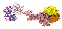
A model of α,β-hemoglobin/haptoglobin hexamer complex. There are 2 α,β-hemoglobin dimers depicted: one space filling model (yellow/orange), and one ribbon model (purple/blue). Each is bound by a haptoglobin molecule (both haptoglobin molecules are shown in pink, with one as a space filling model and one as a ribbon model).

Haptoglobin (abbreviated as Hp) is the protein that in humans is encoded by the HP gene. [5] [6] In blood plasma, haptoglobin binds with high affinity to free hemoglobin [7] released from erythrocytes, and thereby inhibits its deleterious oxidative activity. Compared to Hp, hemopexin binds to free heme. [8] The haptoglobin-hemoglobin complex will then be removed by the reticuloendothelial system (mostly the spleen).

Contents

In clinical settings, the haptoglobin assay is used to screen for and monitor intravascular hemolytic anemia. In intravascular hemolysis, free hemoglobin will be released into circulation and hence haptoglobin will bind the hemoglobin. This causes a decline in haptoglobin levels.

The protein was discovered as a "plasma substance" in 1938 by French biochemists Max-Fernand Jayle and Michel Polonovski. [9] [10]

Function

Hemoglobin that has been released into the blood plasma by damaged red blood cells has harmful effects. The HP gene encodes a preproprotein that is processed to yield both alpha and beta chains, which subsequently combines as a tetramer to produce haptoglobin. Haptoglobin functions to bind the free plasma hemoglobin, which allows degradative enzymes to gain access to the hemoglobin while at the same time preventing loss of iron through the kidneys and protecting the kidneys from damage by hemoglobin. [11]

The cellular receptor target of Hp is the monocyte/macrophage scavenger receptor, CD163. [7] Following Hb-Hp binding to CD163, cellular internalization of the complex leads to globin and heme metabolism, which is followed by adaptive changes in antioxidant and iron metabolism pathways and macrophage phenotype polarization. [7]

Hp has hemoglobin-independent immunomodulatory functions. It dampens lipopolysaccharide-induced cytokine expression. [12] Lipopolysaccharides directly bind to Hp, which, due to the high abundance of Hp in serum, results in their buffering and shielding from toll-like receptor 4. Functionally, this results in delayed activation of the NF-κB pathway. [13]

Difference from hemopexin

When hemoglobin is released from RBCs within the physiologic range of haptoglobin, the potential deleterious effects of hemoglobin are prevented. During hyper-hemolytic conditions or with chronic hemolysis, haptoglobin is depleted and hemoglobin readily distributes to tissues where it might be exposed to oxidative conditions. In such conditions, heme can be released from ferric (Fe3+-bound) hemoglobin. The free heme can then accelerate tissue damage by promoting peroxidative reactions and activation of inflammatory cascades. Hemopexin (Hx) is another plasma glycoprotein that is able to bind heme with high affinity. It sequesters heme in an inert non-toxic form and transports it to the liver for catabolism and excretion. [7]

Synthesis

Haptoglobin is produced mostly by hepatic cells but also by other tissues such as skin, lung and kidney. In addition, the haptoglobin gene is expressed in murine and human adipose tissue. [14]

Haptoglobin had been shown to be expressed in adipose tissue of cattle as well. [15]

Structure

Haptoglobin, in its simplest form, consists of two alpha and two beta chains, connected by disulfide bridges. The chains originate from a common precursor protein, which is proteolytically cleaved during protein synthesis.

Hp exists in two allelic forms in the human population, so-called Hp1 and Hp2, the latter one having arisen due to the partial duplication of Hp1 gene. Three genotypes of Hp, therefore, are found in humans: Hp1-1, Hp2-1, and Hp2-2. Hp of different genotypes have been shown to bind hemoglobin with different affinities, with Hp2-2 being the weakest binder. Allele 2 encodes for two multimerization domains. This results in oligomer formation in carriers of allele 2. [5] The frequency of the short allele varies between 7% and 70% depending on racial origin. [16] It is unclear which evolutionary advantage is conferred by the longer allele; strikingly, a similar partial duplication independently arose much earlier in a precursor of ruminants. Ruminants exclusively express oligomeric haptoglobin. [17]

In other species

Hp has been found in all mammals studied so far, some birds, e.g., cormorant and ostrich but also, in its simpler form, in bony fish, e.g., zebrafish. Hp is absent in at least some amphibians ( Xenopus ) and neognathous birds (chicken and goose).

Clinical significance

Mutations in this gene or its regulatory regions cause ahaptoglobinemia or hypohaptoglobinemia. This gene has also been linked to diabetic nephropathy, [18] the incidence of coronary artery disease in type 1 diabetes, [19] Crohn's disease, [20] inflammatory disease behavior, primary sclerosing cholangitis, susceptibility to idiopathic Parkinson's disease, [21] and a reduced incidence of Plasmodium falciparum malaria. [22]

Since the reticuloendothelial system will remove the haptoglobin-hemoglobin complex from the body, [8] haptoglobin levels will be decreased in case of intravascular hemolysis or severe extravascular hemolysis. In the process of binding to free hemoglobin, haptoglobin sequesters the iron within hemoglobin, preventing iron-utilizing bacteria from benefiting from hemolysis. It is theorized that, because of this, haptoglobin has evolved into an acute-phase protein. HP has a protective influence on the hemolytic kidney. [23] [24]

The different haptoglobin phenotypes differ in their antioxidant, scavenging, [25] and immunomodulatory properties. This aspect of haptoglobin may gain importance in immune suppressed conditions (such as liver cirrhosis) and the various phenotypes may result in different susceptibility levels towards bacterial infections. [26]

Some studies associate certain haptoglobin phenotypes with the risk of developing hypertension in diabetes [27] and schizophrenia. [28]

Test protocol

Measuring the level of haptoglobin in a patient's blood is ordered whenever a patient exhibits symptoms of anemia, such as pallor, fatigue, or shortness of breath, along with physical signs of hemolysis, such as jaundice or dark-colored urine. The test is also commonly ordered as a hemolytic anemia battery, which also includes a reticulocyte count and a peripheral blood smear. It can also be ordered along with a direct antiglobulin test when a patient is suspected of having a transfusion reaction or symptoms of autoimmune hemolytic anemia. Also, it may be ordered in conjunction with a bilirubin.

Interpretation

A decrease in haptoglobin can support a diagnosis of hemolysis, especially when correlated with a decreased hemoglobin, and hematocrit, and also an increased reticulocyte count. Low haptoglobin levels occur regardless of the site and mechanism of haemolysis (intravascular and splenic/"extravascular") [29]

If the reticulocyte count is increased, but the haptoglobin level is normal, this argues against haemolysis, and suggests a bone marrow response to blood loss. If there are symptoms of anemia but both the reticulocyte count and the haptoglobin level are normal, the anemia is most likely not due to hemolysis. Haptoglobin levels that are decreased but do not accompany signs of anemia may indicate advanced liver damage, as the liver is the major site of production of haptoglobin.

As haptoglobin is an acute-phase protein, any inflammatory process (infection, injury, allergy, etc.) may increase the levels of plasma haptoglobin, but patients with haemolysis usually have low haptoglobin regardless of the presence of inflammation [30]

See also

Related Research Articles

<span class="mw-page-title-main">Hemoglobin</span> Metalloprotein that binds with oxygen

Hemoglobin is a protein containing iron that facilitates the transport of oxygen in red blood cells. Almost all vertebrates contain hemoglobin, with the exception of the fish family Channichthyidae and the tissues of some invertebrate animals. Hemoglobin in the blood carries oxygen from the respiratory organs to the other tissues of the body, where it releases the oxygen to enable aerobic respiration which powers the animal's metabolism. A healthy human has 12 to 20 grams of hemoglobin in every 100 mL of blood. Hemoglobin is a metalloprotein, a chromoprotein, and globulin.

<span class="mw-page-title-main">Red blood cell</span> Oxygen-delivering blood cell and the most common type of blood cell

Red blood cells (RBCs), scientific name erythrocytes (from Greek erythros 'red' and kytos 'hollow vessel', with -cyte translated as 'cell' in modern usage), also referred to as red cells, red blood corpuscles (in humans or other animals not having nucleus in red blood cells) or haematids, are the most common type of blood cell and the vertebrate's principal means of delivering oxygen (O2) to the body tissues—via blood flow through the circulatory system. Erythrocytes take up oxygen in the lungs, or in fish the gills, and release it into tissues while squeezing through the body's capillaries.

<span class="mw-page-title-main">Hemolysis</span> Rupturing of red blood cells and release of their contents

Hemolysis or haemolysis, also known by several other names, is the rupturing (lysis) of red blood cells (erythrocytes) and the release of their contents (cytoplasm) into surrounding fluid. Hemolysis may occur in vivo or in vitro.

<span class="mw-page-title-main">Hemoglobinopathy</span> Any of various genetic disorders of blood

Hemoglobinopathy is the medical term for a group of inherited blood disorders and diseases that primarily affect red blood cells. They are single-gene disorders and, in most cases, they are inherited as autosomal co-dominant traits.

<span class="mw-page-title-main">Hereditary spherocytosis</span> Medical condition

Hereditary spherocytosis (HS) is a congenital hemolytic disorder, wherein a genetic mutation coding for a structural membrane protein phenotype leads to a spherical shaping of erythrocytic cellular morphology. As erythrocytes are sphere-shaped (spherocytosis), rather than the normal biconcave disk-shaped, their morphology interferes with these cells' abilities to be flexible during circulation throughout the entirety of the body - arteries, arterioles, capillaries, venules, veins, and organs. This difference in shape also makes the red blood cells more prone to rupture under osmotic and/or mechanical stress. Cells with these dysfunctional proteins are degraded in the spleen, which leads to a shortage of erythrocytes resulting in hemolytic anemia.

<span class="mw-page-title-main">Paroxysmal nocturnal hemoglobinuria</span> Medical condition

Paroxysmal nocturnal hemoglobinuria (PNH) is a rare, acquired, life-threatening disease of the blood characterized by destruction of red blood cells by the complement system, a part of the body's innate immune system. This destructive process occurs due to deficiency of the red blood cell surface protein DAF, which normally inhibits such immune reactions. Since the complement cascade attacks the red blood cells within the blood vessels of the circulatory system, the red blood cell destruction (hemolysis) is considered an intravascular hemolytic anemia. There is ongoing research into other key features of the disease, such as the high incidence of venous blood clot formation. Research suggests that PNH thrombosis is caused by both the absence of GPI-anchored complement regulatory proteins on PNH platelets and the excessive consumption of nitric oxide (NO).

<span class="mw-page-title-main">Hemolytic anemia</span> Medical condition

Hemolytic anemia or haemolytic anaemia is a form of anemia due to hemolysis, the abnormal breakdown of red blood cells (RBCs), either in the blood vessels or elsewhere in the human body (extravascular). This most commonly occurs within the spleen, but also can occur in the reticuloendothelial system or mechanically. Hemolytic anemia accounts for 5% of all existing anemias. It has numerous possible consequences, ranging from general symptoms to life-threatening systemic effects. The general classification of hemolytic anemia is either intrinsic or extrinsic. Treatment depends on the type and cause of the hemolytic anemia.

<span class="mw-page-title-main">Hemopexin</span>

Hemopexin, also known as beta-1B-glycoprotein, is a glycoprotein that in humans is encoded by the HPX gene and belongs to the hemopexin family of proteins. Hemopexin is the plasma protein with the highest binding affinity for heme.

Autoimmune hemolytic anemia (AIHA) occurs when antibodies directed against the person's own red blood cells (RBCs) cause them to burst (lyse), leading to an insufficient number of oxygen-carrying red blood cells in the circulation. The lifetime of the RBCs is reduced from the normal 100–120 days to just a few days in serious cases. The intracellular components of the RBCs are released into the circulating blood and into tissues, leading to some of the characteristic symptoms of this condition. The antibodies are usually directed against high-incidence antigens, therefore they also commonly act on allogenic RBCs. AIHA is a relatively rare condition, with an incidence of 5–10 cases per 1 million persons per year in the warm-antibody type and 0.45 to 1.9 cases per 1 million persons per year in the cold antibody type. Autoimmune hemolysis might be a precursor of later onset systemic lupus erythematosus.

<span class="mw-page-title-main">Thrombotic microangiopathy</span> Medical condition

Thrombotic microangiopathy (TMA) is a pathology that results in thrombosis in capillaries and arterioles, due to an endothelial injury. It may be seen in association with thrombocytopenia, anemia, purpura and kidney failure.

Paroxysmal cold hemoglobinuria (PCH) or Donath–Landsteiner hemolytic anemia (DLHA) is an autoimmune hemolytic anemia featured by complement-mediated intravascular hemolysis after cold exposure. It can present as an acute non-recurrent postinfectious event in children, or chronic relapsing episodes in adults with hematological malignancies or tertiary syphilis. Described by Julius Donath (1870–1950) and Karl Landsteiner (1868–1943) in 1904, PCH is one of the first clinical entities recognized as an autoimmune disorder.

Cold agglutinin disease (CAD) is a rare autoimmune disease characterized by the presence of high concentrations of circulating cold sensitive antibodies, usually IgM and autoantibodies that are also active at temperatures below 30 °C (86 °F), directed against red blood cells, causing them to agglutinate and undergo lysis. It is a form of autoimmune hemolytic anemia, specifically one in which antibodies bind red blood cells only at low body temperatures, typically 28–31 °C.

The Kidd antigen system are proteins found in the Kidd's blood group, which act as antigens, i.e., they have the ability to produce antibodies under certain circumstances. The Jk antigen is found on a protein responsible for urea transport in the red blood cells and the kidney. They are important in transfusion medicine. People with two Jk(a) antigens, for instance, may form antibodies against donated blood containing two Jk(b) antigens. This can lead to hemolytic anemia, in which the body destroys the transfused blood, leading to low red blood cell counts. Another disease associated with the Jk antigen is hemolytic disease of the newborn, in which a pregnant woman's body creates antibodies against the blood of her fetus, leading to destruction of the fetal blood cells. Hemolytic disease of the newborn associated with Jk antibodies is typically mild, though fatal cases have been reported.

<span class="mw-page-title-main">CD163</span> Protein-coding gene in the species Homo sapiens

CD163 is a protein that in humans is encoded by the CD163 gene. CD163 is the high affinity scavenger receptor for the hemoglobin-haptoglobin complex and in the absence of haptoglobin - with lower affinity - for hemoglobin alone. It also is a marker of cells from the monocyte/macrophage lineage. CD163 functions as innate immune sensor for gram-positive and gram-negative bacteria. The receptor was discovered in 1987.

<span class="mw-page-title-main">EIF2AK1</span> Protein-coding gene in the species Homo sapiens

Eukaryotic translation initiation factor 2-alpha kinase 1 is an enzyme that in humans is encoded by the EIF2AK1 gene.

<span class="mw-page-title-main">Hemoglobinemia</span> Abnormally increased hemoglobin in blood plasma

Hemoglobinemia is a medical condition in which there is an excess of hemoglobin in the blood plasma. This is an effect of intravascular hemolysis, in which hemoglobin separates from red blood cells, a form of anemia.

Intravascular hemolysis describes hemolysis that happens mainly inside the vasculature. As a result, the contents of the red blood cell are released into the general circulation, leading to hemoglobinemia and increasing the risk of ensuing hyperbilirubinemia.

Hemoglobin H (Hb H)Disease, also called alpha-thalassemia intermedia, is a disease affecting hemoglobin, the oxygen carrying molecule within red blood cells. It is a form of Alpha-thalassemia which most commonly occurs due to deletion of 3 out of 4 of the α-globin genes.

Hemolytic jaundice, also known as prehepatic jaundice, is a type of jaundice arising from hemolysis or excessive destruction of red blood cells, when the byproduct bilirubin is not excreted by the hepatic cells quickly enough. Unless the patient is concurrently affected by hepatic dysfunctions or is experiencing hepatocellular damage, the liver does not contribute to this type of jaundice.

<span class="mw-page-title-main">Haptoglobin-related protein</span> Blood protein in primates

Haptoglobin-related protein (Hpr) is a serum protein that binds to haemoglobin of red blood cells and is present only in primates. It acts as a molecule of innate immunity in association with apolipoprotein L1 -containing high-density lipoprotein (HDL) particles. In humans, together with related serum protein, haptoglobin, it acts as a cell-killing agent as part of the trypanolytic factor against the protozoan parasite Trypanosoma brucei thereby providing natural resistance to African sleeping sickness. It is produced from the gene HPR that is located on the long arm of chromosome 16 within the HP gene cluster.

References

  1. 1 2 3 GRCh38: Ensembl release 89: ENSG00000257017 - Ensembl, May 2017
  2. 1 2 3 GRCm38: Ensembl release 89: ENSMUSG00000031722 - Ensembl, May 2017
  3. "Human PubMed Reference:". National Center for Biotechnology Information, U.S. National Library of Medicine.
  4. "Mouse PubMed Reference:". National Center for Biotechnology Information, U.S. National Library of Medicine.
  5. 1 2 Dobryszycka W (September 1997). "Biological functions of haptoglobin--new pieces to an old puzzle". European Journal of Clinical Chemistry and Clinical Biochemistry. 35 (9): 647–654. PMID   9352226.
  6. Wassell J (2000). "Haptoglobin: function and polymorphism". Clinical Laboratory. 46 (11–12): 547–552. PMID   11109501.
  7. 1 2 3 4 Schaer DJ, Vinchi F, Ingoglia G, Tolosano E, Buehler PW (28 October 2014). "Haptoglobin, hemopexin, and related defense pathways-basic science, clinical perspectives, and drug development". Frontiers in Physiology. Frontiers Media SA. 5: 415. doi: 10.3389/fphys.2014.00415 . PMC   4211382 . PMID   25389409.
  8. 1 2 "Intravascular hemolysis". eClinpath. Retrieved 8 May 2019.
  9. Shih AW, McFarlane A, Verhovsek M (April 2014). "Haptoglobin testing in hemolysis: measurement and interpretation". American Journal of Hematology. 89 (4): 443–447. doi: 10.1002/ajh.23623 . PMID   24809098.
  10. "Haptoglobins". New England Journal of Medicine. 266 (11): 569–570. 15 March 1962. doi:10.1056/NEJM196203152661115. ISSN   0028-4793.
  11. "Entrez Gene: HP".
  12. Arredouani MS, Kasran A, Vanoirbeek JA, Berger FG, Baumann H, Ceuppens JL (February 2005). "Haptoglobin dampens endotoxin-induced inflammatory effects both in vitro and in vivo". Immunology. 114 (2): 263–271. doi:10.1111/j.1365-2567.2004.02071.x. PMC   1782073 . PMID   15667571.
  13. Zein L, Grossmann J, Swoboda H, Borgel C, Wilke B, Awe S, Nist A, Stiewe T, Stehling O, Freibert S, Adhikary T, Chung H (2022). "Haptoglobin buffers lipopolysaccharides to safeguard against aberrant NFκB activation". BioRXiv (Preprint). doi:10.1101/2022.11.22.516855. S2CID   253803085.
  14. Trayhurn P, Wood IS (September 2004). "Adipokines: inflammation and the pleiotropic role of white adipose tissue". The British Journal of Nutrition. 92 (3): 347–355. doi: 10.1079/BJN20041213 . PMID   15469638.
  15. Saremi B, Al-Dawood A, Winand S, Müller U, Pappritz J, von Soosten D, et al. (May 2012). "Bovine haptoglobin as an adipokine: serum concentrations and tissue expression in dairy cows receiving a conjugated linoleic acids supplement throughout lactation". Veterinary Immunology and Immunopathology. 146 (3–4): 201–211. doi:10.1016/j.vetimm.2012.03.011. PMID   22498004.
  16. Carter K, Worwood M (April 2007). "Haptoglobin: a review of the major allele frequencies worldwide and their association with diseases". International Journal of Laboratory Hematology. 29 (2): 92–110. doi: 10.1111/j.1751-553X.2007.00898.x . PMID   17474882.
  17. Wicher KB, Fries E (October 2007). "Convergent evolution of human and bovine haptoglobin: partial duplication of the genes". Journal of Molecular Evolution. 65 (4): 373–379. Bibcode:2007JMolE..65..373W. doi:10.1007/s00239-007-9002-3. PMID   17922076. S2CID   9958602.
  18. Asleh R, Levy AP (2005). "In vivo and in vitro studies establishing haptoglobin as a major susceptibility gene for diabetic vascular disease". Vascular Health and Risk Management. 1 (1): 19–28. doi: 10.2147/vhrm.1.1.19.58930 . PMC   1993923 . PMID   17319095.
  19. Sadrzadeh SM, Bozorgmehr J (June 2004). "Haptoglobin phenotypes in health and disorders". American Journal of Clinical Pathology. 121 (Suppl): S97-104. doi:10.1309/8GLX5798Y5XHQ0VW. PMID   15298155.
  20. Papp M, Lakatos PL, Palatka K, Foldi I, Udvardy M, Harsfalvi J, et al. (May 2007). "Haptoglobin polymorphisms are associated with Crohn's disease, disease behavior, and extraintestinal manifestations in Hungarian patients". Digestive Diseases and Sciences. 52 (5): 1279–1284. doi:10.1007/s10620-006-9615-1. PMID   17357835. S2CID   35999438.
  21. Costa-Mallen P, Checkoway H, Zabeti A, Edenfield MJ, Swanson PD, Longstreth WT, et al. (March 2008). "The functional polymorphism of the hemoglobin-binding protein haptoglobin influences susceptibility to idiopathic Parkinson's disease". American Journal of Medical Genetics. Part B, Neuropsychiatric Genetics. 147B (2): 216–222. doi:10.1002/ajmg.b.30593. PMID   17918239. S2CID   21568460.
  22. Prentice AM, Ghattas H, Doherty C, Cox SE (December 2007). "Iron metabolism and malaria". Food and Nutrition Bulletin. 28 (4 Suppl): S524–S539. doi: 10.1177/15648265070284S406 . PMID   18297891.
  23. Pintera J (1968). "The protective influence of haptoglobin on hemoglobinuric kidney. I. Bioch- emical and macroscopic observations". Folia Haematologica. 90 (1): 82–91. PMID   4176393.
  24. Miederer SE, Hotz J (December 1969). "[Pathogenesis of kidney hemolysis]". Bruns' Beiträge für Klinische Chirurgie (in German). 217 (7): 661–665. PMID   5404273.
  25. Olaniyan OO, Odewusi OO, Osadolor HB (2021). "Oxidative protein modification and chromosomal instability among type 2 diabetics in Osogbo, Nigeria. ". Alexandria Journal of Medicine (TAJM). 57: 168–176. doi: 10.1080/20905068.2021.1935123 .
  26. Vitalis Z, Altorjay I, Tornai I, Palatka K, Kacska S, Palyu E, et al. (April 2011). "Phenotypic polymorphism of haptoglobin: a novel risk factor for the development of infection in liver cirrhosis". Human Immunology. 72 (4): 348–354. doi:10.1016/j.humimm.2011.01.008. PMID   21262313.
  27. Odewusi Odeyinka O, Ogberetitinor Ofejiro B, Ijeh Wendy A, Obadire Olalere S, Olaniyan Olayinka O (July–September 2023). "Evaluation Of Neuronal Inflammation And Oxidative Dna Damage In Different Haptoglobin Phenotypes Of A Nigerian Type-2 Diabetes Population" (PDF). Journal of Krishna Institute of Medical Science University. 12 (3): 50–62.
  28. Gene Overview of All Published Schizophrenia-Association Studies for HP Archived 21 February 2009 at the Wayback Machine - SzGene database at Schizophrenia Research Forum.
  29. Körmöczi GF, Säemann MD, Buchta C, Peck-Radosavljevic M, Mayr WR, Schwartz DW, et al. (March 2006). "Influence of clinical factors on the haemolysis marker haptoglobin". European Journal of Clinical Investigation. 36 (3): 202–209. doi:10.1111/j.1365-2362.2006.01617.x. PMID   16506966. S2CID   39884908.
  30. Körmöczi GF, Säemann MD, Buchta C, Peck-Radosavljevic M, Mayr WR, Schwartz DW, et al. (March 2006). "Influence of clinical factors on the haemolysis marker haptoglobin". European Journal of Clinical Investigation. 36 (3): 202–209. doi:10.1111/j.1365-2362.2006.01617.x. PMID   16506966. S2CID   39884908.

Further reading